Acceleration of the recognition rate between grafted ligands and receptors with magnetic forces J. Baudry*, C. Rouzeau, C. Goubault, C. Robic, L. Cohen-Tannoudji, A. Koenig, E. Bertrand, and J. Bibette Laboratoire Colloı¨des et Mate´riaux Divise´s, Unite´ Mixte de Recherche 7612, Universite´ Pierre et Marie Curie–Centre National de la Recherche Scientifique–Ecole Supe´rieure de Physique et de Chimie Industrielles, 10 Rue Vauquelin, F-75005 Paris, France Communicated by Paul M. Chaikin, New York University, New York, NY, September 12, 2006 (received for review February 1, 2006)
When ligands and receptors are both attached on surfaces, because of the restriction of configurational freedom, their recognition kinetics may be substantially reduced as compared with freely diffusing species. In nature, this reduction may influence the efficiency of the capture and adhesion of circulating cells. Here we show that similar consequences are observed for colloids grafted with biomolecules that are used as probes for diagnostics. We exploit Brownian magnetic colloids that self-assemble into linear chains to show also that the resulting one-dimensional confinement considerably accelerates the recognition rate between grafted receptors and their ligands. We propose that because confinement significantly augments the colliding frequency, it also causes a large increase in the attempt frequency of the recognition. This work gives the basis of a rapid, homogeneous, and highly sensitive bioanalysis method. association rate 兩 bioassay 兩 magnetic colloids 兩 specific adhesion
D
iagnostics techniques are generally based on building a specific immunocomplex structure in which the antigen to be detected is recognized by two antibodies (1–8). Early immunochemistry was based on precipitation of large complexes made of antibodies and antigens. Following the same track, the use of Brownian particles significantly improved the detection sensitivity, because of the increase in scattered light when aggregation between grafted colloids takes place. As the most simple but very generic example, let us consider an antigen Ag having two different epitopes for two antibodies, A and B. To reveal the presence of such antigen, particles grafted with A and B antibodies are mixed with the sample to be analyzed. The formation of small clusters is then expected, at a rate depending on many factors. Change in light scattering due to the presence of these small clusters will reveal the existence of sandwich-like structures: A–Ag–B (latex agglutination immunoassay). These homogeneous assays, as opposed to heterogeneous assays in which washing steps are necessary before detection, are today by far the most simple and straightforward assays. They were introduced ⬎40 years ago (2), and today several hundred different tests based on this principle can be found on the market, mainly for infectious disease detection and protein quantification, as long as the antigen concentration to be detected is ⲏ1 nmol兾liter (1). Meanwhile, many diagnostic assays require sensitivity in the picomolar range. They are presently performed by using a heterogeneous approach (such as ELISA) (1). For that range of concentration, if we were still to consider the same homogeneous approach, it would in principle take too long. Indeed, decreasing the number of antigens to be detected implies also decreasing the number of particles in solution,† and thus the particles’ colliding frequency. Therefore, at these very low antigen concentrations, the encounter frequency between species becomes a critical issue to consider. In this work, we demonstrate conditions that accelerate specific recognition between grafted ligands and receptors, giving the basis of a rapid, homogeneous, and highly sensitive bioanalysis method. Under a homogeneous magnetic field, Brownian magnetic colloids can transport and hold the reactants in the 16076 –16078 兩 PNAS 兩 October 31, 2006 兩 vol. 103 兩 no. 44
vicinity, considerably increasing the colliding frequency. Because the used particles are superparamagnetic with a high susceptibility, the resulting magnetic colloidal forces induce a fast chaining process (9): the time scale for bringing two colloidal particles at contact in the presence of a magnetic field H and at an initial volume fraction , is given by
⫽
6 , 02H2
[1]
where is the viscosity of the surrounding fluid, 0 is the vacuum magnetic permeability, and is the magnetic susceptibility of the particles (10). For typical experiments (B ⫽ 0H ⫽ 20 mT, ⫽ 0.95, ⫽ 0.03%, and ⫽ 0.001 Pa䡠s), the time to nucleate chains is ⬍1 s. These chains persist as long as the field is maintained and allows for rapid formation of ligands–receptors– ligands links between pairs of particles within the chain. To quantify the influence of this one-dimensional confinement on the recognition rate, we detect the resultant colloidal doublets that remain after the field is switched off, as illustrated in Fig. 1. As a quantitative demonstration, we use ovalbumin as a model ligand. Colloidal magnetic particles 200 nm in diameter (Ademtech, Pessac, France) are grafted with polyclonal IgG rabbit anti-ovalbumin antibodies as a model for receptors (Bertin Technologies, Montigny-le-Bretonneux, France), with ⬇30 antibodies per particle. The colloids volume fraction is 0.03% and corresponds to a particle concentration Cp of 120 pmol兾liter. A final concentration Cova of ovalbumin is adjusted, each sample is first incubated for 1 min at 25°C, and then a homogeneous field of 20 mT is applied for 5 min. The same experiment is also performed with zero field. Optical density, defined as OD ⫽ ⫺logI兾I0, where I and I0 are, respectively, the transmitted and incident light intensity, is measured at a wavelength of 700 nm before and after the field is applied. The difference is plotted in Fig. 2 as a function of Cova. In the absence of field, the signal is low, reflecting the very limited number of persisting doublets and therefore the inefficiency of free Brownian collisions on that time scale. By contrast, in the presence of a 20-mT field, a turbidity difference can be measured down to picomolar concentrations. For Cova much less than Cp, only doublets of particles can form, allowing a straightforward determination of the absolute concentration of links Clink from this turbidity difference (see Methods). The slope of Clink as a function of Cova in the presence of 20-mT magnetic field is found to be close to 1 (Fig. 3), which demonstrates that essentially all of the antigens are Author contributions: J. Bibette designed research; J. Baudry, C. Rouzeau, C.G., C. Robic, L.C.-T., A.K., and E.B. performed research; J. Baudry, C. Rouzeau, C.G., C. Robic, L.C.-T., A.K., and E.B. analyzed data; and J. Baudry and J. Bibette wrote the paper. The authors declare no conflict of interest. *To whom correspondence should be addressed. E-mail:
[email protected]. †In
the regime of low antigen concentration, the particle number is large compared with the antigen number, so only doublets of particles can form. The light scattered by nonaggregated beads will add to the signal originating from the doublets. Turbidity measurements typically resolve better than a 1% intensity variation, so the concentration of beads in solution must not exceed 100 times the concentration of antigen.
© 2006 by The National Academy of Sciences of the USA
www.pnas.org兾cgi兾doi兾10.1073兾pnas.0607991103
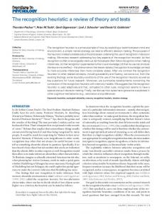
Doublets concentration Clink (pmol/l)
Antigen Capture
+
(Target Protein) Antibody LASER
40
30
20
10
0
Turbidimetric Detection
Lines Formation under Magnetic Field
Fig. 1. The magnetic agglutination assay method. (Step 1) Magnetic particles grafted with polyclonal antibodies (or two different monoclonal antibodies) are mixed with the sample, which contains the antigens. Antigens are first captured by grafted beads. (Step 2) The application of a magnetic field induces a magnetic dipole in each bead, allowing an almost instantaneous formation of chains. Essentially all particles that have captured an antigen will form a link with its neighbor. After typically only a few minutes, the magnetic field is switched off, and because of Brownian motion only doublets that are linked remain. (Step 3) Doublets are detected by a turbidimetric measurement.
recruited within the links in 5 min. In the absence of field, the slope is ⬇10⫺2, which shows that in the case of freely diffusing particles, the agglutination rate is 100 times slower. Therefore, in 5 min without field, less than one-hundredth of the total ovalbumin antigens is recruited within particles links. We have repeated the same experiment at various magnetic field from 0 up to 60 mT. Below the onset of chaining (10 mT), we find almost the same slope as for zero field, whereas above this threshold, we
Turbidity variation (∆OD)
0.8 0.6 0.4 0.2
Fig. 3. Doublets concentration vs. ovalbumin concentration. Circles, B ⫽ 20 mT. In the presence of field, the slope is close to 1, indicating that all antigens participate to doublets. In the absence of field, the slope is ⬇10⫺2 (data not shown). This result suggests a rapid and homogeneous diagnostic test. The limit of detection of this technique can be inferred from Clink at Cova ⫽ 0. Indeed, the zero-dose value of Clink is measuring the noise background due to nonspecific doublets that form during the 5-min assay under field. We get Clink ⫽ 1–3 pmol兾liter as compared with Cp ⫽ 120 pmol兾liter, so approximately one-hundredth of the particles are linked because of nonspecific interactions. This intercept sets a lower limit of detection of ⬇1 pmol兾liter for this approach as compared with 1 nmol兾liter for standard agglutination homogeneous immunoassays.
systematically find the characteristic rapid behavior depicted in Fig. 2. Discussion and Conclusion When ligands and receptors are freely diffusing in solution, the rate of recognition is almost universal because it is diffusion limited and not affected by the molecular details of the association process (11). However, because of the anisotropic nature of the lock-and-key association, this recognition rate naturally depends on both translational and rotational diffusion. By using Schmoluchowski theory, it is possible to estimate the diffusion time needed to form a complex C from spherical species A and B. If we assume that one species concentration is large compared with the other ([A] ⬎⬎ [B]), we get 1兾 ⫽ k[A], where k ⫽ 4DR, D is the relative diffusion coefficient (D ⫽ DA ⫹ DB), and R is the sum of the two spheres’ radii (R ⫽ RA ⫹ RB). However, only a small patch is reactive on both proteins, and in addition species must collide with the proper orientation. As a consequence of these two intrinsic constraints, the Schmoluchowski rate k is slowed down by a factor f, the so-called steric factor, which can be estimated from geometrical assumption (12). If A is uniformly reactive, and B is anisotropic (rotational diffusion coefficient Dr, reactive patch of radius rp with 0 ⫽ rp兾RB), the steric factor reduces to (12)
0.0
f⫽ 0 50 100 150 Ovalbumin concentration
200 (pmol/l)
Fig. 2. Optical density difference measured with a UV兾vis spectrophotometer (Lambda 35; PerkinElmer, Wellesley, MA), before and after the application of the magnetic field B, vs. ovalbumin concentration. Open circles, B ⫽ 20 mT; filled squares, B ⫽ 0 mT. The sample contains ovalbumin in a phosphate buffer (12 mM; pH 7.5) with 0.2% Tween 20. The sample is first incubated for 1 min at 25°C, and then magnetic field is applied for 5 min.
Baudry et al.
兾冑2 ⫹ sin共 0兾2兲cos共 0兾2兲 兾冑 2 ⫹ cot共 0兾2兲
,
[2]
where
⫽ 冑1 ⫹ DrR2兾D.
[3]
We can directly apply this diffusion theory to estimate the duration time of the two distinct steps leading to doublet formation. In our experiment, the first step consists of the PNAS 兩 October 31, 2006 兩 vol. 103 兩 no. 44 兩 16077
APPLIED PHYSICAL SCIENCES
B
20 40 60 Ovalbumin concentration Cova (pmol/l)
BIOPHYSICS
0
capture of the antigen on the beads. For free antigen in solution (RB ⫽ 5 nm, rp ⫽ 0.25 nm) and homogeneously reactive spheres (RA ⫽ 100 nm, concentration 10⫺10 M), k ⫽ 3 ⫻ 1010 M⫺1䡠s⫺1, and f ⫽ 0.2, which leads to 1 of ⬇2 s. The second step consists of bridging one colloidal particle, which has already captured one antigen, with another particle. In the absence of field, only free diffusion is involved, and 2 is estimated to be ⬇1 min (homogeneously reactive particles at concentration 10⫺10 M with radius RB ⫽ 100 nm and rp ⫽ 0.25 nm, so k ⫽ 6 ⫻ 109 M⫺1䡠s⫺1 and f ⫽ 3 ⫻ 10⫺2). Note that these two predicted time scales are certainly underestimated and should be considered as upper limits, because the assumption of uniformly reactive spheres is too optimistic. We have indeed measured 30 antibodies per particle, which is far below the estimated monolayer threshold of ⬇1,000 per particle (‡, 13). In our experiment, the first step requires an incubation time of ⬍1 min, in good agreement with the previous diffusion-based estimation. By contrast, in the absence of field, the second step would require ⬎8 h, as deduced from the slope of Fig. 3. We can therefore conclude that the previous diffusion-based model does not hold once grafted biomolecules are concerned. Some repulsive colloidal forces could be responsible for this slowdown; however, because we do not find any role of the field strength above the onset of chaining, we can rule out this contribution as a major effect. We believe that the immobilization of the biomolecules onto colloidal surfaces causes restrictions to their dynamics, which drops further down the steric factor as compared with a nongrafted case. To improve the rate of colloidal bridging, we must therefore augment the attempt frequency. One simple way consists of increasing the particles’ colliding frequency by locally increasing the particle concentration; this result is readily obtained from chaining magnetic colloids with magnetic field. As deduced from our experiment, the measured
agglutination rate is increased by a factor of 100 above the onset of chaining. This effect shifts the detection threshold of the latex agglutination immunoassay homogeneous bioassay down to 1 pmol兾liter, making these simple methods as sensitive as heterogeneous approaches. However, although confinement has significantly increased the agglutination rate, it also raises the delicate question of the coupling between translation and rotation in such confined geometry and therefore the ultimate limitation of this approach. This letter gives the basis of a previously unreported homogeneous assay for protein detection with high sensitivity. Indeed, because magnetic colloids can immediately self assemble under field, they are very efficient carriers, considerably reducing the diffusion time; most importantly, magnetic forces can hold colloids and reactants in vicinity, which increases the sampling frequency, allowing a more rapid reaction. These results also demonstrate the relatively slower process of recognition when both ligand and receptor are grafted onto colloidal surfaces. Methods Turbidity measurement relies on the fact that a doublet scatters more light than two separate beads. If ␣ is the ratio of the total scattered light intensity by one doublet to the total scattered light intensity by one particle, the optical density difference before and after the magnetic field is applied will be ODa ⫺ ODb ⫽ ha2Qscat(␣ ⫺ 2)n2兾2.3, where h is the optical path length, a is the particle radius, Qscat is the scattering efficiency of one particle, and n2 is the density number of doublets. Qscat ⫽ 0.23 and ␣ ⫽ 2.45 are numerically computed by using Mie Theory (14) using freely available software (ftp:兾兾ftp.eng.auburn.edu兾 pub兾dmckwski兾scatcodes兾index.html) with the measured particle optical index (1.78 ⫹ 0.02i at 633 nm) and the measured particle diameter (200 nm).
as shown by Berg and Purcell (13), a sphere partially covered by receptors behaves almost like a uniformly reactive sphere. Indeed, before diffusion moves the reactants out, they try many orientations during repeated encounters. The same argument explains why the steric factor f is higher than a purely geometrical estimation.
We thank P. Fannin and A. Franceschini for fruitful discussions and I. Genois for technical help. This work was supported by Stago Diagnostica Company and the Bioengineering Program of the French Ministry of Research.
1. Price CP, Newman DJ (1997) Principles and Practice of Immunoassay (MacMillan Reference, London). 2. Singer JM, Plotz CM (1956) Am J Med 21:888–892. 3. Van Weemen BK, Schuurs AHW (1971) FEBS Lett 15:232–236. 4. Lee GU, Metzger S, Natesan M, Yanavich C, Dufrene YF (2000) Anal Biochem 287:261–271. 5. Thomas NE, Coakley WT (1996) Ultrasound Med Biol 22:1277–1284. 6. Ullman E, Kirakossian H, Singh S, Wu Z, Irvin B, Pease J, Switchenko A, Irvine J, Dafforn A, Skold C, Wagner D (1994) Proc Natl Acad Sci USA 91:5426–5430.
7. Benecky MJ, Post DR, Schmitt SM, Kochar MS (1997) Clin Chem 43:1764– 1770. 8. Nam JM, Thaxton CS, Mirkin CA (2003) Science 301:1884–1886. 9. Calderon FL, Stora T, Monval OM, Poulin P, Bibette J (1994) Phys Rev Lett 72:2959–2962. 10. Promislow JHE, Gast AP, Fermigier M (1995) J Chem Phys 102:5492–5498. 11. Northrup SH, Erickson HP (1992) Proc Natl Acad Sci USA 89:3338–3342. 12. Berg OG (1985) Biophys J 47:1–14. 13. Berg HC, Purcell EM (1977) Biophys J 20:193–219. 14. Mackowski DW, Mishchenko MI (1996) J Opt Soc Am A 13:2266–2278.
‡Nevertheless,
16078 兩 www.pnas.org兾cgi兾doi兾10.1073兾pnas.0607991103
Baudry et al.