BAF300 freeze-etch apparatus equipped with a revolving specimen holder. ... for the construction of the revolving specimen stage for our freeze-etching ap-.
The EMBO Joumnal Vol.1 No.5 pp.541-547, 1982
Dimeric arrangement and structure of the membrane-bound acetycholine receptor studied by electron microscopy Horst-Peter Zingsheim*, Dorothea-Charlotte Neugebauerl, Joachim Frank2, Wolfgang Hiinicke, and Francisco Jose Barrantes Max-Planck-Institut fUir Biophysikalische Chenie, (Karl-FriedrichBonhoeffer-Institut), D-3400 Gwttingen-Nikolausberg, FRG Communicated by K. Weber Received on 29 April 1982
The acetylcholine receptor protein (AChR) from the electric organ of Torpedo marmorata is studied in its membranebound form by electron microscopy and single-particle image averaging. About half the molecule protrudes from the membrane surface by S5 nm. The low-resolution 3-D structure of this hydrated portion, including its handedness, can be deduced from averaged axial and lateral projections and from freeze-etched membrane surfaces. In native membrane fragments, a dimeric form of the AChR is observed and the relative orientation of the AChR monomers within the dimer is established. The dimers disappear upon disulfide reduction of the membrane preparations, whereas the average axial projections of the AChR monomer remain unaffected. Since the existence of disulfide bonds linking AChR monomers between their respective b-subunits is well documented, the approximate position of the b-subunit within the low-resolution structure of the AChR molecule can be deduced from the structure of the dimers. Key words: cholinergic receptor/post-synaptic membrane/ membrane proteins/Torpedo marmorata/image processing Introduction The acetylcholine receptor (AChR) from Torpedo electric organ is a membrane protein with an apparent mol. wt. of 250 000, composed of four types of subunits (ca, ,, -y, 6; apparent mol. wt. 40 000, 50 000, 60 000, 65 000, respectively; composition 2a, (3, -y, 6). The a-subunits carry the binding sites for a-toxins, agonists, nicotinic affinity labels, and inhibitors (for reviews, see Heidmann and Changeux, 1978; Karlin, 1980; Hucho, 1981). Predominantly dimeric forms of AChR have been isolated from unmodified membranes. Dimers arise by disulfide-linkage primarily via the respective b-subunits of the monomers (Chang and Bock, 1977; Hamilton et al., 1977; Sobel et al., 1977; Suarez-Isla and Hucho, 1977). Disulfide reduction converts all oligomeric forms to monomers, a change that is accompanied by cleavage of the 6-6-bonds, yielding single 6-subunits (Chang and Bock, 1977; Hamilton et al., 1977, 1979; Karlin, 1980). The functional significance of the dimers remains obscure. No differences in pharmacological properties and ion transport have been detected between monomers and dimers in reconstituted membranes (Anholt et al., 1980; Boheim et al., 1981). Since the AChR arrangement in sub-synaptic membrane regions is remarkably stable (reviews by Bar-
'Present address: Zoologisches Institut der Universitat Moinster, Hufferstr. 1, D-4400 Munster, FRG. 2Division of Laboratories and Research, New York State Department of Health, Albany, NY 12201, USA. *To whom reprint requests should be sent. © IRL Press Limited, Oxford,
England. 0261-4189/82/0105-0541$2.00/0.
rantes, 1979; Fambrough, 1979), one might conjecture that this is partiaUly mediated by covalent intermolecular linkages. Other factors likely to contribute to this stability include the v-peptide, a non-receptor peptide of mol. wt. -43 000 (Rousselet et al, 1979; Barrantes et al., 1980; Lo et al., 1980). Here we report structural data on the arrangement of AChR in the membrane. The study is based on electron microscopy and single-particle image averaging (Frank et al., 1978; Zingsheim et al., 1980a, 1980b). Images of individual particles are aligned by correlation techniques (Frank, 1980) to maximize the overlap of reproducible, structure-related features. Aligned images are averaged to enhance these features and to suppress random variations. This method reveals the existence of membrane-bound dimers, the orientation of the AChR monomer within the dimer, and the approximate location of the b-subunit in the AChR monomer.
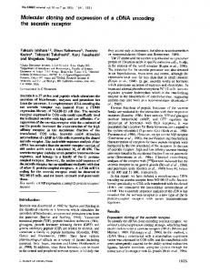
Results Identification ofAChR dimers in the intact membrane Single-particle image averaging was applied to electron micrographs of negatively-stained AChR-rich membrane vesicles (see Materials and methods). The neighbourhoods of individual AChR molecules were initially screened off with circular masks of 6 nm diameter in order to prevent them from affecting the rotation and translation search on which particle alignment is based. However, these regions were aligned and included in the final averages. Although the AChR particles in unmodified membranes appear randomly packed to the eye (Figure la), image averaging reveals that nearest-neighbour particles are associated pair-wise. This is evident in Figure lb which represents contours of equal optical density within an averaged field of 25.6 x 25.6 nm. In this image, fine detail was suppressed by lowpass filtering to a resolution of 6.4 nm, while the salient features, i.e., the gross distribution of stain-excluding mass, have been retained. Adjacent to the central density minimum (indicating stain exclusion) there is a second minimum generated by the superposition of neighbouring AChR particles. These particles, which originally fell outside the circular masks, are located at defined positions relative to the central ones. Both minima are of similar size and contrast. Their separation is 9 nm, corresponding to the nearest-neighbour distance between AChR molecules in native membranes (Barrantes et al., 1980). Upon disulfide reduction of the membranes (using 10 mM dithiothreitol (DTT), see Materials and methods) the second minimum disappears (Figure le). Density contour maps of averaged fields measuring 12.8 x 12.8 nm are shown in Figure lc,f. These correspond to the central portions of the 25.6 x 25.6 nm fields (Figure lb,e), but have been low-pass filtered to a resolution of 1.8 nm. They represent axial views of the AChR structure projected along a direction perpendicular to the membrane plane. Their characteristic shape, resembling a horseshoe, remains essentially unaffected by disulfide reduction. The choice of the indicated resolutions for low-pass filtering was largely governed by an angular uncertainty of about 150 in the rotational alignment step. As a consequence of this error, the superimposed features are progressively blurred -
-
541
H.-P.Zingsheim et al.
Fig. 1. Electron microscopy and image averaging show the dimer arrangement and structure of the membrane-bound AChR. The top row (a,b,c) relates to non-reduced (native) membranes. (a) is a micrograph of a negatively-stained membrane sheet at a magnification of 500 000 x. Dark regions are due to stain; stain-excluding regions are bright. AChR particles are visualized as 'rosettes' of - 7 nm diameter with a densely stained central pit. Image averaging was applied to small fields centered around individual partides. Only these central partides, with their surroundings screened off by a circular mask of 6 nm diameter wee used to find the relative orientations between the partides. Each individual image is represented by optical densities dk on a square grid of m2 image elements (ik = 1.in). In the average image the optical density djk in any image element is the mean of N realizations dlk(n = 1,.. N
dik = (1/N)Z dik()
n= 1 In (b) an average over N = 20 individual images is plotted as a contour map of equal values of dik, but after low-pass filtering to a resolution of 6.4 nm, thus suppressing all fine detail. The width of the field is 25.6 x 25.6 nm. Relative density minima (stain-exclusion) are marked by -; density maxima (stain) are marked by +. Shaded areas indicate the most significant density increments in the stain-excluding regions. The presence of the second density minimum adjacent to the central one indicates the association of the AChR partides into dimers. The dashed square (12.8 x 12.8 nm) in the center is presented as a density contour plot in (c), but at a resolution of 1.8 nm. This reveals the typical horseshoe-shaped structure of membrane-bound AChR in axial projection. The bottom row (d,e,i) relates to membranes subjected to disulfide reduction using 10 mM DTT. (d) is a micrograph (magnification 500 000 x). (e) represents a 25.6 x 25.6 nm average over 20 individual images at 6.4 nm resolution. It shows that the dimer arrangement disappears upon disulfide reduction. The 12.8 x 12.8 nm field at 1.8 nm resolution (f) shows that disulfide reduction does not significantly affect the typical horseshoe shape.
as their distance from the center increases. The density minima in the stain-excluding region of the central particle are located at a radius of 2.2 nm (see also Figure lc,f). Here, the azimuthal superposition error is z0.6 nm, compatible with 1.8 nm resolution (Figure lc,f). However, at a radius of 9 nm the error is 4 2.2 nm, precluding the identification of the stain-filled central pit in the second particle. Therefore the 25.6 x 25.6 nm fields (Figure lb,e) are presented at a resolution of 6.4 nm. The averaged neighbourhood features in non-reduced membranes vary somewhat between preparations, but the second density minimum is consistendy observed in the same
542
position. Analysis of non-reduced membrane samples after removal of the v-peptide by alkaline washing (Neubig et al., 1979; Barrantes et al., 1980) also reveals a nearest-neighbour peak in the same position (data not shown). Surface relief of the membrane-bound A ChR particle The surface ['ES' surfaces, Branton et al. (1975) ] of AChR-rich membranes can be visualized after freeze-etching (Cartaud et al., 1978, 1981; Heuser and Salpeter, 1979). However, the anisotropy of unidirectional shadowing, the random azimuthal orientation of the AChR particles in the membrane, and the variation of the local shadowing angle on the curved membrane faces preclude single-particle averag-
=^:3.Str>,-i ^Ss.tY'6:^-.so"t riC-sx*;s.,.y^_vet~enwf-oS. S*-:
*.
Arrangement and structure of AChIR in the membrane
..
...
...P.E.
Wfx~A a A,:
90Vo of their AChR exposed right-side out (see Materials and methods). Average lateral view of the AChR particle In order to ascertain that the metal deposited by rotary shadowing is indeed piled up on a surface relief structure, it is possible to draw on independent information on the 3-D structure of the AChR. This is available from lateral views of the AChR protein, observed along the edges of negatively stained AChR-rich membranes (Cartaud et al., 1978; Schiebler and Hucho, 1978; Klymkowsky and Stroud, 1979; Figure 3a,b). Alignment (omitting the '1 800-decision', see Materials and methods) and averaging of such views leads to the result shown in Figure 3c. This may be symmetrized with respect to an axis perpendicular to the membrane surface (Figure 3d) because information on the azimuthal orientation of the particles is lacking. The symmetrized result can be 544
regarded as a cylindrically-averaged projection. It shows that the protruding stain-excluding mass is 7 nm wide and extends -4.5 nm from the membrane, thus accommodating about half of the total mass of the monomeric AChR protein. It is distributed around a central cavity which is --4.5 nm deep and 2.5 nm wide. -
-
Discussion The use of membrane specimens provides a frame of reference not found in studies of detergent-solubilized material. Complications due to the action of detergents or due to uncertainties with respect to the direction of view (cf., Cartaud et al., 1981; Wise et aL, 1981; Holtzman et al., 1982) are avoided. An aspect of general interest is that a study of intermolecular relationships at moderate resolutions was possible by non-crystallographic image averaging without using any a priori structural information on the oligomeric organization of the membrane-bound AChR. Obviously, the reliability of the image averaging procedure, in particular of the rotational alignment step, is a crucial prerequisite. Three methods were used to ascertain the validity of this procedure as applied to images of membranebound AChR: a comparison of averages obtained from independent sets of images; a non-parametric statistical test; and the behaviour of the optical density variance of the averages as a function of the total number of images averaged. For a quantitative comparison of two independently obtained averages, the phase residual of their Fourier transforms (Frank et al., 1981b) was computed after masking off the neighbourhood of the particle. The differential phase residual for spacings above 1.5 nm remains below 450 if averages over --20 particles are compared. The non-crystallographic test developed on the basis of non-parametric statistical methods established that significant azimuthal density variations (at the 95%7o level) within distances