■ REVIEW
Active Vision in Parietal and Extrastriate Cortex ELISHA P. MERRIAM and CAROL L. COLBY Department of Neuroscience and Center for the Neural Basis of Cognition University of Pittsburgh
Vision is an active process. We do not see the world directly; rather, we construct a representation of it from sensory inputs in combination with internal, nonvisual signals. In the case of spatial perception, our representation of the visual scene must take into account our own movements. This allows us to perceive the world as stationary despite the constant eye movements that produce new images on the retina. How is this perceptual stability achieved? Our central hypothesis is that a corollary discharge of the eye movement command updates, or remaps, an internal representation when the eyes move. In support of this hypothesis, the authors review evidence that parietal cortex and extrastriate visual areas in both monkeys and humans participate in spatial updating. These findings shed new light on the neural circuitry involved in producing a stable and coherent perception of visual space. NEUROSCIENTIST 11(5):484–493, 2005. DOI: 10.1177/1073858405276871 KEY WORDS Spatial updating, Parietal cortex, Extrastriate cortex fMRI, Visual perception
We think of perception as leading to action. Visual signals arriving in cortex are analyzed and processed through multiple stages, objects are recognized and locations identified, a decision of some kind is made, and an action is generated. This process is conceived of as information moving forward through a system in which motor output represents the end of the process. Equally important, however, may be the reverse process by which information about motor output is fed back to earlier stages, allowing action to influence perception. The idea that action has an impact on perception is an old one. More than a century ago, Helmholtz (1866/1924) proposed that the reason the world appears to stay still when the eyes move is because the “effort of will” involved in the generation of a saccade simultaneously adjusts perception to take that eye movement into account. A simple experiment convinces most observers that Helmholtz’s account is correct. When the retina is displaced by pressing on the eye, the world does seem to move. In contrast, we are oblivious to the changes in the retinal image that occur with each glance. This perceptual stability has long been understood to reflect the fact that what we see is not a direct impression of the external world but a construction, or internal representation, of it. It is this internal representation that is adjusted, or updated, in conjunction with eye movements. Considerable interest has focused in recent years on brain mechanisms of spatial representation, particularly in parietal cortex. In this review, we focus on studies of
updating spatial representations in humans and monkeys. Monkey Lateral Intraparietal Area Neural activity in the lateral intraparietal area (LIP) reflects multiple aspects of the monkey’s environment and behavior (Andersen and others 1997; Colby and Goldberg 1999). Nearly all neurons in area LIP respond to visual stimuli presented in the receptive field (RF) while the monkey fixates on a central point. About half the population also exhibits saccade-related activity, which occurs before, during, and/or after a saccade directed toward the RF. Many other factors, including anticipation, memory, orbital position, decision processes, motor planning, and attention, also have a significant impact on LIP neuron activity (Gnadt and Mays 1995; Snyder and others 2000; Shadlen and Newsome 2001; Toth and Assad 2002; Glimcher 2003). How are we to understand this complex range of activities? The simplest interpretation is that LIP neurons encode salient attended locations. When an LIP neuron fires, it means that an event occurred, or is expected to occur, at a particular location. This event can be the sudden appearance of a stimulus in the RF, or generating a saccade toward it, or directing attention to that location, or remembering that a stimulus appeared there. What is important is that the location has been made salient by its actual or potential relevance for the animal’s behavior. This interpretation raises the issue of how salient spatial locations are represented in area LIP.
This work was supported by NSF IGERT DGE-9987588, NIH NS047493, and NIH EY12032.
Spatial Updating in Area LIP
Address correspondence to: Elisha P. Merriam, Department of Neuroscience, 446 Crawford Hall, University of Pittsburgh, Pittsburgh, PA 15213 (e-mail:
[email protected]).
We have investigated what happens to visual information in the lateral intraparietal area in conjunction with eye
484
THE NEUROSCIENTIST Copyright © 2005 Sage Publications ISSN 1073-8584
Active Vision in Parietal and Extrastriate Cortex
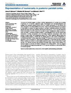
Fig. 1. Remapping of a stimulus trace. A, Visual response. During fixation, the neuron responds to a stimulus in the receptive field (RF). B, Remapped response. During fixation, a stimulus is presented briefly (50 ms) at a location outside the RF. At the same time, a saccade target appears (FP2). The monkey makes a saccade to the target. The saccade brings the RF to the previously stimulated location and the neuron fires even though the stimulus is no longer present. C, Stimulus-only control. The neuron does not respond to a stimulus presented outside the RF during fixation. D, Saccade-only control. The neuron does not respond to a saccade toward FP2 in the absence of a stimulus. Stim = stimulus; H = horizontal; V = vertical. Adapted from Duhamel and others (1992a).
movements (Fig. 1). We discovered that when the eyes move so that the RF of an LIP neuron lands on a recently stimulated screen location, the neuron fires as though a stimulus were still present, even though the screen is blank (Duhamel and others 1992a; Colby and others 1995). The stimulus trace response cannot be accounted for by other visual or motor factors. Control tasks indicate that the simple onset of the stimulus outside the RF does not drive the cell (Fig. 1C and D). Likewise, the saccade to the new fixation point does not by itself activate the cell. Both the saccade and the stimulus are necessary for producing the stimulus trace response. The critical factor is the position of the stimulus relative to the final eye position. The stimulus must occur at a location that is the correct distance and direction from the final position of the fovea, that is, it must correspond to the retinotopic location of the RF following the eye movement. This response to the memory trace of a previous stimulus indicates that LIP neurons participate in updating an internal representation of visual space. We call this process “remapping” to emphasize that visual information is being shifted from the coordinates of the initial eye position to the coordinates of the next eye position. Remapping may thus contribute to maintaining the spatial alignment between the external world and its internal representation. In area LIP, neurons have retinotopic RFs that move with the eye. The conclusion from our remapping studies, however, is that LIP neurons contribute to an eyecentered, and not just a retinotopic, representation of space. The distinction is this. A simple retinotopic repre-
Volume 11, Number 5, 2005
sentation would reflect only the retinal location at which the stimulus originally appeared. In an eye-centered representation, the location of the stimulus is still represented with respect to its distance and direction from the fovea, but this representation is updated when the eyes move, so that stimuli are always represented with respect to the current position of the fovea. The effect of this updating is to produce a representation in which the memory trace of a stimulus is shifted to activate neurons whose retinotopic RFs currently contain the stimulated location. Remapping in the Human Parietal Cortex Humans, like monkeys, must maintain an accurate and stable perception of the world when they move their eyes. Several lines of evidence suggest that humans and monkeys use the same updating mechanisms for generating this stable percept. Behavioral studies have demonstrated that both species have similar abilities in eye movement tasks that require updating (Hallett and Lightstone 1976; Baizer and Bender 1989). Furthermore, neuropsychological and lesion evidence suggest that the parietal lobe is crucial for these abilities in both species. For example, parietal lobe lesions interfere with performance on the double-step saccade task, which is thought to depend on updated spatial information (Duhamel and others 1992b; Heide and others 1995; Li and Andersen 2001). These lesion findings imply that remapping also occurs in the human parietal cortex. We hypothesized that we could visualize remapping in humans using functional magnetic resonance imaging (fMRI) (Merriam and others 2003). In this section, we
THE NEUROSCIENTIST
485
Fig. 2. Human functional magnetic resonance imaging (fMRI) remapping paradigm and predicted results. A, The stimulus appears in the left visual field (LVF) for 2 s, activating right hemisphere occipital and parietal cortex (blue circle). After two sends, the stimulus disappears and a tone cues the subject to make a leftward eye movement. This saccade brings the screen location of the now-extinguished stimulus (dotted circle) into the right visual field (RVF). We predicted that remapping of the stimulus trace would cause activation to shift from the right to the left hemisphere (red hatched circle). B, Time course of expected activation. The shaded region in each panel indicates the time that the stimulus is on, and the vertical line at 2 s indicates the time of the auditory cue to make an eye movement. Activation in the right hemisphere, due to the stimulus, was expected to follow the standard hemodynamic time course (blue curve). Activation in the left hemisphere, due to the remapped stimulus trace, was expected to occur with a similar time course but shifted by 2 s because the cue to make an eye movement occurs 2 s after stimulus onset. We also expected the remapped response to be smaller in amplitude than the visual response. Adapted from Merriam and others (2003). Reprinted from Neuron, vol. 39, Merriam and others, “Spatial Updating in Human Parietal Cortex,” pages 361–73, 2003, with permission from Elsevier.
review functional imaging evidence that the human parietal cortex is involved in remapping. To observe remapping in humans, we designed an imaging experiment that followed the task parameters of the monkey experiments as closely as possible. The updating task used in the imaging experiment is described in Figure 2. Each trial begins when the subject fixates one of two crosses on the screen. The subject’s task is to maintain fixation on the first cross until a tone occurs, which is the instruction to make a saccade to the second cross. On some trials, a stimulus appears at the center of the screen, far from the location of gaze. The stimulus flickers at high contrast in an otherwise dark visual environment, making it a highly salient stimulus. However, as in the single-unit experiments, the stimulus is totally irrelevant to performance of the task. The tone that cues the saccade occurs just as the stimulus disappears. Thus, only a memory trace of the stimulus can be remapped. In our experiment, the saccade target is positioned so that an eye movement to it brings the stimulus
486
THE NEUROSCIENTIST
location into the opposite visual field (Fig. 2A). Gaze is initially directed to the right fixation cross, and the stimulus appears in the left visual field. Concurrent with stimulus offset, a tone instructs the subjects to move their eyes to the left fixation cross. As a result of this eye movement, the stimulus location is now in the right visual field. The predictions from this experiment are straightforward. First, the stimulus should activate visually responsive cortical areas in the contralateral hemisphere. Lowlevel visual areas, such as V1 and V2, should become active because the stimulus has high contrast. Extrastriate and parietal areas should also become active because the sudden onset and high-frequency flicker render the stimulus salient. Second, following the eye movement, the stimulus trace should activate visual areas ipsilateral to the stimulus. We use the term remapped response to describe this ipsilateral activation in order to emphasize that it is not driven by direct visual stimulation. Third, this remapped response should be larger than the responses elicited by either an ipsilateral stimulus alone or an ipsiversive saccade alone. Both ipsilateral stimuli and ipsiversive saccades may induce some activation; receptive fields in extrastriate and parietal cortices can be large, and they sometimes encroach a few degrees into the ipsilateral side of space. There may also be some activation associated with the saccade because of retinal stimulation during the movement itself. Nonetheless, these sources of activity should be smaller than the remapped response. Fourth, remapped responses should have a characteristic shape and time course that distinguish them from visual responses. Specifically, remapped responses should occur later in time than visual responses, because the cue that triggers the eye movement occurs after the stimulus has been on the screen for two seconds. Remapped responses should also be lower in amplitude than visual responses; at the single neuron level, responses to memory traces are about half as large as responses to actual stimuli. These four predictions were confirmed in our human imaging data. Our main result is illustrated in Figure 3. A left visual field, stimulus strongly activated the right hemisphere, as shown in Figure 3C. This right hemisphere activation reflects visual activity that is directly driven by the stimulus. We also observed activation in the ipsilateral parietal lobe (Fig. 3C, left hemisphere). We interpret this ipsilateral activation as a response to the remapped trace of the stimulus. The shape of this ipsilateral activation suggests that it is indeed related to remapping. As described above, the remapped activation should occur later than the visual activation. This pattern of activity is present in the responses from both hemispheres shown in Figure 4, and was observed in a population of 16 hemispheres (Merriam and others 2003). In addition to occurring later, the remapped activation should also be smaller in amplitude than the visual activation. This pattern is visible in one of the two hemispheres shown in Figure 4 and is present in the majority of cases reported in Merriam and others (2003).
Active Vision in Parietal and Extrastriate Cortex
Comparison of Monkey and Human Remapping Two main features of the fMRI task are notably different from the monkey version, described above in Figure 1. First, the fMRI task is several orders of magnitude slower. In the original single-step task in monkey, a stimulus flashes briefly outside of the RF of the neuron being recorded. The stimulus appears for only 50 ms, just after the saccade has been cued, but before the eyes actually begin to move (Fig. 1). Because of the high temporal fidelity of single-unit recordings, Duhamel and others (1992a) were able to determine the precise onset time of the remapped response. In the fMRI version of the task, the stimulus appear and remain on the screen for two seconds prior to the eye movement. This allowed for a clear dissociation between visual and remapped activity: visual activity began two seconds prior to the onset of remapped activity. These parameters were chosen to accommodate the relatively poor temporal resolution of fMRI. Second, the geometry of the task differed from the original version. In the single-step task used in the physiology experiments, a saccade moves the RF onto a recently stimulated screen location. The single-unit experiments thus require reasonably detailed information regarding the spatial extent of visual RFs; the magnitude and direction of the eye movement are chosen depending on each neuron’s particular RF size and location. However, the measure of neural activity reflected in fMRI data is at a much coarser spatial scale, and the concept of a visual “receptive field” does not apply in the context of fMRI. To overcome this limitation, we arranged the location of the stimulus and eye movement direction such that the visual and remapped responses were located in opposite hemispheres. However, despite these differences between the single-unit and fMRI versions of the task, the essential finding is the same in both studies. Area LIP in Humans
Fig. 3. Region of interest in parietal cortex. A, Posterior view of both hemispheres of a single subject rendered at the outermost layer of gray matter. The regions of interest (ROIs) are shown in blue. B, Partially unfolded view of the same two hemispheres. Blue shading indicates the location of the ROI. Shades of gray indicate the curvature of the cortical surface: dark gray indicates concave areas, and light gray indicates convex areas. C, Activation from a single subject on updating trials in which a left visual field stimulus was followed by a leftward saccade. This condition elicited activation in contralateral (right) hemisphere occipital and parietal areas, as expected. Activation was also observed in the ipsilateral (left) parietal lobe, indicating that the visually evoked activation was remapped. Adapted from Merriam and others (2003). Reprinted from Neuron, vol. 39, Merriam and others, “Spatial Updating in Human Parietal Cortex,” pages 361–73, 2003, with permission from Elsevier.
Volume 11, Number 5, 2005
Many previous studies have demonstrated that contralateral visual stimuli activate the striate and extrastriate visual cortex (Sereno and others 1995; DeYoe and others 1996; Engel and others 1997). A similar contralateral bias exists in parietal cortex as well (Sereno and others 2001). Consistent with these data, we found strong and extensive activation contralateral to the stimulus location in the single-step task. We also discovered that the conjunction of a stimulus and a saccade can produce strong ipsilateral activation and that this activation bears many similarities to neuronal remapping data. Our findings indicate a broad equivalence of function between monkey and human parietal cortex. Whether there are human equivalents of the physiologically defined parietal areas is currently a matter of much interest. Sereno and others (2001) identified a topographic area in a medial side branch of the human intraparietal sulcus and suggested that this area could be a human “homologue” of the
THE NEUROSCIENTIST
487
Fig. 4. Visual and remapped responses from the left hemisphere of a single subject. A, Time course of activation evoked by the visual and remapped responses from parietal cortex. The Magnetic Resonance (MR) time course over the 15-s task epoch represents the average of 72 trials. The shaded gray bar indicates the time when the stimulus was present, and the vertical line at 2 s shows the time of the auditory cue to make a saccade. The remapped response (red line) occurs later and is smaller than the visual response (blue line). B, BOLD-image raster plots of the visual responses from the same hemispheres for 72 successive trials. Activation on individual trials is plotted along the y-axis, with percent signal change represented in pseudocolor plotted over time (x-axis). C, BOLD-image raster plots of the remapped responses for 72 successive trials. D, Eye position recorded in 36 trials of the same task performed outside the scanner. Adapted from Merriam and others (2003). Reprinted from Neuron, vol. 39, Merriam and others, “Spatial Updating in Human Parietal Cortex,” pages 361–73, 2003, with permission from Elsevier.
monkey area LIP. In that experiment, subjects made saccades to a series of targets located in sequence around the visual field. The targets were presented only briefly, and subjects had to remember their locations for several seconds before each eye movement. Sereno and others observed a traveling wave of activity across a region of the posterior parietal lobe as subjects remembered consecutive target locations, implying that neurons in this area of the human brain are topographically organized for delayed saccades to remembered locations. The existence of a topographic map is strong evidence that an activated region corresponds to a discrete cortical area.
488
THE NEUROSCIENTIST
However, further studies are needed to determine the degree to which this particular area displays physiological response properties similar to monkey area LIP. Impact of Action on Representation in the Extrastriate Visual Cortex Extrastriate visual cortex contains a hierarchy of visual areas, connected by feed-forward and feed-back projections (Maunsell and Van Essen 1983). This hierarchy, identified initially on the basis of laminar patterns of connectivity, also reflects the increasing complexity of
Active Vision in Parietal and Extrastriate Cortex
response properties as visual signals are transformed beyond V1. Substantial evidence has accumulated for the impact of top-down processes related to attention and working memory on neural activity at various levels of the hierarchy (Robinson and others 1978; Moran and Desimone 1985; Mountcastle and others 1987; Motter 1993, 1994; Steinmetz and others 1994; McAdams and Maunsell 1999; Reynolds and others 1999; Seidemann and Newsome 1999; Recanzone and Wurtz 2000; Pasternak and Zaksas 2003). Much less is known about the impact of motor action on visual processing in extrastriate areas. We have investigated the extent to which extrastriate visual cortex participates in remapping. We began with area V3A because it is the extrastriate area most closely connected with LIP. As a first step, we asked whether area V3A neurons exhibit extraretinal signals that are so prominent in area LIP. We recorded from area V3A while monkeys performed standard fixation and memory-guided saccade tasks. We found that activity in V3A is subject to significant modulation by extraretinal factors including attention, anticipation, memory, and saccadic eye movements (Nakamura and Colby 2000). We concluded that area V3A is not purely visual in function but also participates in cognitive functions, and could contribute to the updating of spatial representations. Neurons in Extrastriate Cortex Remap Stimulus Traces Neurons in LIP, Frontal Eye Field (FEF), and Superior Colliculus (SC) update the trace of a visual stimulus in conjunction with eye movements. We asked whether remapping is limited to these oculomotor and quasi-oculomotor areas, or whether it can be observed in areas thought to be primarily visual in function. We began in area V3A with our standard remapping task. We found that more than half of V3A neurons (52%) responded in this task, as illustrated in Figure 5. Remapped responses in area V3A are as robust as those in area LIP, and V3A neurons were not active in the control conditions. We concluded that remapping is not limited to neurons in oculomotor and closely allied brain regions but rather is present as well in dorsal stream visual areas. Receptive Fields in Extrastriate Cortex Are Dynamic What happens to the RF of a neuron during remapping? We asked whether V3A neurons remap stimulus traces by shifting the RF or whether the RF expands at the time of a saccade. We did this by testing the neurons’ sensitivity at two locations, the old (initial) receptive field and the new (future) receptive field. We presented the stimuli at four different times relative to the saccade in order to understand how the RF changes over time. All eight conditions were randomly interleaved. We found that neural responses fell along a continuum. Some, like the neuron illustrated in Figure 6, appear to shift the location of the RF around the time of an intended saccade: they
Volume 11, Number 5, 2005
simultaneously become less responsive at the initial location and much more responsive at the future location of the RF. In contrast, other neurons appear to undergo a momentary expansion of the RF immediately before a saccade. The neuron illustrated in Figure 7 exhibited predictive remapping: it began to respond even before the onset of the saccade that would move the RF onto the stimulated screen location (panel B). When tested in all eight conditions, the neuron exhibited a dual responsiveness (Fig. 8). In conditions 1 and 2, the neuron responded to a stimulus presented at either the old or the new RF. This dual responsiveness continued until saccade onset, when it ceased abruptly (time condition 3). These data indicate that for some cells there is a temporary expansion of the effective RF around the time of a saccade. Many previous experiments have demonstrated that RFs are dynamic. The present results indicate that intended motor action is one of the factors that contributes to this plasticity. Neurons in V3 and V2, But Not V1, Remap Stimulus Traces Finally, we asked whether neurons at even earlier stages of the visual system hierarchy would remap stimulus traces. Using the same tasks and conditions, we tested neurons in the V3, V2, and in striate cortex. We found that many neurons in area V3 respond in the single-step task, but that the proportion drops off rapidly in V2. In striate cortex, only 1 neuron out of 64 tested showed evidence of remapping (Fig. 9). Two other trends are clear. First, the proportion of neurons that remap predictively decreased markedly at lower levels of the hierarchy. Second, the mean latency of the remapped response relative to saccade onset was much longer at lower levels. All of these findings suggest that earlier stages of the visual system are connectionally or computationally further from the source of the central signal that drives remapping. The next critical question for future research is whether remapping is in fact an entirely top-down process, in which the computation is carried out in LIP (or elsewhere), or whether it proceeds in parallel at multiple levels of the visual system. Active Vision in Parietal and Extrastriate Cortex In the dorsal stream, we found that there is robust remapping in single neurons in areas V3A, V3, and V2 (Nakamura and Colby 2002). This is significant because it provides support for the idea that extrastriate cortex is not simply engaged in passive elaboration of retinal signals, but rather that an active process is guiding the acquisition and maintenance of stimulus representations in extrastriate cortex (Maunsell 1995). The function of such active processes in extrastriate cortex may be to narrow the task of stimulus representation to only those locations or stimulus features that are currently of importance for the organism. Psychophysical work on integration of information across saccades indicates that
THE NEUROSCIENTIST
489
Fig. 5. Remapping in extrastriate area V3A. The cartoons show the location of stimuli on the screen. Time lines show horizontal (H) and vertical (V) eye position (calibration bar, 5 deg) and the timing of stimulus events. Rasters from 10 correct trials are aligned on the specified event and summed to generate histograms. A, Fixation task: a 3-deg bar at the optimal orientation is flashed in the receptive field (RF) while the monkey fixates. B, Single-step task: while the monkey fixates FP1, the stimulus is flashed outside the RF for 50 ms; it is extinguished before the saccade to FP2. The neuron fires after the saccade brings the RF onto the stimulated location, even though the stimulus is already gone. C, Stimulus-alone control: presentation of the stimulus outside the RF does not drive the neuron in the absence of a saccade. D, Saccade-alone control: the saccade alone does not drive the neuron in the absence of a stimulus. Stim = stimulus. Adapted from Nakamura and Colby (2002).
Fig. 6. V3A neuron responses to stimuli presented in the old or new receptive field (RF) at four different timings (same neuron as in Fig. 1). Histograms are aligned on stimulus onset. Inverted triangles above each raster indicate the mean time of saccade onset. Excitability at the new RF increases while excitability at the old RF decreases even before the saccade (time 2). Stim = stimulus; H = horizontal. Adapted from Nakamura and Colby (2002).
490
THE NEUROSCIENTIST
Active Vision in Parietal and Extrastriate Cortex
Fig. 7. V3A neuron that responds to a stimulus flashed outside the receptive field (RF) even before the saccade that will bring the RF to the stimulus location. Same format as Figure 6. A, Fixation task. B, Single-step task, time 1 condition in which the visual stimulus is flashed in the new RF long before the saccade. C, The stimulus alone does not drive the neuron in the absence of a saccade. D, The saccade alone does not drive the neuron. Stim = stimulus; H = horizontal; V = vertical.
Fig. 8. Responses to stimuli presented in the old or new receptive field (RF) (same neuron as in Fig. 3). Before the saccade, this neuron responded to stimuli presented in either the old or the new RF even when the stimulus is presented long before the saccade (bottom row, time 1). Stim = stimulus; H = horizontal. Adapted from Nakamura and Colby (2002).
rather little information is maintained from one fixation to the next (Hayhoe and others 1991; Irwin 1991; Lachter and Hayhoe 1995; Irwin and Andrews 1996). Perceptual stability may result not from fusing complete
Volume 11, Number 5, 2005
images acquired in separate glances but from integrating information about only a few selected objects or potential targets. Furthermore, the spatial reference frame in which this limited information is integrated may reflect
THE NEUROSCIENTIST
491
Fig. 9. Neurons in V3A, V3, and V2 remap stimulus traces. A, Percentage of neurons in each area that respond to a stimulus flashed in the new receptive field (RF). Filled bars show percentage of neurons that remap after saccade onset. Shaded bars indicate percentage that remap predictively, responding before saccade onset. B, Perisaccadic change in responsiveness to a stimulus presented in the new RF. For each neuron, there are four data points, corresponding to the four times at which the stimulus appeared relative to saccade onset (horizontal axis): blue, time 1; black, time 2; red, time 3; and green, time 4. Negative values on the horizontal axis indicate that the stimulus was flashed before saccade onset. Firing rates for each neuron were normalized to that neuron’s firing rate in a fixation task. Normalized firing rate (response to stimulus trace at the new RF at time X, divided by response at time 4) is plotted against the time of stimulus presentation relative to saccade onset. Neurons are judged to have no response if the activity does not change significantly relative to the baseline (100 ms before stimulus onset) and are plotted as zero. Adapted from Nakamura and Colby (2002).
the demands of the specific task. Psychophysical results indicate that eye-centered (updated retinotopic), objectcentered, and other spatial reference frames are used to integrate transsaccadic information (Karn and others 1993; Cave and others 1994; Pelz and Hayhoe 1995; Hikosaka and others 1996). They also suggest that target selection and transsaccadic integration may take place at relatively early stages of the visual system (Hikosaka and others 1993, 1996; Shimojo and others 1996). The visual response properties of neurons in extrastriate and parietal cortex have commonly been studied dur-
492
THE NEUROSCIENTIST
ing fixation or under anesthesia. Perception, however, normally takes place in the context of frequent eye movements. Understanding the dynamic nature of RFs around the time of saccades provides important information about how we perceive the visual world in the natural environment. Specifically, predictive remapping allows spatial processing to proceed in advance of a saccade, as if the saccade had already taken place. Furthermore, it permits the maintenance of spatially accurate representations across saccades. This mechanism may be useful in con-
Active Vision in Parietal and Extrastriate Cortex
structing a stable image of the visual world despite eye movements. References Andersen RA, Snyder L, Bradley D, Xing J. 1997. Multimodal representation of space in the posterior parietal cortex and its use in planning movements. Annu Rev Neurosci 20:303–30. Baizer JS, Bender DB. 1989. Comparison of saccadic eye movements in humans and macaques to single-step and double-step target movements. Vision Res 29(4):485–95. Cave K, Pinker S, Thomas CE, Heller L, Wolfe JM, Lin H. 1994. The representation of location in visual images. J Cog Psychol 26:1–32. Colby C, Duhamel J, Goldberg M. 1995. Oculocentric spatial representation in parietal cortex. Cereb Cortex 5(5):470–81. Colby CL, Goldberg ME. 1999. Space and attention in parietal cortex. Annu Rev Neurosci 22:319–49. DeYoe EA, Carman GJ, Bandettini P, Glickman S, Wieser J, Cox R, and others. 1996. Mapping striate and extrastriate visual areas in human cerebral cortex. Proc Natl Acad Sci U S A 93(6):2382–6. Duhamel JR, Colby CL, Goldberg ME. 1992a. The updating of the representation of visual space in parietal cortex by intended eye movements. Science 255(5040):90–2. Duhamel JR, Goldberg ME, Fitzgibbon EJ, Sirigu A, Grafman J. 1992b. Saccadic dysmetria in a patient with a right frontoparietal lesion. the importance of corollary discharge for accurate spatial behaviour. Brain 115(Pt 5):1387–402. Engel SA, Glover GH, Wandell BA. 1997. Retinotopic organization in human visual cortex and the spatial precision of functional mri. Cereb Cortex 7(2):181–92. Glimcher PW. 2003. The neurobiology of visual-saccadic decision making. Annu Rev Neurosci 26:133–79. Gnadt JW, Mays LE. 1995. Neurons in monkey parietal area LIP are tuned for eye-movement parameters in three-dimensional space. J Neurophysiol 73(1):280–97. Hallett PE, Lightstone AD. 1976. Saccadic eye movements to flashed targets. Vision Res 16(1):107–14. Hayhoe M, Lachter J, Feldman J. 1991. Integration of form across saccadic eye movements. Perception 20(3):393–402. Heide W, Blankenburg M, Zimmermann E, Kompf D. 1995. Cortical control of double-step saccades: implications for spatial orientation. Ann Neurol 38(5):739–48. Helmholtz H. 1866/1924. Treatise on physiological optics. New York: Dover. Hikosaka O, Miyauchi S, Shimojo S. 1993. Focal visual attention produces illusory temporal order and motion sensation. Vision Res 33(9):1219–40. Hikosaka O, Miyauchi S, Takeichi H, Shimojo S. 1996. Multimodal spatial attention visualized by motion illusion. In: McClelland J, Inui T, editors. Attention and performance XVI: information integration in perception and communication. Cambridge, MA: MIT Press. p 157–77. Irwin DE. 1991. Information integration across saccadic eye movements. Cognit Psychol 23:420–56. Irwin DE, Andrews RV. 1996. Integration and accumulation of information across saccadic eye movements. In: McClelland J, Inui T, editors. Attention and performance XVI: information integration in perception and communication. Cambridge, MA: MIT Press. p 125–56. Karn KS, Moller P, Hayhoe MM. 1993. Precision of the eye position signal. In: d’Ydewalle G, Van Rensbergen J, editors. Perception and cognition. Amsterdam: Elsevier. p 71–81. Lachter J, Hayhoe M. 1995. Capacity limitations in memory for visual locations. Perception 24(12):1427–41. Li CS, Andersen RA. 2001. Inactivation of macaque lateral intraparietal area delays initiation of the second saccade predominantly from contralesional eye positions in a double-saccade task. Exp Brain Res 137(1):45–57.
Volume 11, Number 5, 2005
Maunsell JH. 1995. The brain’s visual world: representation of visual targets in cerebral cortex. Science 270(5237):764–9. Maunsell JH, Van Essen DC. 1983. The connections of the middle temporal visual area (MT) and their relationship to a cortical hierarchy in the macaque monkey. J Neurosci 3(12):2563–86. McAdams CJ, Maunsell JH. 1999. Effects of attention on orientationtuning functions of single neurons in macaque cortical area V4. J Neurosci 19(1):431–41. Merriam EA, Genovese CR, Colby CL. 2003. Spatial updating in human parietal cortex. Neuron 39(2):361–73. Moran J, Desimone R. 1985. Selective attention gates visual processing in the extrastriate cortex. Science 229(4715):782–4. Motter BC. 1993. Focal attention produces spatially selective processing in visual cortical areas V1, V2, and V4 in the presence of competing stimuli. J Neurophysiol 70(3):909–19. Motter BC. 1994. Neural correlates of attentive selection for color or luminance in extrastriate area V4. J Neurosci 14(4):2178–89. Mountcastle VB, Motter BC, Steinmetz MA, Sestokas AK. 1987. Common and differential effects of attentive fixation on the excitability of parietal and prestriate (V4) cortical visual neurons in the macaque monkey. J Neurosci 7(7):2239–55. Nakamura K, Colby CL. 2000. Visual, saccade-related, and cognitive activation of single neurons in monkey extrastriate area V3A. J Neurophysiol 84(2):677–92. Nakamura K, Colby CL. 2002. Updating of the visual representation in monkey striate and extrastriate cortex during saccades. Proc Natl Acad Sci U S A 99(6):4026–31. Pasternak T, Zaksas D. 2003. Stimulus specificity and temporal dynamics of working memory for visual motion. J Neurophysiol 90(4):2757–62. Pelz JB, Hayhoe MM. 1995. The role of exocentric reference frames in the perception of visual direction. Vision Res 35(16):2267–75. Recanzone GH, Wurtz RH. 2000. Effects of attention on MT and MST neuronal activity during pursuit initiation. J Neurophysiol 83(2):777–90. Reynolds JH, Chelazzi L, Desimone R. 1999. Competitive mechanisms subserve attention in macaque areas V2 and V4. J Neurosci 19(5):1736–53. Robinson DL, Goldberg ME, Stanton GB. 1978. Parietal association cortex in the primate: sensory mechanisms and behavioral modulations. J Neurophysiol 41(4):910–32. Seidemann E, Newsome W. 1999. Effect of spatial attention on the responses of area MT neurons. J Neurophysiol 81(4):1783–94. Sereno MI, Dale AM Reppas JB, Kwong KK, Belliveau JW, Brady TJ, and others. 1995. Borders of multiple visual areas in humans revealed by functional magnetic resonance imaging. Science 268(5212): 889–93. Sereno MI, Pitzalis S, Martinez A. 2001. Mapping of contralateral space in retinotopic coordinates by a parietal cortical area in humans. Science 294(5545):1350–4. Shadlen MN, Newsome WT. 2001. Neural basis of a perceptual decision in the parietal cortex (area LIP) of the rhesus monkey. J Neurophysiol 86(4):1916–36. Shimojo S, Tanaka Y, Hikosaka O, Miyauchi S. 1996. Vision, attention and action: inhibition and facilitation in sensory-motor links revealed by the reaction time and the line motion. In: McClelland J, Inui T, editors. Attention and performance XVI: information integration in perception and communication. Cambridge, MA: MIT Press. p 597–630. Snyder LH, Batista AP, Andersen RA. 2000. Intention-related activity in the posterior parietal cortex: a review. Vision Res 40(10–12): 1433–41. Steinmetz MA, Connor CE, Constantinidis C, McLaughlin JR. 1994. Covert attention suppresses neuronal responses in area 7a of the posterior parietal cortex. J Neurophysiol 72(2):1020–3. Toth LJ, Assad JA. 2002. Dynamic coding of behaviourally relevant stimuli in parietal cortex. Nature 415(6868):165–8.
THE NEUROSCIENTIST
493