|
|
Received: 8 January 2016 Revised: 12 April 2016 Accepted: 8 July 2016
DOI: 10.1002/brb3.550
ORIGINAL RESEARCH
Activity in primary motor cortex during action observation covaries with subsequent behavioral changes in execution Nadav Aridan1 | Roy Mukamel1,2 1 School of Psychological Sciences, Tel-Aviv University, Tel-Aviv, Israel
Abstract
2
Introduction: Observing someone else perform a movement facilitates motor plan-
Sagol School of Neurosciences, Tel-Aviv University, Tel-Aviv, Israel Correspondence Roy Mukamel, School of Psychological Sciences, Tel-Aviv University, Tel-Aviv, Israel. Email:
[email protected]
ning, execution, and motor memory formation. Rate, an important feature in the execution of repeated movements, has been shown to vary following movement observation although the underlying neural mechanisms are unclear. In the current study, we examined how the rate of self-paced index finger pressing is implicitly modified following passive observation of a similar action performed at a different rate. Methods: Fifty subjects performed a finger pressing sequence with their right hand at their own pace before and after passive observation of either a 1-min video depicting the task performed at 3 Hz by someone else or a black screen. An additional set of 15 subjects performed the task in an MRI scanner. Results: Across all 50 subjects, the spontaneous execution rate prior to video observation had a bimodal distribution with modes around 2 and 4 Hz. Following video observation, the slower subjects performed the task at an increased rate. In the 15 subjects who performed the task in the MRI scanner, we found positive correlation between fMRI signal in the left primary motor strip during passive video observation and subsequent behavioral changes in task performance rate. Conclusion: We conclude that observing someone else perform an action at a higher rate implicitly increases the spontaneous rate of execution, and that this implicit induction is mediated by activity in the contralateral primary motor cortex. KEYWORDS
functional magnetic resonance imaging, imitation, mirror neurons, motor system
1 | INTRODUCTION
Action observation can also induce implicit changes in behavior such as a higher tendency to adopt the gestures and mannerisms of interacting
Passively observing actions performed by others influences subse-
partners (a phenomenon known as the Chameleon effect; Chartrand
quent actions performed by the observer. Such influences can take
& Bargh, 1999; Ferguson & Bargh, 2004) or prime subsequent actions
various forms including changes in reaction time, implicit or explic-
(Edwards, Humphreys, & Castiello, 2003). Furthermore, action obser-
it imitation, and skill learning. For example, subjects are slower to
vation has been shown to introduce gains in skill learning—even in
respond to a visual cue if it depicts an action that is incongruent with
the absence of physical practice (Cross, Kraemer, Hamilton, Kelley, &
the response—even though the content of the observed action is irrel-
Grafton, 2009; Mattar & Gribble, 2005).
evant to the task (Brass, Bekkering, & Prinz, 2001; Craighero, Bello,
At the neural level, the mirror neuron system (MNS) has been sug-
Fadiga, & Rizzolatti, 2002; Stürmer, Aschersleben, & Prinz, 2000).
gested to support such phenomena. This system comprises neurons
This is an open access article under the terms of the Creative Commons Attribution License, which permits use, distribution and reproduction in any medium, provided the original work is properly cited. Brain and Behavior 2016; e00550 wileyonlinelibrary.com/journal/brb3
© 2016 The Authors. Brain and Behavior | 1 published by Wiley Periodicals, Inc.
|
Aridan and Mukamel
e00550 (2 of 9)
active during action execution that also respond during passive
would correspond to the degree of subsequent implicit behavioral
observation of similar actions (Rizzolatti & Craighero, 2004). Such
change.
neurons were originally described in the ventral premotor cortex of the macaque monkey (Gallese, Fadiga, Fogassi, & Rizzolatti, 1996; di Pellegrino, Fadiga, Fogassi, Gallese, & Rizzolatti, 1992) and later also in the parietal cortex (Fogassi et al., 2005; Rozzi, Ferrari, Bonini, Rizzolatti, & Fogassi, 2008) and motor cortices (Dushanova & Donoghue, 2010;
2 | MATERIALS AND METHODS 2.1 | Subjects
Tkach, Reimer, & Hatsopoulos, 2007; Vigneswaran, Philipp, Lemon, &
Fifty right-handed healthy volunteers participated in the behavioral
Kraskov, 2013). In humans, there is evidence for a network of regions
study (34 female, mean age 23.7 years, range 18–29 years) and anoth-
with mirroring properties including the inferior parietal lobule, inferior
er 15 right-handed healthy volunteers participated in the fMRI study
frontal gyrus, ventral premotor cortex, and also regions that are less
(10 female, mean age 25.0 years, range 22–30 years). All subjects had
typically associated with the motor pathway (Gazzola & Keysers, 2009;
normal or corrected-to-normal vision, provided written informed con-
Molenberghs, Cunnington, & Mattingley, 2012; Mukamel, Ekstrom,
sent to participate in the study, and were compensated for their time.
Kaplan, Iacoboni, & Fried, 2010). These neural circuits in humans have
The study was approved by the ethics committee at Tel-Aviv University
been suggested to be important for imitation, action understanding,
and Helsinki committee at Tel-Aviv Sourasky Medical Center.
and learning by observation (Iacoboni, 2009; Keysers, Kaas, & Gazzola, 2010; Rizzolatti & Sinigaglia, 2010). Indeed, observing an action for the explicit purpose of subsequent imitation evokes stronger activation in the MNS compared with observation for a different purpose such as visual discrimination (Buccino et al., 2004; Grèzes, Costes, & Decety, 1999; Suchan, Melde, Herzog, Hömberg, & Seitz, 2008).
2.2 | Behavioral study 2.2.1 | Procedure Subjects performed a repeated serial button-pressing task using their
The role of the MNS in explicit learning by observation has been
right index finger (execution task). They were instructed to sequen-
examined in several imaging studies. Cross et al. (2009) report that
tially press four color-marked keys back and forth for 60 s at their own
observing a dance sequence that was trained—either physically or by
pace using their right index finger (sequence: 1-2-3-4-3-2…). Each key
observation—engages common regions within the left inferior pari-
produced a unique 90 ms duration note (E3, F3, G3, or A4) using “Midi-
etal lobule and right premotor cortex. Importantly, observation of
ox” v7.0.2 software (http://www.midiox.com) and only one key could
untrained dance sequences elicited lower responses in these regions.
be pressed at a given time. Subjects performed the sequence for a
These results suggest a common substrate for observational and
few seconds before the beginning of the experiment in order to famil-
physical training in these regions. Few studies also examined the link
iarize them with the task and to verify they understood it correctly.
between neural activity during action observation (training phase) and
Following the initial execution task, subjects were randomly assigned
subsequent physical performance on the task. Frey and Gerry (2006)
to one of two groups. One group passively observed a 60-s video of
report a correlation between fMRI activity in the right intraparietal
the same serial button-pressing task performed by someone else at a
cortex during action observation with subsequent performance accu-
rate of 3 Hz (“experiment group”; N = 25). Mean interpress interval
racy in a problem-solving task. Along similar lines, Krüger et al. (2014)
(IPI) depicted in the video was 333 ± 11 ms. The other group observed
report correlation between fMRI activity during action observation in
a black screen (“control group”; N = 25) for the same time period.
the right medial superior parietal lobule (SPL) and left parietal oper-
Subjects were instructed to fixate on the center of the screen and
culum with subsequent imitation accuracy. Other studies, using tran-
refrain from moving during the observation task. Finally, both groups
scranial magnetic stimulation (TMS), point to a role of primary motor
performed the execution task again (Fig. 1A). Importantly, during
cortex (M1) in learning by observation (Avanzino et al., 2015; Brown,
action observation, subjects were not instructed to attend a particu-
Wilson, & Gribble, 2009).
lar feature of the video and participants in both groups did not know
While these studies support a role of regions within the human
that they would be instructed to perform the execution task a second
MNS in explicit learning and imitation, much less is known about the
time. Individual subject performance was measured as the median of
role of the MNS in implicit imitation and whether its activation is auto-
the interbutton press intervals (mIPI; in milliseconds) throughout each
matic or requires explicit awareness. At the behavioral level it has been
execution session separately. Changes in performance rate were cal-
demonstrated that observing an action performed at a certain rate
culated for each subject as the difference in this measure between the
implicitly influences the spontaneous execution rate of a subsequent
first and second execution tasks (ΔmIPI = mIPI_before − mIPI_after).
action performed by the observer (Bove et al., 2009). The aim of this study was to further explore this phenomenon and probe candidate brain regions involved in such implicit imitation of rate using whole- brain fMRI. To this end, subjects performed a serial button-pressing task at their own pace before and after observing a video of someone
2.3 | fMRI study 2.3.1 | Procedure
else performing the same task. We hypothesized that across subjects,
Subjects performed the serial button-pressing task using their right
activity levels in mirror neuron regions during action observation
hand (similar to the task described in the behavioral study procedure)
|
e00550 (3 of 9)
Aridan and Mukamel
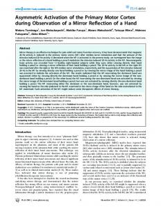
F I G U R E 1 (A) Behavioral experiment design. Subjects performed a repeated serial button-pressing task for 60 s at their own pace. This was followed by observation of a 60-s video of someone else performing the task at a rate of 3 Hz (experiment group) or observation of a black screen (control group) for a similar duration. Finally, both groups performed the execution task a second time. (B) fMRI experiment design. Subjects performed 10 consecutive blocks of a repeated serial button-pressing task at their own pace. Each 9-s execution block was followed by 6 s of silent rest. The execution blocks were followed by 10 consecutive observation blocks of a video depicting someone else performing the task at a rate of 4 Hz. Finally, subjects performed the execution task again
while lying in an fMRI scanner (Fig. 1B). They were instructed to
slices were acquired, providing whole-brain coverage (slice thickness,
produce the same sequence of button presses as in the behavioral
4 mm; slice gaps, 0 mm; in-plane resolution, 1.72 × 1.72 × 4 mm; TR,
study (sequence: 1-2-3-4-3-2…), back and forth at their own pace.
3,000 ms; TE, 35 ms; flip angle, 90°; field of view, 220 × 220 mm2;
As the subjects could not see their fingers, they used four different
matrix size, 128 × 128). For anatomical reference, a whole-brain high-
fingers (all digits except thumb), one finger for each key. Each key
resolution T1-weighted scan (voxel size, 1 × 1 × 1 mm) was acquired
produced a different 70-ms duration pure tone of 400, 500, 600, or
for each subject.
700 Hz and auditory feedback of the generated tones was provided
Functional magnetic resonance imaging data analysis was per-
via MR-compatible “Optoacoustics” headphones (OptoActive). Only
formed using “Brain Voyager QX” v. 2.8 software package (Brain
one key could be pressed at a given time. The subjects performed the
Innovation, Maastricht, the Netherlands). Preprocessing of func-
sequence for a few seconds in the scanner before the beginning of the
tional data included cubic spline slice time correction, trilinear/
experiment in order to familiarize them with the task and verify they
sinc three-dimensional (3D) motion correction, temporal high-pass
understood it correctly. Each 9 s experimental block was followed by
filtering of 0.006 Hz, and spatial smoothing using a Gaussian filter
6 s of silent rest. Subjects performed 10 such execution blocks con-
(FWHM = 6 mm). Both anatomical and functional images were trans-
secutively, followed by an observation task. In the observation task,
formed into the standardized coordinate system of Talairach (Talairach
subjects observed a video of someone else performing the same
& Tournoux, 1988). Experimental timeline was convolved with a stan-
button-pressing task at a rate of 4 Hz (10 repetitions of 9 s obser-
dard hemodynamic response function (implemented in BVQX) and
vation followed by 6 s of rest). Subjects were visually monitored to
data analysis was performed using the general linear model (GLM).
verify they did not move during the observation task. Finally, subjects
In order to examine in which brain regions the activity patterns
performed the execution task again. The entire run lasted 7.5 min. As
during passive action observation covary with the subsequent behav-
in the behavioral study, for each subject, the behavioral change in per-
ioral change in execution rate, we performed a whole-brain regression
formance rate was calculated as the difference between the median
analysis using the change in task execution rate for each participant
interpress interval (mIPI; in milliseconds) before and after video obser-
(ΔmIPI = mIPI before observation − mIPI after observation), as a
vation (ΔmIPI = mIPI_before − mIPI_after).
regressor against the activation levels of each voxel across all subjects during the observation session (β-value of each subject during
2.3.2 | fMRI data acquisition and preprocessing
passive observation relative to baseline resting periods). The regression was corrected for multiple comparisons using q(FDR) rest ∩ obser-
fore labeled subjects as “slow” or “fast”, according to their initial spon-
vation task > rest). To examine the correlation between the fMRI sig-
taneous rate (below or above the median: 3.1 Hz for the experiment
nal during action observation in these ROIs and subsequent changes
group and 3.7 Hz for the control group) and examined them separate-
in execution rate, we performed a regression analysis. We used the
ly. Initial spontaneous rates were not significantly different between
same behavioral measure as in the whole-brain analysis as a regressor
groups (“slow” experimental group vs. control and “fast” experimen-
against the activation levels. The neural measure this time was the
tal vs. control). We found a significant three-way interaction in the
mean β-value across all voxels in the ROI during the observation ses-
pressing rate between group type (slow/fast), observation condition
sion relative to baseline. The regression was performed for each ROI
(experiment/control), and time (before/after), F1,46 = 6.8, p rest). Figure 4A displays the mul-
neous execution rate of the task across all 50 subjects (obtained from
tisubject map of this contrast. The identified mirror ROIs included
the first execution session) had a bimodal distribution with modes of 2
the following regions (number of voxels and Talairach coordinates):
and 4.2 Hz, with a median of 3.5 Hz. Therefore, in the fMRI study, we
left premotor cortex (555 voxels, x = −57.4, y = −7.6, z = 40.6), left
decided to use videos depicting the button-pressing task at a slightly
SPL (879 voxels, x = −34.3, y = −56.3, z = 58.1), right supplementa-
higher rate of 4 Hz in order to enhance the behavioral effect in the
ry motor area (SMA; 244 voxels, x = 4.9, y = −7.0, z = 65.2), and the
smaller group of fMRI subjects.
right premotor cortex (272 voxels, x = 54.0, y = −10.0, z = 45.8). Two additional clusters were found in the right and left superior temporal
3.2 | fMRI study
cortices, corresponding to auditory cortex, and are likely to be due to the auditory feedback which was present both in the execution and
The performance rate of the majority of fMRI subjects (12 of the
observation tasks. None of the ROIs showed a significant correlation
15) exhibited a spontaneous execution rate during the first session
between the mean β-values across voxels obtained during the passive
below 4 Hz (the rate displayed in the subsequent observation ses-
observation session and the subsequent change in task performance
sion). Repeated measures analysis of variance (ANOVA) of the per-
rate (ΔmIPI) across subjects.
formance rate in each block of the first execution session showed no significant difference in performance rate across the 10 blocks (F1.8,25.7 = 1.25, p = .3, Greenhouse–Geisser corrected) suggesting
4 | DISCUSSION
that the performance rate of the subjects was stable throughout the scan. Compatible with the results from the behavioral study, subjects’
In the current study, we examined whether passive action observa-
performance rate increased following the observation session (mean
tion, without the explicit purpose of future imitation, induces implicit
mIPI across subjects: before = 390 ms, after = 296 ms, p rest). The identified ROIs included the left and right premotor cortex, left SPL, and right SMA. (B) For each individual subject, fMRI β-values of all voxels within an ROI were averaged and plotted against the behavioral change in execution rate (ΔmIPI = mIPI before − mIPI after). None of the ROIs exhibited a significant correlation (r values: left premotor cortex = 0.19, left SPL = 0.30, right SMA = −0.15, right premotor cortex = −0.09)
with similar frequency modes (Collyer, Broadbent, & Church, 1994).
others. For example, squeeze force (Obhi & Hogeveen, 2010) and grip
Other studies (Bove et al., 2009; McAuley, Jones, Holub, Johnston,
force (Salama, Turner, & Edwards, 2011) have been shown to modulate
& Miller, 2006; Vanneste, Pouthas, & Wearden, 2001) point to 2 Hz
according to the force of observed actions. Hand movement velocity is
as a common natural spontaneous rate across subjects. It is possible
also implicitly influenced by the velocity of observed movement (Bisio,
that the additional 4 Hz frequency mode obtained from our subjects
Stucchi, Jacono, Fadiga, & Pozzo, 2010), and the rate of a repetitive
(and also in the study by Collyer et al., 1994) represents a harmony
movement is another movement parameter that has been demonstrat-
of the more commonly reported 2 Hz frequency. We note that our
ed to change following observation (Avanzino et al., 2015; Bove et al.,
behavioral task was slightly different than the simple index finger
2009). In the case of movement rate, the study by Bove and colleagues
tapping used in previous studies. Our behavioral task also included a
reported higher execution rates in subject groups who observed a
spatial component as subjects were asked to press four different but-
video depicting a high execution rate, and lower execution rates in
tons back and forth with their index finger. Nonetheless, across the
subject groups who observed a video depicting a low execution rate
different variations in the task, it seems that 2 Hz is a common natural
(relative to the spontaneous rate of a control group who observed a
rate. Interestingly, it has been shown that a 3-day physical training
neutral stimulus). In our pre/post within-subject design, we found that
period can modulate this spontaneous rate in an attractor fashion—
using a video depicting an execution rate of 3 or 4 Hz (behavioral or
with training at a higher rate resulting in increased spontaneous rate
fMRI studies, respectively) resulted in an increased execution rate in
and training at a lower rate resulting in decreased spontaneous rate
subjects who had a lower execution rate, but no decreased perfor-
in a subsequent test (Hammerbeck, Yousif, Greenwood, Rothwell, &
mance rate in those who were originally faster. It is an open question
Diedrichsen, 2014). In our short-term experimental design (ten 9 s
whether showing a very low performance rate (e.g., 1 Hz as used by
blocks in the fMRI study), we did not see significant within session
Bove and colleagues) would have resulted in a decreased execution
learning effects.
rate in the faster group. At least for the subjects with a spontaneous
Implicit induction of various movement parameters has also been
rate above 3 or 4 Hz in our study, showing an action performed at
demonstrated following passive observation of actions performed by
lower rate did not result in a reduction of their subsequent execution
|
e00550 (7 of 9)
Aridan and Mukamel
rate. The question why some subjects have a slower spontaneous tap-
elicit significant neural responses in frontal regions (Simon & Mukamel
ping rate than others and why they are more susceptible to changes
2016). The current design does not allow determining which particular
in spontaneous rate through visual induction deserves further study.
element of the observed action underlies these changes in behavior,
Differences in spontaneous rate that lasted even up to 2 days follow-
however previous studies point to the importance of the presence
ing the initial observation session have been reported, suggesting that
of a biological agent in such effects (Avanzino et al., 2015; Kilner,
such effects are not temporally confined to the immediate timeframe
Paulignan, & Blakemore, 2003).
following action observation (Bove et al., 2009). In the current study,
Although the primary motor cortex (M1) is not classically consid-
the execution session immediately followed the observation session,
ered an integral part of the core parietofrontal MNS (Iacoboni, 2005;
thus our behavioral and fMRI results pertain to the immediate effects
Rizzolatti & Sinigaglia, 2010), there is accumulating evidence for M1
of action observation.
activity during action observation. Electrophysiological studies in
We examined the neural correlates of this implicit induction of
monkeys demonstrated the existence of cells with mirroring proper-
execution rate using fMRI. Our whole-brain analysis demonstrates
ties in this region (Dushanova & Donoghue, 2010; Tkach et al., 2007;
that the level of activity in the left motor strip elicited during action
Vigneswaran et al., 2013; Waldert, Vigneswaran, Philipp, Lemon, &
observation correlates with subsequent changes in the execution rate
Kraskov, 2015). TMS studies further support the role of M1 in obser-
across subjects. The initial or final execution rates alone (rather than
vational learning. Action observation has been shown to modulate
the difference) did not correlate with fMRI activity. The subjects in
TMS-evoked excitability in M1 (Avanzino et al., 2015; Celnik et al.,
both our experiments were visually monitored for movements, thus
2006; Stefan et al., 2005) and repetitive TMS to M1 has been shown
covert imitation during the observation task is unlikely to explain our
to interfere with the behavioral effects of observational learning
result. We also specifically examined regions within the MNS as it has
(Brown et al., 2009). Interestingly, the study by Vigneswaran et al.
been implicated to play a functional role in automatic/implicit imita-
(2013) reports pyramidal tract neural activity in the primate prima-
tion. To this end, we defined the MNS by using a GLM conjunction
ry motor cortex that is facilitated during action execution and sup-
analysis (execution > rest ∩ observation > rest). Adopting this, more
pressed during action observation. Along similar lines, a neuroimaging
statistically lenient ROI approach did not yield additional regions
study in human reports increased fMRI BOLD signal during action
exhibiting a significant correlation with subsequent behavioral chang-
execution and reduced signal during action observation (Gazzola &
es. However, this negative result should be taken with caution due
Keysers, 2009). Such lower activity levels in M1 during action obser-
to our limited sample size which may have resulted in low statistical
vation might explain why this region is less consistently reported in the
power to detect such an effect in these regions.
context of human mirroring studies (Caspers, Zilles, Laird, & Eickhoff,
Previous studies examined the relationship between neural activ-
2010).
ity during action observation, and subsequent performance level on a
The motor strip we detected using a whole-brain regression
task similar to the one observed. Frey and Gerry (2006) had subjects
analysis with behavior did not pass statistical threshold in our
observe a problem-solving procedure and report that fMRI activity
multisubject GLM observation/execution conjunction analysis.
levels in the right intraparietal sulcus during observation correlated
Therefore, it was not defined as part of the MNS network or exam-
with subsequent performance accuracy. Using a bimanual imitation
ined in the ROI analysis. Closer inspection of this region revealed
task, Krüger et al. (2014) report a positive correlation with behavior
that this was mainly due to the greater signal variability across
in the right SPL and the left parietal operculum and a negative cor-
subjects during action observation. In some subjects, action obser-
relation with behavior in the left IPL and the right vPMC. The above-
vation elicited strong signals that passed the statistical threshold
mentioned brain regions have been previously implicated in the MNS
for a single-subject GLM observation/execution conjunction anal-
(Iacoboni et al., 1999; Molenberghs et al., 2012). It should be noted
ysis, while in others it did not. The fact that this variability across
that both studies used a masked ROI analysis approach that did not
subjects correlated with their subsequent behavioral changes (as
include M1. Indeed, TMS stimulation of M1 has been shown to dis-
seen in the whole-brain regression) suggests that this activity level
rupt positive/negative effects of explicit learning by observation of
in M1 during observation has functional significance. This variabil-
congruent/incongruent actions, respectively (Brown et al., 2009). In
ity across subjects might also explain why M1 is less frequent-
our study, subjects were not instructed to attend any particular aspect
ly reported in imaging studies in the context of mirroring (when
of the observed stimulus and did not know that they would be asked
defined by observation tasks). Conversely, activation in the classical
to perform the execution task a second time. Furthermore, there
parietofrontal mirror neuron regions during action observation is
was no element of performance level (whether explicit or implicit) in
more robust and consistent across subjects although, at least in our
our task (i.e., performance-wise, subjects had no particular incentive
study, activity level in these regions did not correlate with subse-
to tap at a particular rate). The changes in behavior we report are a
quent implicit behavioral changes. Other studies using an explicit
result of implicit mimicry/contagion of the observed action and our
imitation/learning task report correlation with behavior within the
whole-brain fMRI results demonstrate that these implicit behavioral
parietofrontal MNS but their ROI masks did not include M1. Taken
changes correlate with activity level in M1 during passive observation.
together, these studies suggest an interesting dissociation between
This result is in agreement with our recent finding that visual presen-
the functional properties of mirroring activity across different
tation of actions that are not consciously perceived, is sufficient to
regions within the MNS. An intriguing scheme integrating our
|
e00550 (8 of 9)
results with the literature is one in which action observation elicits activity in the classical parietofrontal MNS, and the degree of its translation to behavior (at least during implicit imitation) depends on the relay of this information to M1. Subjects in which this relay is strong manifest higher levels of M1 activation during action observation and also stronger subsequent behavioral changes. The fact that our correlation with behavior during observation was found in M1 contralateral to the passively observed hand supports this view, although it deserves further study. To conclude, we demonstrate that following exposure to a video depicting someone else perform a button-pressing task, subjects tend to implicitly shift their spontaneous execution rate toward the higher rate of the observed action. The degree of this behavioral shift correlates with the degree of activation elicited during action observation in the contralateral motor cortex.
ACKNOWLE DG ME NTS The authors thank the laboratory members for fruitful comments on the manuscript. The study was supported by the I-CORE Program of the Planning and Budgeting Committee and the Israel Science Foundation (Grant No. 51/11), the Human Frontiers Science Project Organization (HFSPO) (CDA00078/2011-C), and the Israel Science Foundation (Grants No. 1771/13 and 2043/13) to R. M.
F UNDI NG I NFORM ATI O N This study was supported by the Israel Science Foundation (Grant/ Award Number: 1771/13 and 2043/13), the Human Frontiers Science Project Organization (HFSPO) (Grant/Award Number: CDA00078/2011-C), and the Israeli Centers for Research Excellence (Grant/Award Number: 51/11).
CO NFLI CT OF I NTE RE S T None declared.
REFERENCES Avanzino, L., Lagravinese, G., Bisio, A., Perasso, L., Ruggeri, P., & Bove, M. (2015). Action observation: Mirroring across our spontaneous movement tempo. Scientific Reports, 5, 10325. Bisio, A., Stucchi, N., Jacono, M., Fadiga, L., & Pozzo, T. (2010). Automatic versus voluntary motor imitation: Effect of visual context and stimulus velocity. PLoS One, 5, 1–8. Bove, M., Tacchino, A., Pelosin, E., Moisello, C., Abbruzzese, G., & Ghilardi, M. F. (2009). Spontaneous movement tempo is influenced by observation of rhythmical actions. Brain Research Bulletin, 80, 122–127. Brass, M., Bekkering, H., & Prinz, W. (2001). Movement observation affects movement execution in a simple response task. Acta Psychologica, 106(1–2), 3–22. Brown, L. E., Wilson, E. T., & Gribble, P. L. (2009). Repetitive transcranial magnetic stimulation to the primary motor cortex interferes with motor learning by observing. Journal of Cognitive Neuroscience, 21, 1013– 1022.
Aridan and Mukamel
Buccino, G., Vogt, S., Ritzl, A., Fink, G. R., Zilles, K., Freund, H.-J., & Rizzolatti, G. (2004). Neural circuits underlying imitation learning of hand actions. Neuron, 42(2), 323–334. Caspers, S., Zilles, K., Laird, A. R., & Eickhoff, S. B. (2010). ALE meta-analysis of action observation and imitation in the human brain. NeuroImage, 50(3), 1148–1167. Celnik, P., Stefan, K., Hummel, F., Duque, J., Classen, J., & Cohen, L. G. (2006). Encoding a motor memory in the older adult by action observation. NeuroImage, 29(2), 677–684. Chartrand, T. L., & Bargh, J. A. (1999). The chameleon effect: The perception-behavior link and social interaction. Journal of Personality and Social Psychology, 76, 893–910. Collyer, C., Broadbent, H., & Church, R. (1994). Preferred rates of repetitive tapping and categorical time production. Perception & Psychophysics, 55(4), 443–453. Craighero, L., Bello, A., Fadiga, L., & Rizzolatti, G. (2002). Hand action preparation influences the responses to hand pictures. Neuropsychologia, 40, 492–502. Cross, E. S., Kraemer, D. J. M., Hamilton, A. F. D. C., Kelley, W. M., & Grafton, S. T. (2009). Sensitivity of the action observation network to physical and observational learning. Cerebral Cortex, 19(2), 315–326. Dushanova, J., & Donoghue, J. (2010). Neurons in primary motor cortex engaged during action observation. European Journal of Neuroscience, 31, 386–398. Edwards, M., Humphreys, G., & Castiello, U. (2003). Motor facilitation following action observation: A behavioural study in prehensile action. Brain and Cognition, 53(3), 495–502. Ferguson, M. J., & Bargh, J. A. (2004). How social perception can automatically influence behavior. Trends in Cognitive Sciences, 8(1), 33–39. Fogassi, L., Ferrari, P. F., Gesierich, B., Rozzi, S., Chersi, F., & Rizzolatti, G. (2005). Parietal lobe: From action organization to intention understanding. Science (New York, N.Y.), 308, 662–667. Frey, S. H., & Gerry, V. E. (2006). Modulation of neural activity during observational learning of actions and their sequential orders. The Journal of Neuroscience: The Official Journal of the Society for Neuroscience, 26, 13194–13201. Gallese, V., Fadiga, L., Fogassi, L., & Rizzolatti, G. (1996). Action recognition in the premotor cortex. Brain, 119, 593–609. Gazzola, V., & Keysers, C. (2009). The observation and execution of actions share motor and somatosensory voxels in all tested subjects: Single-subject analyses of unsmoothed fMRI data. Cerebral Cortex, 19, 1239–1255. Grèzes, J., Costes, N., & Decety, J. (1999). The effects of learning and intention on the neural network involved in the perception of meaningless actions. Brain : A Journal of Neurology, 122(Pt 1), 1875–1887. Hammerbeck, U., Yousif, N., Greenwood, R., Rothwell, J. C., & Diedrichsen, J. (2014). Movement speed is biased by prior experience. Journal of Neurophysiology, 111(1), 128–134. Iacoboni, M. (2005). Neural mechanisms of imitation. Current Opinion in Neurobiology, 15, 632–637. Iacoboni, M. (2009). Imitation, empathy, and mirror neurons. Annual Review of Psychology, 60, 653–670. Iacoboni, M., Woods, R. B., Brass, M., Bekkering, H., Mazziotta, J. C., & Rizzolatti, G. (1999). Cortical mechanisms of human imitation. Science, 286, 2526–2528. Keysers, C., Kaas, J. H., & Gazzola, V. (2010). Somatosensation in social perception. Nature Reviews. Neuroscience, 11, 417–428. Kilner, J. M., Paulignan, Y., & Blakemore, S. J. (2003). An interference effect of observed biological movement on action. Current Biology, 13, 522–525. Krüger, B., Bischoff, M., Blecker, C., Langhanns, C., Kindermann, S., Sauerbier, I., & Pilgramm, S. (2014). Parietal and premotor cortices: Activation reflects imitation accuracy during observation, delayed imitation and concurrent imitation. NeuroImage, 100, 39–50. Mattar, A. A. G., & Gribble, P. L. (2005). Motor learning by observing. Neuron, 46(1), 153–160.
Aridan and Mukamel
McAuley, J. D., Jones, M. R., Holub, S., Johnston, H. M., & Miller, N. S. (2006). The time of our lives: Life span development of timing and event tracking. Journal of Experimental Psychology. General, 135(3), 348–367. Molenberghs, P., Cunnington, R., & Mattingley, J. B. (2012). Brain regions with mirror properties: A meta-analysis of 125 human fMRI studies. Neuroscience and Biobehavioral Reviews, 36(1), 341–349. Mukamel, R., Ekstrom, A. D., Kaplan, J., Iacoboni, M., & Fried, I. (2010). Single-neuron responses in humans during execution and observation of actions. Current Biology, 20, 750–756. Obhi, S. S., & Hogeveen, J. (2010). Incidental action observation modulates muscle activity. Experimental Brain Research, 203(2), 427–435. di Pellegrino, G., Fadiga, L., Fogassi, L., Gallese, V., & Rizzolatti, G. (1992). Understanding motor events: A neurophysiological study. Experimental Brain Research, 91(1), 176–180. Rizzolatti, G., & Craighero, L. (2004). The mirror-neuron system. Annual Review of Neuroscience, 27, 169–192. Rizzolatti, G., & Sinigaglia, C. (2010). The functional role of the parieto- frontal mirror circuit: Interpretations and misinterpretations. Nature Reviews. Neuroscience, 11, 264–274. Rozzi, S., Ferrari, P. F., Bonini, L., Rizzolatti, G., & Fogassi, L. (2008). Functional organization of inferior parietal lobule convexity in the macaque monkey: Electrophysiological characterization of motor, sensory and mirror responses and their correlation with cytoarchitectonic areas. European Journal of Neuroscience, 28, 1569–1588. Salama, I. M., Turner, S., & Edwards, M. G. (2011). Automatic priming of grip force following action observation. Quarterly Journal of Experimental Psychology (2006), 64, 833–838. Simon, S., & Mukamel, R. (2016). Power modulation of electroencephalogram mu and beta frequency depends on perceived level of observed actions. Brain and Behavior, doi: 10.1002/brb3.494 Stefan, K., Cohen, L. G., Duque, J., Mazzocchio, R., Celnik, P., Sawaki, L., … Classen, J. (2005). Formation of a motor memory by action observation.
|
e00550 (9 of 9)
The Journal of Neuroscience: The Official Journal of the Society for Neuroscience, 25, 9339–9346. Stürmer, B., Aschersleben, G., & Prinz, W. (2000). Correspondence effects with manual gestures and postures: A study of imitation. Journal of Experimental Psychology: Human Perception and Performance, 26, 1746–1759. Suchan, B., Melde, C., Herzog, H., Hömberg, V., & Seitz, R. J. (2008). Activation differences in observation of hand movements for imitation or velocity judgement. Behavioural Brain Research, 188(1), 78–83. Talairach, J., & Tournoux, P. (1988) Co-planar stereotaxic atlas of the human brain: 3-dimensional proportional system: An approach to cerebral imaging. New York, NY: Thieme Medical. Tkach, D., Reimer, J., & Hatsopoulos, N. G. (2007). Congruent activity during action and action observation in motor cortex. The Journal of Neuroscience: The Official Journal of the Society for Neuroscience, 27, 13241–13250. Vanneste, S., Pouthas, V., & Wearden, J. H. (2001). Temporal control of rhythmic performance: A comparison between young and old adults. Experimental Aging Research, 27(1), 83–102. Vigneswaran, G., Philipp, R., Lemon, R. N., & Kraskov, A. (2013). M1 corticospinal mirror neurons and their role in movement suppression during action observation. Current Biology, 23, 236–243. Waldert, S., Vigneswaran, G., Philipp, R., Lemon, R. N., & Kraskov, A. (2015). Modulation of the intracortical LFP during action execution and observation. Journal of Neuroscience, 35, 8451–8461.
How to cite this article: Aridan, N. and Mukamel, R. (2016), Activity in primary motor cortex during action observation covaries with subsequent behavioral changes in execution. Brain and Behavior, 00: 1–9. e00550, doi: 10.1002/brb3.550