result in the development of the disorder known as hand-arm vibration syndrome (HAVS; (1,3)). One of the ... sample was split into two 0.5 ml samples. One set ...
ACUTE VIBRATION INDUCES OXIDATIVE STRESS AND CHANGES IN TRANSCRIPTION IN SOFT TISSUE OF RAT TAILS Stacey Waugh1, Stephen S. Leonard2, G. Roger Miller1, Kristine M. Krajnak1 1 Engineering and Control Technology Branch, 2Pathology and Physiology Research Branch, National Institute for Occupational Safety and Health, Morgantown, West Virginia, U.S.A.
Introduction Repeated exposure to hand-arm vibration through the use of vibrating hand tools can result in the development of the disorder known as hand-arm vibration syndrome (HAVS; (1,3)). One of the hallmark symptoms of HAVS is cold-induced peripheral vasospasms that result in finger blanching (4). Although the vascular and neural pathology associated with vasospasms has been described, little is known about cellular mechanisms leading to this damage (4). To understand how vibration may alter vascular and neural physiology and anatomy, rats were exposed to a single bout of tail vibration and the molecular responses of neural and vascular tissues were measured to determine if there are immediate or sustained affects of vibration that may underlie longer term changes in physiology. Methods Experiment 1. Male Sprague Dawley rats (n = 32, 6 weeks of age) were housed in AAALAC accredited facilities. All procedures were approved by the NIOSH Animal Care and Use Committee and were in compliance with the CDC guidelines for care and use of laboratory animals. Vibration exposures were performed by restraining rats in Broome-style restrainers, and securing their tails to a vibration platform using 6 mm wide straps that were placed over the tail every 3 cm. Restraint control rats were treated in the same manner, except that the tail platform was mounted on isolation blocks and not on a shaker. The vibration exposure was 125 Hz, 49 m/s2, for 4 h. Rats were euthanized with an overdose of pentobarbital (100 mg/kg) 1 h or 24 hours after the exposure. RTqPCR was used to measure transcript levels for endothelin 1 (ET1), the 3 forms of nitric oxide synthetase (NOS) NOS-1, NOS-2, NOS-3 and norepinephrine receptor subtypes 1D, 2A, 2C, in artery tissue, and to measure calcitonin gene-related peptide (CGRP) and nitric oxide synthase-1 (NOS-1) in ventral nerves. Data were analyzed using 2-way ANOVAs. Experiment 2. Male Sprague Dawley rats (n =24, 6 weeks of age) were maintained as described above. Animals were exposed to a single bout of restraint or vibration. Another group of animals served as cage controls. All animals were euthanized 24 h after the exposure, and tail arteries were isolated and frozen. Reactive oxygen species ROS) were measured using electron spin resonance spectroscopy (ESR). Arteries were homogenized over ice in 1 ml of PBS with protease inhibitor cocktail, using a tissue tearer (Biospecs Products Inc. Racine, WI USA). The sample was split into two 0.5 ml samples. One set had PBS and the spin label hydroxyl-TEMPO [0.1 mM] added while the other set had hydroxyl-TEMPO [1.0 mM] plus the specific hydroxyl radical scavenger, dimethylthiourea (DMTU), added to confirm the presence of the hydroxyl radical. Samples were vortexed and then placed in a flat cell for ESR analysis. The ESR spectrometer settings were: receiver gain, 6.32 x 10²; time constant, 0.02 s; modulation amplitude, 1.0 G; scan time, 20 sec; magnetic field, 3490 ± 100 G (2). Data were analyzed using 1-way ANOVAs.
160
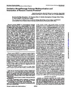
Results B
*
2 1 0 -1 -2 -3 -4 -5
Restraint Control
Fold change in CGRP trancript levels
F o ld c h a n g e in N O S -1 tr a n s c r ip t le v e ls
A
*
3 2 1 0 -1 -2
Vibrated
Restraint Control
Vibrated
ESR Peak Ht (% change from TEMPO alone)
Figures 1A-B. Fold changes in NOS-1(A) and CGRP (B) in the ventral tail arteries rats exposed to a single bout of vibration or restraint. The data are expressed as fold changes in transcript levels (mean ± sem) from the time matched controls. White bars represent transcript levels from tissue collected 1 h after the exposure and black bar represent transcript levels from tissue collected 24 h after the exposure (* different from time matched restraint control, p < 0.05). Exposure to vibration resulted in a reduction in NOS-1 transcript levels (main effect of exposure F (1, 26) = 6.67, P < 0.02) and an increase in CGRP transcript levels in nerve tissue collected 24 h after the exposure (p < 0.05). 100
• • •
*
80 60 40 20 0
Cage
Restraint
Vibration
Figure 2. ROS measured using ESR. Acute vibration exposure resulted in an increase in hydroxyl radicals. The data to the left represent the ESR peak height when both TEMPO and DMTU were added to homogenates from the tail artery. The data are presented as the percent increase in peak height between TEMPO + DMTU and TEMPO alone. (mean ± sem; p < 0.05, different from cage and restraint controls; F(1, 20) = 8.68, p < 0.002).
Discussion Nitric oxide (NO) is a potent vasodilator produced by nerves and arteries. Vibration-induced reductions in the neural form of NOS, NOS-1, may result in reductions in NO synthesis and contribute to a prolonged noradrenergic-induced vasoconstriction. Increases in oxidative stress can result in a reduction in NOS activity. Acute exposure to vibration increase ROS in the arteries. This increase in ROS in arteries (and potentially nerves) may result in a reduction in NOS activity and NO production. The increase in CGRP transcript levels, which are not seen until 24 h after the exposure, may act to relieve the vibration induced vasoconstriction.
References 1. Griffin MJ. Handbook of Human Vibration. San Diego: Academic Press, 1990. 2. Machida Y, et al.,. Am J Physiol Heart Circ Physiol 284: H449-H455, 2003. 3. Palmer KT, et al., Occup Environ Med 58: 279-280, 2001. 4. Stoyneva Z, et al., Cardiovasc Res 57: 615-624, 2003.
161