Human Physiology
Adipocyte Differentiation-related Protein in Human Skeletal Muscle: Relationship to Insulin Sensitivity Susan A. Phillips,*† Charles C. Choe,*‡ Theodore P. Ciaraldi,*‡ Andrew S. Greenberg,§ Alice P. S. Kong,*‡ Sunita C. Baxi,* Louis Christiansen,*‡ Sunder R. Mudaliar,*‡ and Robert R. Henry*‡
Abstract PHILLIPS, SUSAN A., CHARLES C. CHOE, THEODORE P. CIARALDI, ANDREW S. GREENBERG, ALICE P. S. KONG, SUNITA C. BAXI, LOUIS CHRISTIANSEN, SUNDER R. MUDALIAR, AND ROBERT R. HENRY. Adipocyte differentiation-related protein in human skeletal muscle: relationship to insulin sensitivity. Obes Res. 2005;13:1321–1329. Objective: To determine whether adipocyte differentiationrelated protein (ADRP), a lipid droplet–associated protein that binds to and sequesters intracellular fatty acids, is 1) expressed in human skeletal muscle and 2) differentially regulated in human skeletal muscle obtained from obese non-diabetic (OND) and obese diabetic (OD) subjects. Research Methods and Procedures: Ten OND subjects and 15 OD subjects underwent a weight loss or pharmacological intervention program to improve insulin sensitivity. Anthropometric data, hemoglobin A1C, fasting glucose, lipids, and glucose disposal rate were determined at baseline and at completion of studies. Biopsies of the vastus lateralis muscle (SkM) were obtained in the fasting state from OND and OD subjects. Protein expression was determined by Western blotting. Results: ADRP was highly expressed in SkM from OND (4.4 ⫾ 1.54 AU/10 g, protein, n ⫽ 10) and OD (5.02 ⫾ 1.33 AU/10 g, n ⫽ 12) subjects. OND subjects undergoing weight loss had decreased triglyceride levels and improved
Received for review December 23, 2004. Accepted in final form May 16, 2005. The costs of publication of this article were defrayed, in part, by the payment of page charges. This article must, therefore, be hereby marked “advertisement” in accordance with 18 U.S.C. Section 1734 solely to indicate this fact. *Veterans Affairs San Diego Healthcare System, San Diego, California; †Department of Pediatrics and ‡Department of Medicine, University of California, San Diego, La Jolla, California; and §Jean Mayer Human Nutrition Research Center at Tufts University, Boston, Massachusetts. Address correspondence to Robert R. Henry, VA San Diego Healthcare System, 3350 La Jolla Village Drive, San Diego, CA 92161. E-mail:
[email protected] Copyright © 2005 NAASO
insulin action. SkM ADRP content increased with weight loss from 5.14 ⫾ 2.15 AU/10 g to 9.92 ⫾ 1.57 AU/10 g (p ⬍ 0.025). OD subjects were treated with either troglitazone or metformin, together with glyburide, for 3 to 4 months. Both treatments attained similar levels of glycemic control. OD subjects with lower baseline ADRP content (2.85 ⫾ 1.07 AU/10 g, n ⫽ 6) displayed up-regulation of ADRP expression (to 9.27 ⫾ 2.76 AU/10 g, p ⬍ 0.025). Discussion: ADRP is the predominant lipid droplet–associated protein in SkM, and low ADRP expression is upregulated in circumstances of improved glucose tolerance. Up-regulation of ADRP may act to sequester fatty acids as triglycerides in discrete lipid droplets that could protect muscle from the detrimental effects of fatty acids on insulin action and glucose tolerance. Key words: human, intramyocellular lipid, insulin resistance, skeletal muscle, fat metabolism
Introduction
Type 2 diabetes (T2D)1 is characterized by impairment in pancreatic -cell function and resistance to insulin action in multiple tissues, including skeletal muscle. Skeletal muscle insulin resistance is also a feature of obesity, hypertension, and dyslipidemia (1). Emerging evidence suggests that altered lipid metabolism may play an important role in the development of skeletal muscle insulin resistance. Although extramyocellular lipid, predominantly adipose tissue, is the largest lipid depot, intramyocellular lipid, usually in the form of triglyceride (IMTG), seems to play an important role in the development of skeletal muscle insulin resistance. In humans, IMTG concentrations are highly correlated with insulin sensitivity (2–5), so that skeletal muscle
1 Nonstandard abbreviations: T2D, type 2 diabetes; IMTG, intramyocellular lipid, usually in the form of triglyceride; PAT, perilipin, adipocyte differentiation-related protein, and tail interacting protein 47; ADRP, adipocyte differentiation-related protein; TIP47, tail interacting protein 47; GDR, glucose disposal rate; SkM, vastus lateralis muscle.
OBESITY RESEARCH Vol. 13 No. 8 August 2005
1321
ADRP Expression in Human Skeletal Muscle, Phillips et al.
triglyceride levels are inversely related to insulin action (6). In addition, depletion of IMTG by weight loss (7,8) or by pharmacological intervention (8,9) is associated with improvements in insulin sensitivity. However, a paradox exists in the relationship between IMTG and insulin sensitivity, because IMTG is also elevated in highly insulin-sensitive endurance-trained athletes (10). It seems from these studies that, although quantitative measures of IMTG are often indicative of insulin sensitivity, qualitative measures of IMTG may be needed to explain their full metabolic impact. An important qualitative aspect of TG storage is its localization and organization within the cell (11). A growing number of proteins (11,12) have been identified to be associated with intracellular neutral lipid droplets. Among them, a family of three PAT-related proteins, perilipin, adipocyte differentiation-related protein (ADRP/adipophilin), and tail interacting protein 47 (TIP47), has emerged as proteins that share regions of extensive sequence similarity called PAT domains (12). Another lipid droplet–associated molecule, S3–12, lacks a PAT domain, but shares a 33-amino acid motif also found in ADRP, as well as possesses regions of similarity in the COOH terminus with both ADRP and TIP47 (12,13). Among the lipid droplet–associated proteins, perilipin and ADRP are the most extensively studied. Perilipins are adipocyte and steroidogenic cell-specific phosphoproteins that coat neutral lipid droplets (14,15) and regulate lipolytic signaling through interaction with hormone-sensitive lipase (15,16). Ablation of perilipin expression in mice results in impaired glucose tolerance and peripheral insulin resistance (17). Less is known about the perilipin-related protein, ADRP. Although initially discovered as a marker of early adipocyte development (18,19), ADRP is now recognized to be widely expressed in many tissues including rodent skeletal muscle (20,21). A number of findings support the view that ADRP, like perilipin, serves to organize intracellular lipid. For instance, the ADRP protein is localized at the surface of lipid droplets, and its levels are increased significantly on lipid loading of cells (20). ADRP also possesses features such as being posttranslationally acylated (22) and having the capacity to bind long-chain fatty acids (22,23) and unesterified cholesterol (24) that support a role in lipid droplet organization (20,25). This study had several goals. First, given the important association of IMTG content with measures of insulin sensitivity, we sought to determine the relative protein expression of perilipin and ADRP in human skeletal muscle. Second, we sought to determine whether there are differences in the extent of lipid droplet–associated protein expression in skeletal muscle tissues obtained from subjects with T2D. Third, we asked whether improvements in insulin sensitivity, as conferred by weight loss or pharmacological intervention, would alter ADRP expression in skeletal muscle. 1322
OBESITY RESEARCH Vol. 13 No. 8 August 2005
Research Methods and Procedures Human Subjects and Treatment Protocols Subjects were recruited from diabetes clinics and by advertisement. The subjects were classified as diabetic or non-diabetic by their response to a 75-gram oral glucose tolerance test according to American Diabetes Association criteria. Insulin action was determined with a 3-hour hyperinsulinemic (300 mU/m2 per minute) euglycemic (5.0 to 5.5 mM) clamp; the glucose disposal rate (GDR) was measured during the last 30 minutes of the clamp (26). Percutaneous needle biopsies of vastus lateralis muscle (SkM) were performed before the insulin infusion, as previously described (27), and muscle tissue was immediately frozen in liquid nitrogen. Glucose and insulin (28) levels were determined by standard techniques. Characteristics of the subject groups are summarized in Table 1. The Committee on Human Investigation of the University of California, San Diego, approved the experimental protocols. Informed written consent was obtained from all subjects after explanation of the protocol. Weight Loss Protocol Obese non-diabetic subjects (BMI, 30 to 45 kg/m2) were studied. All subjects were weight stable for at least 3 months before study. Subjects entered the Special Diagnostics and Treatment Unit for baseline characterization: oral glucose tolerance test, hyperinsulinemic/euglycemic clamp, and muscle biopsy. Subjects were placed on a very low calorie diet of 600 to 800 kcal/d for up to 24 weeks or until ⬃10% to 15% of initial body weight was lost. Subjects remained as inpatients during the first 4 weeks of diet therapy and were then monitored at weekly visits. Once the weight loss goal was met, subjects were introduced to a weight maintenance diet. Metabolic characterization studies were repeated after subjects were weight stable at their new level for at least 2 to 3 weeks. Drug Treatment Protocol Nine male and female subjects with type 2 diabetes (age range, 30 to 70 years) who were poorly controlled (hemoglobin A1C ⬎ 8.5% and fasting plasma glucose ⬎ 140 mg/dL) on at least half-maximal doses of any sulfonylurea agents were recruited. Except for diabetes, the subjects were healthy and on no other medications known to influence glucose metabolism. After screening, their existing sulfonylurea medication was discontinued, and all subjects were uniformly started on glyburide 10 mg twice a day for at least 4 weeks. After stabilization on sulfonylurea therapy, baseline studies, including biopsies, were performed in all subjects. Subjects were randomized to either the troglitazone or metformin treatment group. Treatment involved troglitazone titration up to 600 mg/d or metformin up to 2550 mg/d over 4 to 6 weeks as required to achieve glycemic goals. After 3 to 4 months of troglitazone or metformin treatment,
ADRP Expression in Human Skeletal Muscle, Phillips et al.
mined using BioRad reagent (Richmond, CA) by spectrophotometric assay according to the manufacturer’s instructions.
Table 1. Clinical characteristics of subjects
Number Sex (M/F) BMI (kg/m2) Fasting glucose (mM) Fasting insulin (pM) Hemoglobin A1C (%) GDR (mg/kg per min) Triglycerides (mg/dL) Free fatty acids (mM)
Nondiabetics
Diabetics
10 4/6 33.8 ⫾ 1.21 5.29 ⫾ 0.11 100 ⫾ 9 5.6 ⫾ 0.1 8.55 ⫾ 0.54 143 ⫾ 15 0.511 ⫾ 0.05
15 13/2 35.1 ⫾ 2.15 11.1 ⫾ 0.86* 230 ⫾ 42* 8.4 ⫾ 0.5* 4.80 ⫾ 0.63* 159 ⫾ 16 0.616 ⫾ 0.07
Glucose, insulin, free fatty acid, and triglyceride levels were measured after an overnight fast. Data are mean ⫾ SE. * p ⬍ 0.05 vs. non-diabetic group.
subjects were readmitted for repeat studies. Subjects were counseled to consume a fixed calorie diet for the duration of the study protocol. Data resulting from this protocol were presented in a series of earlier publications (29 –31). The effects of the two treatment protocols on clinical parameters are summarized in Table 2. Preparation of Muscle Lysates Fifty to 100 mg of muscle was homogenized using a polytron at setting 6 to 7 for 1 minute on ice in 500 L buffer A (20 mM Tris, pH 7.5, 5 mM EDTA, 10 mM Na4P2O7, 100 mM NaF, 2 mM Na3VO4) containing 1% NP-40, 1 mM phenylmethylsulfonyl fluoride, 10 mg/mL aprotinin, and 10 g/mL leupeptin. Tissue lysates were solubilized by vortexing for 30 seconds at 4 °C and centrifuging for 10 minutes at 14,000g. The supernatants were stored at ⫺80 °C until analysis. Protein content was deter-
Electrophoresis and Western Blotting Procedures for the electrophoresis, transfer, and Western blotting of proteins were similar to standard methods (32). Samples were prepared in Laemmli’s sample buffer. Detection was by enhanced chemiluminescence, followed by densitometric analysis. Quantitation of the blots was performed using ScanAnalysis software (Biosoft, Cambridge, United Kingdom). Analysis was repeated several times for each individual, and consistent results were obtained. Human skeletal muscle protein from a single subject was included in each gel as an internal standard to permit the correction of variability among blots and allow for normalization of multiple analyses. Materials Bovine serum albumin (Cohn fraction V) was obtained from Roche (Indianapolis, IN). Rabbit antihuman ADRP and rabbit antihuman perilipin antibodies have been described previously (33). Horseradish peroxidase– conjugated anti-rabbit IgGs were purchased from Amersham (Arlington Heights, IL), and SuperSignal-enhanced chemiluminescence substrate was from Pierce (Rockford, IL). Electrophoresis and protein assay reagents were purchased from BioRad. All other chemicals were reagent grade and purchased from Sigma (St. Louis, MO). Statistical Analysis Data are presented as the mean ⫾ SE. Statistical analyses were preformed using GraphPad Prism version 4.0 statistical program (GraphPad Software, San Diego, CA). Statistical significance was tested with the unpaired or paired Student’s t test when appropriate.
Table 2. Effect of weight loss and pharmacological interventions on hemoglobin A1c levels and GDR in non-diabetic and diabetic subjects
Weight loss Treatment of diabetes
Intervention
Change in hemoglobin A1C (%)
Change in GDR (mg/kg per min)
Change in triglycerides (mg/dL)
Caloric restriction SU ⫹ troglitazone SU ⫹ metformin
⫺0.09 ⫺1.08 ⫺2.05
⫹1.42 ⴙ2.61* ⫹1.17
⫺59.0 ⫾ 30.6 ⫺22.2 ⫾ 26.0 ⫺40.7 ⫾ 10.7*
Results presented as percent change from pretreatment for each individual, mean ⫾ SE. * p ⬍ 0.05 vs. baseline.
OBESITY RESEARCH Vol. 13 No. 8 August 2005
1323
ADRP Expression in Human Skeletal Muscle, Phillips et al.
Results Expression of Lipid Droplet–associated Molecules in Human Adipose and Skeletal Muscle Tissues Because the expression of lipid droplet–associated proteins in human tissues has not been well characterized, protein extracts were prepared from skeletal muscle tissue and freshly isolated subcutaneous adipocytes. To control for different levels of adipose tissue infiltration of muscle, skeletal muscle tissue was obtained by biopsy from BMImatched diabetic and non-diabetic subjects. Protein expression was determined by Western blot analysis. As expected, perilipin (67 kDa) was highly expressed in human adipocytes and in fully differentiated 3T3-L1 murine adipocytes but was not detected in skeletal muscle tissue (Figure 1A). These data are consistent with previous data showing that perilipin is not expressed in human skeletal muscle (34). The lack of a perilipin signal in skeletal muscle indicates that there was negligible adipose tissue infiltration of this tissue. ADRP protein, however, was abundant in skeletal muscle, with much lower levels in adipocytes and in the adipocyte cell line (Figure 1B). These results suggest that the predominant lipid droplet–associated protein in skeletal muscle is ADRP, although this does not rule out the presence of other lipid-associated proteins besides perilipin. Modulation of ADRP Expression in Insulin-resistant States To evaluate the potential involvement of skeletal muscle ADRP in metabolic regulation, we choose to study its expression in diabetic and non-diabetic subjects who were matched for obesity (Table 1). Diabetic subjects differed significantly from non-diabetic subjects in all measures of insulin sensitivity including hemoglobin A1C, fasting glucose and insulin levels, and GDR (Table 1). There were no statistically significant differences in the levels of circulating free fatty acids or in circulating triglyceride levels (Table 1). While not as insulin resistant as the type 2 diabetic subjects, the obese non-diabetic subjects displayed lower insulin sensitivity (8.56 ⫾ 0.54 mg/kg per minute) compared with a group of age-matched, lean non-diabetic subjects (35). Skeletal muscle ADRP protein expression was similar in diabetic and non-diabetic subjects (Figure 2), suggesting that adiposity may be a major determinant of ADRP expression in skeletal muscle of insulin-resistant subjects. To further explore the potential role of ADRP in metabolic regulation and insulin sensitivity, we studied the effect of two interventions, weight loss and insulin sensitizer pharmacotherapy, that are known to improve insulin sensitivity on skeletal muscle ADRP expression (Table 2). In the first intervention, seven non-diabetic weight stable subjects with a BMI between 35– 45 kg/m2 were placed on a very low calorie diet for up to 24 weeks or until they achieved a 10% to 15% reduction in initial body mass. Once weight loss was 1324
OBESITY RESEARCH Vol. 13 No. 8 August 2005
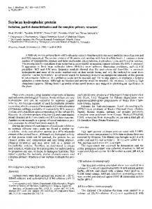
Figure 1: Perilipin and ADRP protein expression in human skeletal muscle tissue and human fat cell extracts, and in 3T3-L1 adipocytes. (A) Representative Western blot for perilipin. (B) Western blot for ADRP. For both perilipin and ADRP, gels were loaded with human skeletal muscle lysates (20 g protein per lane) obtained from three subjects, human fat cell lysates (5 g protein per lane), and differentiated 3T3-L1 adipocyte lysates (1 g protein per lane). The membranes were probed first for perilipin and stripped and reprobed for ADRP.
achieved, they were placed on a weight maintenance diet. ADRP expression studies were completed at baseline and after subjects were weight stable at their new level for at least 2 to 3 weeks. The averaged ADRP expression increased 192% over baseline (5.14 ⫾ 2.15 vs. 9.92 ⫾ 1.57 AU/10 g protein) with weight loss (Figure 3B). All of the subjects except one showed increases in ADRP expression with weight loss; it is interesting to note that this subject had the highest ADRP content in the baseline state (Figure 3C). In a separate intervention, nine type 2 diabetic subjects were treated with known insulin sensitizers (troglitazone or metformin) in combination with a sulfonylurea for up to 12 weeks. Studies of ADRP expression were done both at baseline and at the end of the treatment period. The average expression of ADRP increased by 73% (5.00 ⫾ 1.85 vs. 8.67 ⫾ 2.15 AU/10 g protein) after insulin sensitizer treatment (Figure 4B). This increase in ADRP expression over baseline levels was observed in all but one subject (Figure 4C). Interestingly, the subjects who failed to increase ADRP expression had baseline levels of ADRP that were already greater than those achieved by the majority of subjects after treatment. To gain further insight on factors affecting ADRP expression in our study, we sought to determine what factor or factors common to all three study interventions of weight loss and metformin and troglitazone treatment might explain the observed changes in ADRP. Correlation analysis was preformed, but no significance was shown between changes in ADRP expression and the degree of improvement in hemoglobin A1c, free fatty acid levels, or triglyceride levels, although the change in triglycerides approached statistical significance.
ADRP Expression in Human Skeletal Muscle, Phillips et al.
Figure 2: Skeletal muscle ADRP protein expression in non-diabetic and diabetic subjects. All subjects underwent an overnight fast before undergoing biopsy of the SkM. Muscle lysates (10 g) were separated by SDS/PAGE on 10% gels and immunoblotted with anti-ADRP antibody. Results are means ⫾ SE for 10 to 12 subjects/group.
Discussion Obesity, even in the absence of T2D, is strongly associated with resistance to insulin-stimulated glucose uptake in skeletal muscle. Although the mechanisms underlying this association are not entirely clear, recent studies suggested that regional differences in adipose tissue distribution may contribute significantly to this association. In addition, evidence is mounting that lipid storage outside of adipose tissue, including IMTG, also contributes importantly to the development of insulin resistance. Multiple (36) independent studies have shown a significant correlation between increased levels of IMTG in both obese and diabetic subjects and measures of insulin resistance (4 – 6,37,38). Consistent with these findings are observations that interventions known to improve insulin sensitivity, such as weight loss and treatment with thiazolidinediones can also decrease levels of IMTG (7,39,40). The association between IMTG and insulin resistance, however, is not absolute—a paradox exists in endurance-trained athletes (10). In these highly insulin-sensitive subjects, elevated IMTG content actually predicts increased insulin sensitivity (41– 43). To address the paradoxical relationship between IMTG and insulin sensitivity, the effects of two insulin-sensitizing treatments, weight loss and exercise, with opposing actions on IMTG accumulation, were assessed in diabetic subjects. Consistent with previous reports, the combination of diet and exercise resulted in significant improvements in insulin sensitivity (44). Although no changes in IMTG were observed, lipid droplet size decreased significantly and was
Figure 3: The effect of weight loss on skeletal muscle ADRP expression in obese non-diabetic subjects. (A) Representative autoradiograph of ADRP results for two subjects pre- and post-diet is shown. (B) Quantitation of ADRP protein content in skeletal muscle. Results are means ⫾ SE (n ⫽ 7). (C) Individual patterns of responses of skeletal muscle ADRP protein content to weight loss. * p ⬍ 0.05 vs. pre-diet.
correlated directly with improvements in insulin sensitivity (44). These results suggest that qualitative aspects of lipid storage such as droplet size, number, and location may be relevant to understanding the relationship between insulin sensitivity and IMTG. An important role in the qualitative organization of intracellular lipid droplets is played by perilipin, ADRP, and other droplet-associated proteins. Studies in human and rodent tissues strongly support a tissue-specific role for perilipin and S3–12 in the organization and metabolism of lipid droplets within adipocyte and steroidogenic cells OBESITY RESEARCH Vol. 13 No. 8 August 2005
1325
ADRP Expression in Human Skeletal Muscle, Phillips et al.
Figure 4: Response of ADRP protein expression in skeletal muscle to diabetes treatment. (A) Representative Western blots of ADRP protein in skeletal muscle before and after treatment. (B) Quantitation of skeletal muscle ADRP before (pre) and after (post) diabetes treatment. Results are means ⫾ SE; n ⫽ 9. (C) Individual patterns of responses of skeletal muscle ADRP protein to metformin (‚) or troglitazone (F) therapy. * p ⬍ 0.05 vs. pretreatment.
(14,21,45). Perilipin mRNA and protein are exclusively expressed in adipocytes and steroidogenic cells, and lipolytic signals are modulated by perilipin phosphorylation and interaction with hormone sensitive lipase (46). In contrast, data suggest that ADRP, which serves to organize neutral lipid stores, may play a broader role as part of a coordinated cellular response to lipogenic signals (47). For example, in db/db mice ADRP is among a number of genes in the kidney involved in lipid transport, oxidation, and storage that are differentially regulated by peroxisome proliferator activated receptor ␣ to favor lipid accumulation (47). Studies in human kidney cells have, likewise, shown ADRP to be part of a larger programmed cellular response to perox1326
OBESITY RESEARCH Vol. 13 No. 8 August 2005
isome proliferator activated receptor ␣ regulation of fatty acid metabolism (48). ADRP mRNA is widely expressed (20), and a number of studies have shown colocalization of ADRP and lipid droplets (13,46,49). Importantly, ADRP is acylated (22) and capable of specifically binding long-chain fatty acids (50), properties that could explain its association with lipid droplets (22). Important questions remain, however, regarding the role of these proteins in skeletal muscle, where the organization and amount of IMTG are key determinants of insulin sensitivity. For example, published data are unavailable on the level of perilipin and/or ADRP protein expression in human skeletal muscle and their relationship to insulin sensitivity. The primary finding of this study was that the lipid droplet–associated protein ADRP is highly expressed in human skeletal muscle. Although originally described as an adipocyte-specific gene product (19), recent studies, in agreement with our findings, have clearly shown ADRP mRNA to be expressed in a wide range of rodent tissues, including skeletal muscle (20). Significant posttranslational regulation of ADRP protein expression, however, makes protein expression data crucial to drawing inferences regarding function (20). We also report here the novel finding that ADRP in skeletal muscle is up-regulated in response to improvements in insulin action caused by weight loss and insulin sensitizer pharmacotherapy. The relationship between ADRP expression and insulin sensitivity is not absolute; we were unable to detect any difference at baseline between diabetic and non-diabetic subjects (GDR ⫽ 4.8 ⫾ 0.63 vs. 8.55 ⫾ 0.54 mg/kg per min). It is important to note that these subjects were matched for adiposity and that circulating free fatty acid or triglyceride levels did not differ significantly between the groups. Perhaps, as suggested by in vitro and animal studies, free fatty acids play a role in the regulation or modulation of baseline ADRP expression. Our studies further showed that, although a link between ADRP expression and insulin sensitivity is not detectable at baseline, it becomes readily apparent with insulin-sensitizing interventions. Irrespective of baseline value, the relative expression of ADRP increased in all but one non-diabetic weight loss subjects and two troglitazone-treated subjects. Despite the absence of clinical characteristics capable of distinguishing these non-responders from responders, they did share the highest baseline expression levels of ADRP. One can postulate that, in these subjects, there may be a plateau in the response of ADRP protein expression to changes in insulin sensitivity. Together these findings suggest a link between qualitative aspects of IMTG storage, e.g., its subcellular organization by ADRP, and insulin sensitivity. Recently published studies exploring the paradoxical relationship between IMTG and insulin sensitivity may lend further insight to our findings. Here, subjects undergoing combined insulin sensitizing weight loss and exercise interventions failed to
ADRP Expression in Human Skeletal Muscle, Phillips et al.
manifest measurable changes in IMTG content. On histological evaluation of skeletal muscle, however, significant decreases in lipid droplet size were noted, and these decreases correlated directly with improvements in insulin sensitivity (44). The absence of change in IMTG content together with increases in droplet number would mean a greater droplet surface area available for “coating” with ADRP. These findings are also consistent with reports on IMTG content in T2D subjects showing that high IMTG is not invariably detrimental to insulin action. For example, diabetic subjects treated with troglitazone had significant improvements in insulin sensitivity despite unchanged IMTG content (51). Taken together, these findings suggest that improvements in insulin sensitivity resulting from weight loss, exercise, and pharmacotherapy may be coupled to qualitative aspects of lipid storage, such as the role of lipid droplet–associated proteins. How might increases in ADRP expression be linked to improvements in insulin sensitivity? One possibility is that sequestration of IMTG by ADRP could potentially protect the cell against the damaging effects of fatty acid oxidation. In the insulin-resistant subjects studied, reduced expression of ADRP may prevent protective “coating” of lipid droplets and make them more accessible to cytosolic lipases. Resulting elevations in intracellular lipid breakdown products, such as diacylglycerol and ceramide, could lead to observed impairments in insulin action (5). Alternatively, unregulated delivery of free fatty acids to the mitochondria could lead to increased reactive oxygen species and oxidative damage. Another potential mechanism linking increased expression of ADRP to improved insulin sensitivity may be enhanced skeletal muscle oxidative capacity. General reductions in oxidative enzyme capacity (9,52,53) and specific defects in mitochondrial content and oxidative capacity of skeletal muscle have been reported in subjects with T2D (54,55). The shown activity by ADRP as a fatty acid transporter (22,50) and its likely role in carrier-mediated fatty acid influx (49) suggest that ADRP may facilitate the delivery of lipids to mitochondria for more efficient oxidation (56). Further support for this hypothesis comes from fluorescence microscopy showing sequences within the carboxyl terminus of localized ADRP to mitochondria (57). In conclusion, we report here that ADRP is the predominant lipid droplet–associated protein expressed in human skeletal muscle and that increased ADRP expression results from weight loss and pharmacotherapy, interventions associated with improved insulin sensitivity. We conclude that regulation of ADRP may represent an important protective mechanism for the organization of intramyocellular lipid.
Acknowledgments This work was supported by the Department of Veterans Affairs, the VA San Diego Healthcare System, grants from the American Diabetes Association (T.P.C.), American Diabetes Association Mentor-Based Fellowship (R.R.H.),
American Diabetes Association Grant (A.S.G.), U.S. Department of Agriculture, Agricultural Research Service under contract 53-3KO6-5-10 (A.S.G.), Pfizer Parke-Davis Co., NIH Grants KO8 DK-61987 (S.A.P.), RO1 DK-58291 (R.R.H.), and 2 RO1 DK50647 (A.S.G.), and Grant MO1 RR-00827 from the General Clinical Research Branch, Division of Research Resources, NIH.
References 1. Reaven GM. Banting lecture 1988. Role of insulin resistance in human disease. Diabetes. 1988;37:1595– 607. 2. Krssak M, Falk Petersen K, Dresner A, et al. Intramyocellular lipid concentrations are correlated with insulin sensitivity in humans: A 1h NMRnmr spectroscopy study. Diabetologia. 1999;42:113– 6. 3. Jacob S, Machann J, Rett K, et al. Association of increased intramyocellular lipid content with insulin resistance in lean nondiabetic offspring of type 2 diabetic subjects. Diabetes. 1999;48:1113–9. 4. Phillips DI, Caddy S, Ilic V, et al. Intramuscular triglyceride and muscle insulin sensitivity: Evidence for a relationship in nondiabetic subjects. Metabolism. 1996;45:947–50. 5. Perseghin G, Scifo P, De Cobelli F, et al. Intramyocellular triglyceride content is a determinant of in vivo insulin resistance in humans: a 1h–13c nuclear magnetic resonance spectroscopy assessment in offspring of type 2 diabetic parents. Diabetes. 1999;48:1600 – 6. 6. Pan DA, Lillioja S, Kriketos AD, et al. Skeletal muscle triglyceride levels are inversely related to insulin action. Diabetes. 1997;46:983– 8. 7. Goodpaster BH, Theriault R, Watkins SC, Kelley DE. Intramuscular lipid content is increased in obesity and decreased by weight loss. Metabolism. 2000;49:467–72. 8. Oakes ND, Camilleri S, Furler SM, Chisholm DJ, Kraegen EW. The insulin sensitizer, brl 49653, reduces systemic fatty acid supply and utilization and tissue lipid availability in the rat. Metabolism. 1997;46:935– 42. 9. Shimabukuro M, Koyama K, Chen G, et al. Direct antidiabetic effect of leptin through triglyceride depletion of tissues. Proc Natl Acad Sci U S A. 1997;94:4637– 41. 10. Goodpaster BH, He J, Watkins S, Kelley DE. Skeletal muscle lipid content and insulin resistance: Evidence for a paradox in endurance-trained athletes. J Clin Endocrinol Metab. 2001;86:5755– 61. 11. Murphy DJ. The biogenesis and functions of lipid bodies in animals, plants and microorganisms. Prog Lipid Res. 2001; 40:325– 438. 12. Lu X, Gruia-Gray J, Copeland NG, et al. The murine perilipin gene: the lipid droplet-associated perilipins derive from tissue-specific, mRNA splice variants and define a gene family of ancient origin. Mamm Genome. 2001;12:741–9. 13. Wolins NE, Rubin B, Brasaemle DL. Tip47 associates with lipid droplets. J Biol Chem. 2001;276:5101– 8. 14. Londos C, Brasaemle DL, Gruia-Gray J, et al. Perilipin: Unique proteins associated with intracellular neutral lipid droplets in adipocytes and steroidogenic cells. Biochem Soc Trans. 1995;23:611–5. OBESITY RESEARCH Vol. 13 No. 8 August 2005
1327
ADRP Expression in Human Skeletal Muscle, Phillips et al.
15. Egan JJ, Greenberg AS, Chang MK, Wek SA, Moos MC Jr, Londos C. Mechanism of hormone-stimulated lipolysis in adipocytes: translocation of hormone-sensitive lipase to the lipid storage droplet. Proc Natl Acad Sci U S A. 1992;89: 8537– 41. 16. Londos C, Gruia-Gray J, Brasaemle DL, et al. Perilipin: possible roles in structure and metabolism of intracellular neutral lipids in adipocytes and steroidogenic cells. Int J Obes Relat Metab Disord. 1996;20(Suppl 3):S97–101. 17. Tansey JT, Sztalryd C, Gruia-Gray J, et al. Perilipin ablation results in a lean mouse with aberrant adipocyte lipolysis, enhanced leptin production, and resistance to diet-induced obesity. Proc Natl Acad Sci U S A. 2001;98:6494 –9. 18. Jiang HP, Harris SE, Serrero G. Molecular cloning of a differentiation-related mRNA in the adipogenic cell line 1246. Cell Growth Differ. 1992;3:21–30. 19. Jiang HP, Serrero G. Isolation and characterization of a full-length cDNA coding for an adipose differentiation-related protein. Proc Natl Acad Sci U S A. 1992;89:7856 – 60. 20. Brasaemle DL, Barber T, Wolins NE, Serrero G, Blanchette-Mackie EJ, Londos C. Adipose differentiationrelated protein is an ubiquitously expressed lipid storage droplet-associated protein. J Lipid Res. 1997;38:2249 – 63. 21. Dalen KT, Schoonjans K, Ulven SM, et al. Adipose tissue expression of the lipid droplet-associating proteins s3–12 and perilipin is controlled by peroxisome proliferator-activated receptor-gamma. Diabetes. 2004;53:1243–52. 22. Heid HW, Schnolzer M, Keenan TW. Adipocyte differentiation-related protein is secreted into milk as a constituent of milk lipid globule membrane. Biochem J. 1996;320:1025–30. 23. Gao J, Ye H, Serrero G. Stimulation of adipose differentiation related protein (adrp) expression in adipocyte precursors by long-chain fatty acids. J Cell Physiol. 2000;182:297–302. 24. Frolov A, Petrescu A, Atshaves BP, et al. High density lipoprotein-mediated cholesterol uptake and targeting to lipid droplets in intact l-cell fibroblasts. A single- and multiphoton fluorescence approach. J Biol Chem. 2000;275:12769 – 80. 25. Heid HW, Moll R, Schwetlick I, Rackwitz HR, Keenan TW. Adipophilin is a specific marker of lipid accumulation in diverse cell types and diseases. Cell Tissue Res. 1998;294: 309 –21. 26. Thorburn AW, Gumbiner B, Bulacan F, Wallace P, Henry RR. Intracellular glucose oxidation and glycogen synthase activity are reduced in non-insulin-dependent (type II) diabetes independent of impaired glucose uptake. J Clin Invest. 1990;85:522–9. 27. Henry RR, Abrams L, Nikoulina S, Ciaraldi TP. Insulin action and glucose metabolism in nondiabetic control and niddm subjects. Comparison using human skeletal muscle cell cultures. Diabetes. 1995;44:936 – 46. 28. Desbuquois B, Aurbach GD. Use of polyethylene glycol to separate free and antibody-bound peptide hormones in radioimmunoassays. J Clin Endocrinol Metab. 1971;33:732– 8. 29. Ciaraldi TP, Kong AP, Chu NV, et al. Regulation of glucose transport and insulin signaling by troglitazone or metformin in adipose tissue of type 2 diabetic subjects. Diabetes. 2002;51: 30 – 6. 30. Kim YB, Ciaraldi TP, Kong A, et al. Troglitazone but not metformin restores insulin-stimulated phosphoinositide 1328
OBESITY RESEARCH Vol. 13 No. 8 August 2005
31.
32.
33.
34.
35.
36.
37.
38.
39.
40.
41.
42.
43.
44.
45.
3-kinase activity and increases p110beta protein levels in skeletal muscle of type 2 diabetic subjects. Diabetes. 2002; 51:443– 8. Chu NV, Kong AP, Kim DD, et al. Differential effects of metformin and troglitazone on cardiovascular risk factors in patients with type 2 diabetes. Diabetes Care. 2002;25:542–9. Laemmli UK. Cleavage of structural proteins during the assembly of the head of bacteriophage t4. Nature. 1970;227: 680 –5. Zhang HH, Halbleib M, Ahmad F, Manganiello VC, Greenberg AS. Tumor necrosis factor-alpha stimulates lipolysis in differentiated human adipocytes through activation of extracellular signal-related kinase and elevation of intracellular camp. Diabetes. 2002;51:2929 –35. Hagstrom-Toft E, Qvisth V, Nennesmo I, et al. Marked heterogeneity of human skeletal muscle lipolysis at rest. Diabetes. 2002;51:3376 – 83. Kim YB, Kotani K, Ciaraldi TP, Henry RR, Kahn BB. Insulin-stimulated protein kinase c lambda/zeta activity is reduced in skeletal muscle of humans with obesity and type 2 diabetes: reversal with weight reduction. Diabetes. 2003;52: 1935– 42. Kelley DE, Goodpaster BH. Skeletal muscle triglyceride. An aspect of regional adiposity and insulin resistance. Diabetes Care. 2001;24:933– 41. Goodpaster BH, Thaete FL, Kelley DE. Thigh adipose tissue distribution is associated with insulin resistance in obesity and in type 2 diabetes mellitus. Am J Clin Nutr. 2000;71:885–92. Goodpaster BH, Thaete FL, Simoneau JA, Kelley DE. Subcutaneous abdominal fat and thigh muscle composition predict insulin sensitivity independently of visceral fat. Diabetes. 1997;46:1579 – 85. Greco AV, Mingrone G, Giancaterini A, et al. Insulin resistance in morbid obesity: reversal with intramyocellular fat depletion. Diabetes. 2002;51:144 –51. Jucker BM, Schaeffer TR, Haimbach R, et al. Reduction of intramyocellular lipid following short-term rosiglitazone treatment in Zucker fatty rats: an in vivo nuclear magnetic resonance study. Metabolism. 2003;52:218 –25. Thamer C, Machann J, Bachmann O, et al. Intramyocellular lipids: anthropometric determinants and relationships with maximal aerobic capacity and insulin sensitivity. J Clin Endocrinol Metab. 2003;88:1785–91. Hoppeler H, Howald H, Conley K, et al. Endurance training in humans: aerobic capacity and structure of skeletal muscle. J Appl Physiol. 1985;59:320 –7. Morgan TE, Short FA, Cobb LA. Effect of long-term exercise on skeletal muscle lipid composition. Am J Physiol. 1969;216:82– 6. He J, Goodpaster BH, Kelley DE. Effects of weight loss and physical activity on muscle lipid content and droplet size. Obes Res. 2004;12:761–9. Wolins N, Lozier J, Eggerman TL, Jones E, AguilarCordova E, Vostal JG. Intravenous administration of replication-incompetent adenovirus to rhesus monkeys induces thrombocytopenia by increasing in vivo platelet clearance. Br J Haematol. 2003;123:903–5.
ADRP Expression in Human Skeletal Muscle, Phillips et al.
46. Londos C, Brasaemle DL, Schultz CJ, Segrest JP, Kimmel AR. Perilipins, adrp, and other proteins that associate with intracellular neutral lipid droplets in animal cells. Semin Cell Dev Biol. 1999;10:51– 8. 47. Mishra R, Emancipator SN, Miller C, Kern T, Simonson MS. Adipose differentiation-related protein and regulators of lipid homeostasis identified by gene expression profiling in the murine db/db diabetic kidney. Am J Physiol Renal Physiol. 2004;286:F913–21. 48. Schmuth M, Haqq CM, Cairns WJ, et al. Peroxisome proliferator-activated receptor (ppar)-beta/delta stimulates differentiation and lipid accumulation in keratinocytes. J Invest Dermatol. 2004;122:971– 83. 49. Gao J, Serrero G. Adipose differentiation related protein (adrp) expressed in transfected cos-7 cells selectively stimulates long chain fatty acid uptake. J Biol Chem. 1999;274: 16825–30. 50. Serrero G, Frolov A, Schroeder F, Tanaka K, Gelhaar L. Adipose differentiation related protein: expression, purification of recombinant protein in Escherichia coli and characterization of its fatty acid binding properties. Biochim Biophys Acta. 2000;1488:245–54. 51. Mayerson AB, Hundal RS, Dufour S, et al. The effects of rosiglitazone on insulin sensitivity, lipolysis, and hepatic and
52.
53.
54.
55.
56.
57.
skeletal muscle triglyceride content in patients with type 2 diabetes. Diabetes. 2002;51:797– 802. Ferraro RT, Eckel RH, Larson DE, et al. Relationship between skeletal muscle lipoprotein lipase activity and 24hour macronutrient oxidation. J Clin Invest. 1993;92:441–5. Zurlo F, Nemeth PM, Choksi RM, Sesodia S, Ravussin E. Whole-body energy metabolism and skeletal muscle biochemical characteristics. Metabolism. 1994;43:481– 6. Kelley DE, He J, Menshikova EV, Ritov VB. Dysfunction of mitochondria in human skeletal muscle in type 2 diabetes. Diabetes. 2002;51:2944 –50. Stump CS, Short KR, Bigelow ML, Schimke JM, Nair KS. Effect of insulin on human skeletal muscle mitochondrial atp production, protein synthesis, and mRNA transcripts. Proc Natl Acad Sci U S A. 2003;100:7996 – 8001. Chanderbhan R, Noland BJ, Scallen TJ, Vahouny GV. Sterol carrier protein2. Delivery of cholesterol from adrenal lipid droplets to mitochondria for pregnenolone synthesis. J Biol Chem. 1982;257:8928 –34. Nakamura N, Fujimoto T. Adipose differentiation-related protein has two independent domains for targeting to lipid droplets. Biochem Biophys Res Commun. 2003;306: 333– 8.
OBESITY RESEARCH Vol. 13 No. 8 August 2005
1329