[Downloaded free from http://www.nigeriamedj.com on Sunday, November 18, 2012, IP: 82.128.0.54] || Click here to download free Android application for this journ
CASE REPORT
Advanced sickle cell associated interstitial lung disease presenting with cor pulmonale in a Nigerian Ademola Emmanuel Fawibe, Philip Manman Kolo, James Ayodele Ogunmodede, Olufemi Olumuyiwa Desalu, Kazeem Alakija Salami Department of Medicine, University of Ilorin and University of Ilorin Teaching Hospital, Ilorin, Nigeria
ABSTRACT Address for correspondence: Dr. Ademola Emmanuel Fawibe, P. O. Box 4923, GPO Ilorin, Kwara State, Nigeria. E‑mail:
[email protected]
Previous studies have reported abnormal pulmonary function and pulmonary hypertension among Nigerians with sickle cell disease, but there is no report of interstitial lung disease among them. We report a Nigerian sickle cell patient who presented with computed tomography proven interstitial lung disease complicated by pulmonary hypertension and cor pulmonale. Key words: Chronic lung disease, cor pulmonale, Nigerians, pulmonary hypertension, sickle cell disease
INTRODUCTION The chronic complications of sickle cell disease (SCD) are usually collectively referred to as sickle cell chronic lung disease (SCLD). It is defined by radiologic and clinical features of ventilatory dysfunction and pulmonary hypertension, which may later progress to cor pulmonale.1 SCLD was first reported as a cause of cor pulmonale by Yater and Hansmann in 1936.2 The precise prevalence remains largely unknown due to lack of detailed epidemiological studies. A prevalence rate of 4% was reported by Powars et al.3 We reported a prevalence rate of 18.4% among young adult SCD patients in our center.4 Previous studies have reported abnormal pulmonary function among Nigerians with SCD5,6 but we are not aware of any previous report with computed tomography (CT) evidence of extensive interstitial lung disease. We report a SCD patient who presented with CT proven interstitial lung disease complicated by pulmonary hypertension and cor pulmonale.
Case Report
Mrs. A.O., a 40‑year‑old known SCD patient with hemoglobin SS genotype was referred to the pulmonary division on account of dry cough of 3 weeks, difficulty in breathing Access this article online
Quick Response Code:
Website: www.nigeriamedj.com
DOI: 10.4103/0300-1652.103552
of 2 weeks, and bilateral leg swelling of 4 days. There was low‑grade fever, no weight loss, and no contact with chronic cough index. Difficulty in breathing initially occurred on exertion but later occurred even at rest. There was orthopnea but no paroxysmal nocturnal dyspnea. There was bilateral leg swelling, which worsened progressively. No swelling in other parts of the body. No reduction in her urine volume. She was not a diagnosed asthmatic, hypertensive, or diabetic and she was not previously treated for tuberculosis or any other chronic lung diseases. She was only admitted for childbirth in her adulthood years. She had a single episode of blood transfusion when she was 6 years old. Afterwards, she has been crisis free for more than 20 years. She neither smokes cigarettes nor consumes alcoholic drinks. No history of exposure to cigarette smoke from others. She is married in a monogamous family with two girls and one boy. Her last childbirth was 15 years prior to presentation. She works as a secretary in the registration office of a state polytechnic. No history of exposure to occupational dust or fumes. No history of chronic drug use and no history of allergy.
On general examination, she was a middle aged looking woman, conscious, dyspneic with central cyanosis, afebrile (Temperature=36.5), not pale, anicteric, not dehydrated, grade 3 digital clubbing, bilateral pitting pedal edema up to the lower third of the leg, weight=84 kg, height=1.65 m, and BMI=30.9 kg/m2. Oxygen saturation in room air was 78% and this increased to 92% on 5 L/min of oxygen via nasal prong.
Respiratory examination revealed that she was in respiratory distress as evidenced by respiratory rate of 42/ min, flaring of alae nasi, and use of accessory muscles of respiration, and the trachea was central with symmetrical chest movement. Percussion note was resonant with coarse
Nigerian Medical Journal | Vol. 53 | Issue 2 | April-June | 2012
Page | 105
[Downloaded free from http://www.nigeriamedj.com on Sunday, November 18, 2012, IP: 82.128.0.54] || Click here to download free Android application for this journ
Fawibe, et al.: Sickle cell associated interstitial lung disease in a Nigerian
crepitations over the entire right lung and posterior lower zone of the left lung.
Cardiovascular examination: pulse=100 beats/min, regular with premature beats, full volume; arterial wall was not thickened. No locomotor brachialis. BP was 105/60 mmHg. JVP was raised to the angle of the jaw. She had a right parasternal heave and apex beat was in the 5th left intercostal just lateral to the mid‑clavicular line. Heart sounds were S1, S2, S3 gallop rhythm with loud pulmonary component of second heart sound.
Abdomen – full, moves with respiration, liver was 4 cm below the costal margin, mildly tender, smooth surface, and span of 16 cm. Spleen and kidneys were not enlarged. No demonstrable ascites.
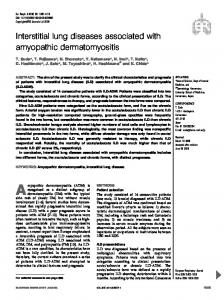
Figure 1: CT cut through the apex of the lungs showing diffuse ground glass opacity with thickening of interlobar septa
Other systems were within normal limits.
Investigations
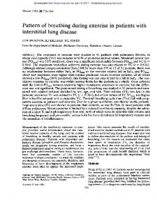
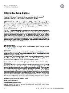
Her packed cell volume was 32% with a white blood cell count of 13.5 ×10⁹/L (neutrophil=52%, lymphocyte=38%, other=9%) and erythrocyte sedimentation rate of 3 mm/h. Sputum examination for acid and alcohol fast bacilli was negative in three samples; sputum culture did not yield any growth. Chest X‑ray showed widespread reticulonodular opacities involving the whole of the right lung field and left upper zone with an area of hyperluscency in the left mid and lower zones. Electrocardiography revealed sinus tachycardia of 120 beats/min, P pulmonale, and right ventricular hypertrophy with ventricular extrasystole. Echocardiography demonstrated tricuspid regurgitation with pulmonary hypertension. Spirometry showed severe restrictive pattern {FVC=1.13 L (36% of predicted); FEV1=0.84 L (35% of predicted) FEV1/FVC ratio=74.5% (92% of predicted)}. Chest CT demonstrated diffuse ground glass opacity with thickening of interlobar septa in the entire right lung field as well as left apex and part of the left lower region. There were multiple bullae with reduced attenuation (suggestive of compensatory emphysema of the remaining lung tissue) in the left lung field, sparing the apex. The pulmonary trunk was prominent and there was cardiomegaly [Figures 1-3].
Figure 2: CT cut through the mid zone of the lung showing diffuse ground glass opacity with thickening of interlobar septa in the right lung field and multiple bullae with reduced attenuation in the left lung field. The pulmonary trunk is prominent and there is cardiomegaly
She was admitted and managed initially in the intensive care unit with intranasal oxygen, cardiopulmonary monitoring, tablet levofloxacin 500 mg daily, intravenous frusemide 60 mg 8 hourly, tablet spironolactone 25 mg twice daily, and tablet aspirin 75 mg daily.
The heart failure gradually improved but she continued to desaturate in room air. She was advised on domiciliary oxygen, which she initially rejected but later purchased a cylinder of oxygen (did not purchase oxygen concentrator because of poor power supply) and pulse oximeter. She was discharged home, on 1–3 L/min of domiciliary oxygen (to be titrated with the pulse oximeter readings), aspirin,
Page | 106
Figure 3: CT cut through the lower zone of the lung and upper abdomen showing ground glass opacity in the right lung field and part of the left lower region
Nigerian Medical Journal | Vol. 53 | Issue 2 | April-June | 2012
[Downloaded free from http://www.nigeriamedj.com on Sunday, November 18, 2012, IP: 82.128.0.54] || Click here to download free Android application for this journ
Fawibe, et al.: Sickle cell associated interstitial lung disease in a Nigerian
and oral diuretics, after 1 month of hospitalization. She was re‑admitted 1 week later with productive cough and features of right heart failure. She was managed with oxygen, diuretics, antibiotics, and aspirin. She improved and was discharged after 3 weeks. She has remained relatively stable at subsequent clinic visits. Her clinic oxygen saturation was 91–94% (off oxygen) during clinic visits.
DISCUSSION
We report a sickle cell patient with CT evidence of extensive interstitial lung disease, abnormal oxygen saturation, and severe restrictive pattern on spirometry as well as clinical, electrocardiographic, and echocardiographic features of pulmonary hypertension and cor pulmonale. These features are in keeping with advanced SCLD in the patient despite being relatively crisis free and without sickle cell‑related hospital admissions for over 20 years prior to presentation. This is likely due to chronic occult pulmonary insults by similar mechanisms to acute chest syndrome. The occult insults, which are too tiny to be discernible at the active stage, accumulate over the years and may later become visible radiologically. As far back as 1970, Lagundoye7 reported generalized loss of translucency of the lung fields on chest radiographs of stable patients with sickle cell anemia and related hemoglobinopathies in Nigeria. Our patient has generalized loss of translucency of the entire right lung field with some involvement of the left lung field. Similarly, the chest CT shows diffuse ground glass opacity with thickening of interlobar septa in the entire right lung field and some involvement of the left lung. There was no diffuse interstitial fibrosis. This is similar to the report of a study on CT scan findings of 29 patients who were 5 to 54 years old in which none of the patients had diffuse interstitial fibrosis or honeycombing.8 Even autopsy studies show little fibrosis.1 Although SCD has been reported as a cause of pulmonary hypertension/cor pulmonale in Nigeria,9 we are not aware of any previous report associating it with CT proven interstitial lung disease in sickle cell patients. SCLD is a major cause of death in adult sickle cell patients. In a longitudinal study, the mean time to death after diagnosis
of patients in advanced SCLD (Stage 3 to Stage 4) was 2.5 years.3 There are no data available to support any specific treatment of interstitial lung disease in sickle cell patients. Little is also known about the factors that determine the development and extent of progression of SCLD. Many of the recent studies on chronic pulmonary complications of SCD are focusing mainly on pulmonary hypertension in sickle cell patients. However, studies are needed to address the pathogenesis and treatment of the interstitial lung disease component of SCLD. While we await further studies and in the absence of proven treatments for SCLD, symptomatic treatment is indicated as we did in our patient.
REFERENCES
1. Weil JV, Catro O, Malik AB, Rodgers G, Bonds DR, Jacobs TP. NHLBI Workshop summary: Pathogenesis of lung disease in sickle hemoglobinopathies. Am Rev Respir Dis 1993;148:249‑56. 2. Yater WM, Hansmann GH. Sickle cell anemia: A new cause of Corpulmonale of right heart failure. Am J med Sci 1936;191:474‑84. 3. Powars D, Weidman JA, Odom‑Maryon T, Niland JC, Johnson C. Sickle cell chronic lung disease: Prior morbidity and the risk of pulmonary failure. Medicine(Baltimore) 1988;67:66‑76. 4. Fawibe AE, Oluboyo PO, Salami AK. Sickle cell chronic lung disease among young adult Nigerians. West Afr J Med 2010;29:30‑3. 5. Jaja SI, Opesanwo O, Mojiminiyi FB, Kehinde MO. Lung function, haemoglobin F and irreversibly sickle cells in sickle cell patients. West Afr J Med 2000;19:225‑9. 6. Fawibe AE, Oluboyo PO, Salami AK. Ventilatory function in young adult Nigerians with sickle cell anaemia. Afr JRespir Med 2008;1:21‑4. 7. Lagundoye SB. Radiological features of sickle cell anaemia and related haemoglobinopathies in Nigeria. Afr J MedSci 1970;1:315‑42. 8. Aquino S, Gamsu G, Fahy J, Claster S, Embury S, Mentzer W, et al. Chronic pulmonary disorders in sickle cell disease: Findings at thin‑section CT. Radiology 1994;193:807‑11. 9. Aliyu ZY, Gordeuk V, Sachdev V, Babadoko A, Mamman AI, Akpanpe P, et al. Prevalence and risk factors for pulmonary artery systolic hypertension among sickle cell disease patients in Nigeria. Am J Hematol 2008;83:485‑90. How to cite this article: Fawibe AE, Kolo PM, Ogunmodede JA, Desalu OO, Salami KA. Advanced sickle cell associated interstitial lung disease presenting with cor pulmonale in a Nigerian. Niger Med J 2012;53:105-7. Source of Support: Nil, Conflict of Interest: None declared.
Announcement
Android App A free application to browse and search the journal’s content is now available for Android based mobiles and devices. The application provides “Table of Contents” of the latest issues, which are stored on the device for future offline browsing. Internet connection is required to access the back issues and search facility. The application is compatible with all the versions of Android. The application can be downloaded from https://market.android.com/details?id=comm.app.medknow. For suggestions and comments do write back to us.
Nigerian Medical Journal | Vol. 53 | Issue 2 | April-June | 2012
Page | 107