Mathiesen, L. R., J. Drucker, D. Lorenz, J. A. Wag- ner, R. J. Gerety, and R. H. Purcell. 1978. Localiza- tion of hepatitis A antigen in marmoset organs during.
INFECTION AND IMMUNITY, Apr. 1980, p. 45-48 0019-9567/80/04-0045/04$02.00/0
Vol. 28, No. 1
Hepatitis A Virus in the Liver and Intestine of Marmosets After Oral Inoculation LARS R. MATHIESEN,'* ANNE MARIE M0LLER,' ROBERT H. PURCELL,2 WILLIAM T.
LONDON,' AND STEPHEN M. FEINSTONE' Enterovirus Department, Statens Seruminstitut, Copenhagen, Denmark' and Hepatitis Viruses Section, National Institute ofAllergy and Infectious Diseases, Bethesda, Maryland 20205' A total of 12 seronegative marmosets (Saguinus mystax) were inoculated orally with hepatitis A virus (HAV) and sacrificed at 3- to 4-day intervals. Tissues from the livers, intestines, mesenteric lymph nodes, and spleens were obtained for immunofluorescence studies, and bile and intestinal contents were obtained for enzyme-linked immunosorbent assay studies. Two marmosets sacrificed on days 34 and 41 after inoculation developed antibody to HAV and demonstrated HAV in their livers but not in any part of their intestinal tissues. None of the remaining marmosets sacrificed from days -3 to 31 survived long enough to develop antibody to HAV, but an additional two marmosets, which were sacrificed on days 21 and 31, demonstrated HAV in their livers and also in bile but not in the intestinal tissues or their contents. The mesenteric lymph nodes and spleens were negative for HAV by immunofluorescence in all of the marmosets. No evidence of HAV replication was demonstrated in any part of the intestine at any time during the incubation period or during acute illness in the marmosets inoculated orally with HAV. The shedding of HAV in stools in the late incubation period can be explained by excretion of HAV from the livers with the bile. In 1958 Ward et al. demonstrated that stools collected during the incubation period of hepatitis A (2 to 3 weeks before jaundice) were infectious (11). They concluded that hepatitis A, like poliomyelitis (1, 10), had an alimentary phase in which virus multiplied and was excreted before any signs of liver dysfunction were present. Since 1973, when Feinstone et al. first reported a serological technique for detection of hepatitis A virus (HAV) and antibody to HAV (antiHAV) (5), several studies have demonstrated that HAV is excreted in stools in the late incubation period (2-4). This was interpreted as additional evidence of an intestinal phase of replication, although the possibility of primary replication in the liver with excretion of HAV in the bile could not be excluded (3). In 1977 we reported the detection of HAV by immunofluorescence in liver biopsies from chimpanzees experimentally infected intravenously (8). HAV could be demonstrated in livers before fecal shedding in stools, but an intestinal biopsy taken during the acute stage was negative for HAV. We therefore suggested that liver and not intestine was the principal site of infection. In a later study with marmosets, which were also inoculated intravenously with HAV, intestinal biopsies obtained during the acute phase were all negative for HAV, whereas HAV could be demonstrated in the livers and, in some of the marmosets, also in the spleens, lymph nodes,
and kidneys (7). These data supported the theory that HAV does not replicate extensively in the intestine, at least during the acute phase of illness after inoculation by the intravenous route. To determine whether there is an intestinal phase of replication at some time after oral inoculation with HAV, we looked for HAV by immunofluorescence in livers and intestinal biopsies with tissues obtained from marmosets inoculated orally with HAV and sacrificed at different times after inoculation. MATERIALS AND METHODS Animals and inoculum. The marmosets (Saguinus mystax) used in this study were jungle caught and were obtained through the Division of Research Resources, National Institutes of Health. Feeding and handling of the marmosets has been described previously (6). A total of 15 animals without anti-HAV were used in this study. Of these, one was killed without inoculation and served as a negative control. The other 14 were inoculated via gastric tubes with 1 ml of a 2% stool suspension containing HAV shown to be infectious for chimpanzees at a dilution of at least 10' when given intravenously (3; Purcell, unpublished data). Clinical specimens. Two of the inoculated marmosets were followed with daily stool collections and twice weekly percutaneous needle liver biopsies and serum samples and served as positive controls to assure that the inoculum was infectious. The remaining 45
46
MATHIESEN ET AL.
INFECTr. IMMUN.
12 marmosets were bled twice weekly but not biopsied; they were sacrificed at intervals of approximately 3 days, and tissues from the peripheral and central parts of the livers, spleens, and mesenteric lymph nodes, as well as cross-sections from the duodena, jejuna, ilea, and transverse colons, were obtained. The tissues were embedded in Tissue Tac, snap frozen in liquid nitrogen, and stored at -70'C. In addition, sera, bile, and contents of the different parts of the intestines were collected and stored at -70'C. Liver enzyme determinations. Alanine aminotransferase and isocitrate dehydrogenase were measured on serum samples obtained twice a week from all of the marmosets, beginning before inoculation. Fluorescent conjugate. Anti-HAV fluorescein isothiocyanate conjugated human convalescent immunoglobulin G has been described previously (7). Immunofluorescence. Cross sections (thickness, 4 ,um) were cut on a cryostat and placed on glass slides pretreated with gelatin. The slides were air dried and stained unfixed by applying 1 drop of fluorescein isothiocyanate conjugate on the tissue; this was followed by incubation for 30 min in a humidified chamber at 200C. Thereafter, the slides were washed three times in phosphate-buffered saline, and a cover slip was applied over mounting media consisting of 90% glycerine in tris(hydroxymethyl)aminomethane buffer
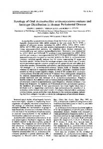
when they were sacrificed but not in the sera obtained before that time. In the remaining marmosets anti-HAV could not be demonstrated at any time (Fig. 1). HAV by immunofluorescence. HAV was found in the livers at necropsy in the marmosets sacrificed on days 21, 31, 34, 41, and 63 but not in the marmosets killed on days -3, 3, 8, 9, 10, 14, 17, 21, and 24 (Fig. 1). In the serial liver biopsies taken biweekly from the two control marmosets, HAV was found in all liver biopsies from day 18 to the death of one animal on day 63, but only in the biopsy taken on day 42 in the other animal (Fig. 1). HAV could not be demonstrated in any of the biopsies from duodena, jejuna, ilea, transverse colons, mesenteric lymph nodes, or spleens from any of the marmosets. HAV by enzyme-linked immunosorbent assay. The marmoset sacrificed on day 41 was the only one in which HAV could be demonstrated in the intestinal contents. This marmoset also demonstrated HAV in its liver, but unfortunately no bile was available. Two of the marmosets demonstrated HAV in (pH 9.5). The slides were examined immediately with a Leitz their bile as well as in their livers when they Ortholux 2 microscope equipped with an HBO 200 W/ were sacrificed on days 21 and 31, but HAV 2 mercury light source and vertical illuminator with a could not be demonstrated in their intestinal model 500 dichroic mirror reflector. The excitation contents. filter combination was KP 490 plus LP 470 plus BG The marmosets sacrificed on days 34 and 63 38. A 2-mm LP 520 filter was used as barrier filter. An did not have HAV in their bile or their intestinal x40 fluorescence objective with an aperture of 1.3 was contents, although they demonstrated HAV in used in combination with x6.3 oculars. Blocking tests. In all biopsies in which HAV was their livers. In the stools collected daily from the two demonstrated, the specificity was confirmed by a blocking test read under code, as previously described positive control marmosets, HAV was demonstrated from days 19 to 36 in one of the animals (7,8). Serological testing. Sera were tested for anti- but could not be demonstrated in stools collected HAV by blocking enzyme-linked immunosorbent as- from days 20 to 96 from the other marmoset. say (9). The bile and a 20% suspension of the stools Unfortunately, stools collected before day 20 and intestinal contents were tested for HAV by en- were not available from the second control marzyme-linked immunosorbent assay (9). moset.
RESULTS Liver enzyme elevations. The two positive control animals had peak elevations of alanine aminotransferase and isocitrate dehydrogenase on days 35 and 46, respectively, and the marmoset killed on day 34 had an elevation on day 32. The marmoset sacrificed on day 41 and the remaining marmosets, which were sacrificed on days 3 through 31, did not show elevation in alanine aminotransferase or isocitrate dehydrogenase (Fig. 1). Seroconversion for anti-HAV. The two positive control marmosets seroconverted for anti-HAV after enzymes became elevated (days 39 and 49) and were positive for anti-HAV in the subsequent sera. The two animals sacrificed on days 34 and 41 showed anti-HAV in their sera
DISCUSSION Previous studies with intravenous inoculations of HAV into marmosets have shown that although not all marmosets have elevated alanine aminotransferase or isocitrate dehydrogenase, they do all seroconvert for anti-HAV, indicating that they are all infected (7). Our results confirm that marmosets are also susceptible to oral inoculation with HAV; all four marmosets followed for 32 days or more seroconverted for anti-HAV and demonstrated HAV in their livers. Two additional marmosets sacrificed on days 21 and 31 demonstrated HAV in the liver biopsies but had not yet seroconverted. In the remaining marmosets, which were sacrificed between days 3 and 27, no evidence of
VOL. 28, 1980
HAV AFTER ORAL INOCULATION
47
POSITIVE CONTROL MARMOSETS NO.14 AND 15 SERUM ICO HAV IN LIVER HAV IN STOOL ANTI -WAV IN1 IN Itpaim brcKnP ANTI-HAV
t
1
SERUMI CD
EJ
HAV IN LIVER HAV IN STOOL ANTI-HAV IN SERUMQ
SEQUENTIALLY SACRIFICED MARMOSETS S 9 10
MARMOSET NO
1
2
3LS
6
11 12
13
SERUM ICD LIVER
0
0
000
0 0
0
0 0
0
+
0
0
0
0
000
0
0
+
0
0
+ +
+
+
0 0
0
0
+
0
0
+ 0
0 Z INTESTINE *. CONTENT 0 >
0 0 0
0 0 000
0 0
BILE
SMALL
INTESRINE Id
2
0
CONTENTS 0
MESENTERIC LYMPH NODE 0 ANTI-HAV IN SERUMI0
-3 0
0 0
7
00
0 0 0 0 0 0 0 0 0000 0 000000
000
0
0
10
0
0
0
20
0
0
0 0 0
+ 0
0
0
0
0
0
0
0 0 0 +
0 +
0
0
0 0
00000 000
14
+
50 d0 30 DAYS AFTER INOCULATION
60
70
FIG. 1. Detection of serum isocitrate dehydrogenase (aCD) elevation, HA V, and anti-HA V in 2 marmosets followed longitudinally and 13 marmosets sacrificed sequentially after oral inoculation with HA V. Superscript I indicates identical results in tissues from central and peripheral parts of the liver. Superscript 2 indicates that tissues and intestinal contents from duodenum, jejunum, and ileum were tested separately.
infection with HAV was demonstrated. We can therefore only assume that all of the marmosets were infected and would have seroconverted and demonstrated HAV in their livers if they had been followed to the acute stage instead of being sacrificed during the incubation period. We were unable to demonstrate HAV by immunofluorescence in any part of the intestinal tissue during the incubation period and the acute stage of illness. Mesenteric lymph nodes and spleens were also negative by immunofluorescence, whereas HAV could be demonstrated readily in the livers during the late incubation period and the acute stage of illness. Specifically, two marmosets had HAV in their livers and bile in the late incubation period but not in their intestinal tissues or contents. Similarly, one animal had HAV in its liver and ileal contents but not in its intestinal tissue. Thus, HAV in the intestinal tract appeared to originate from the liver via the bile. We have demonstrated previously that immunofluorescence is very sensitive for detection of HAV in tissue (7) and that the enzyme-linked immunosorbent assay is equally sensitive for detection of HAV in intestinal contents (9), so it is unlikely that our negative findings in the intestinal tissues and contents are due to insensitivity of the techniques. However, it is difficult to exclude the possibility that there may be a period of intestinal multiplication of HAV, brief both in duration and extent, that precedes multiplication in the liver and indeed is the source of the virus that
seeds the liver via the mesenteric lymphatic or circulatory system. If so, we were unable to detect it with the methods used here. However, HAV produced in the liver and excreted with the bile appears to be more important for the shedding of HAV in stools than HAV derived from the intestine, if in fact it exists. ACKNOWLEDGMENTS Financial support from the Michaelsen Foundation, the Danish Foundation for the Advancement of Medical Science, the P. Carl Petersen Foundation, the Danish Medical Research Council (grant 512-10472), and the Johan and Hanne Weimann's Foundation is gratefully acknowledged. LITERATURE CITED 1. Bodian, D. 1956. Poliovirus in chimpanzee tissues after virus feeding. Am. J. Hyg. 64:181-197. 2. Bradley, D. W., C. R. Gravelle, E. H. Cook, R. M. Fields, and J. E. Maynard. 1977. Cyclic excretion of hepatitis A virus in experimentally infected chimpanzees: biophysical characterization of the associated HAV particles, J. Med. Virol. 1:133-138. 3. Dienstag, J. L., S. M. Feinstone, R. H. Purcell, J. H. Hoofhagle, L F. Barker, W. T. London, H. Popper, J. M. Peterson, and A. Z. Kapikian. 1975. Experimental infection of chimpanzees with hepatitis A virus. J. Infect. Dis. 132:532-545. 4. Dienstag, J. L., A. Z. Kapikian, S. M. Feinstone, R. H. Purcell, J. D. Boggs, and M. E. Conrad. 1975. Faecal shedding of hepatitis A antigen. Lancet i:765767. 5. Feinstone, S. M., A. Z. Kapikian, and R. H. Purcell. 1973. Hepatitis A: detection by immune electron microscopy of a virus-like antigen associated with acute illness. Science 182:1026-1028. 6. Lorenz, D., L Barker, D. Stevens, M. Petersen, and R. Kirschstein. 1970. Hepatitis in the marmoset, Saguinus mystax. Proc. Soc. Exp. Biol. Med. 135:348-354.
48
MATHIESEN ET AL.
7. Mathiesen, L. R., J. Drucker, D. Lorenz, J. A. Wag-
R. J. Gerety, and R. H. Purcell. 1978. Localization of hepatitis A antigen in marmoset organs during acute infection with hepatitis A virus. J. Infect. Dis. 138:369-377. 8. Mathiesen, L. R., S. M. Feinstone, R. H. Purcell, and J. A. Wagner. 1977. Detection of hepatitis A antigen by immunofluorescence. Infect. Immun. 18:524-530. 9. Mathiesen, L. R., S. M. Feinstone, D. C. Wong, P. Skinhoej, and R. H. Purcell. 1978. Enzyme-linked ner,
INFECT. IMMUN. immunosorbent assay for detection of hepatitis A antigen in stools and antibody to hepatitis A antigen in sera: comparison with solid-phase radioimmunoassay, immune electron microscopy, and immune adherence hemagglutination assay. J. Clin. Microbiol. 7:184-193. 10. Sabin, A. B. 1956. Pathogenesis of poliomyelitis. Reappraisal in the light of new data. Science 123:1151-1157. 11. Ward, R., S. Krugman, J. P. Giles, A. M. Jacobs, and 0. Bodansky. 1958. Infectious hepatitis. N. Engl. J. Med. 258:407-416.