Communicated by Alexander G. Bearn, June 9, 1986. ABSTRACT. The antigenic group ..... Humphries, S. E., Berg, K., Gill, L., Cumming, A. M.,. Robertson, F. W. ...
Proc. Natl. Acad. Sci. USA Vol. 83, pp. 7367-7370, October 1986 Genetics
Genetic linkage between the antigenic group (Ag) variation and the apolipoprotein B gene: Assignment of the Ag locus (low density lipoprotein/Ag polymorphism/restriction fragment length polymorphism/haplotypes)
K. BERG*, L. M. POWELL+, S. C. WALLISt, R. PEASEt, T. J. KNOTT+, AND J. SCOTTt *Institute of Medical Genetics, University of Oslo, Norway; and tMedical Research Council, Clinical Research Centre, Harrow, United Kingdom
Communicated by Alexander G. Bearn, June 9, 1986
ABSTRACT The antigenic group (Ag) system of homospecific human serum antigens of low density lipoprotein is detected by antiserum from multiply transfused patients. A complex series of common Ag alleles has been described, but the biochemical nature of this polymorphism is uncertain. Here we report that DNA polymorphisms at the human apolipoprotein B (apoB) locus are very closely linked to alleles of the Ag system. We also show a strong association between Ag(x) and a polymorphism detected with the restriction endonuclease Xba I. We conclude that the immunologically determined Ag system represents protein polymorphism of apoB rather than primary genetic differences in posttranslational processing or lipid binding. These studies therefore demonstrate that the Ag locus is located on the short arm of human chromosome 2 in the region p23-p24 to which the apoB gene has been assigned. Since the Ag(x) antigen is associated with altered plasma lipid levels, this determinant may indicate a functionally important domain of apoB.
to be associated with altered plasma lipid levels in several middle-aged populations (7). Individuals who lacked the Ag(x) trait [Ag(x-)] had higher levels of fasting triglycerides and cholesterol than those with the Ag(x) trait [Ag(x+)]. No direct association between Ag(x) and premature atherosclerosis has been reported (6, 8). However, in pigs, genetic polymorphism of LDL has been strongly correlated with premature atherosclerosis (9). cDNA clones for human apoB have been identified and have been used to detect restriction fragment length polymorphisms (RFLPs) of the apoB gene (10-17). Here we describe close genetic linkage between the Ag variation, expressed as immunological allotypes of LDL and DNA polymorphisms at the apoB locus. We also report a strong association between Ag(x) and a polymorphism detected with the restriction endonuclease Xba I.
MATERIALS AND METHODS Subjects. From families of Norwegian monozygotic twins (18), we selected for DNA study those that had maximum potential linkage information for Ag(x). In most cases, one
Epidemiological studies have shown a strong correlation between high plasma low density lipoprotein (LDL) cholesterol levels and premature coronary heart disease. Plasma LDL is responsible for the transport of cholesterol between peripheral tissues and the liver. LDL concentration is regulated by its rate of production from very low density lipoprotein and clearance by the hepatic LDL receptor. The sole protein component of LDL is apolipoprotein B (apoB), which is essential for the structural integrity of the particle and is the ligand that mediates its clearance by the LDL receptor pathway (1-3). The antigenic group (Ag) system of homospecific human serum alloantigens was detected in 1961 by the use of an antiserum from a multiply transfused patient (4). Subsequently, the Ag system was shown to reside in LDL and a complex series of alleles has since been described (5). These variants have been shown to behave as autosomal dominant or codominant traits in families and to have differences in gene frequency between populations. Linkage between the Ag system and other genetic markers has not been demonstrated and the biochemical nature of this polymorphism has not been elucidated (6). It is not known, therefore, whether the Ag antigenic determinants reside in apoB, the carbohydrate side chains of apoB, or in LDL lipid. Homospecific polymorphism of LDL has also been found in most animal species that have been studied and appears to be a ubiquitous phenomenon (6). Plasma LDL concentration is profoundly affected by hereditary factors. Thus defects of the LDL receptor gene cause familial hypercholesterolemia with a marked increase in LDL and severe premature atherosclerosis (1, 2). A component of the Ag system termed Ag(x) has been reported
analyze the polymorphisms described (11-13). The apoB probe used was that reported by Knott et al. (10). Linkage Analyses. This study comprises only two generation families. They were scored for linkage likelihood between Ag(x) and the apoB gene polymorphisms according to published lod score tables (20). Lod score (log1o of the odds in favor of linkage) is a statistic that assesses the probability of linkage between genetic markers over a range of possible recombination fractions. The maximum lod score indicates the most likely value of the recombination fraction. A lod score of 3 represents odds in favor of linkage of 103:1. Association Tests. Population association between Ag(x) and the various apoB DNA polymorphisms was sought by x2 analyses.
The publication costs of this article were defrayed in part by page charge payment. This article must therefore be hereby marked "advertisement" in accordance with 18 U.S.C. §1734 solely to indicate this fact.
Abbreviations: apoB, apolipoprotein B; LDL, low density lipoprotein; Ag, antigenic group (system of inherited allotypes in LDL); RFLP, restriction fragment length polymorphism; kb, kilobase(s).
7367
parent was Ag(x+) and the other was Ag(x-) and there was at least one Ag(x-) child [to prove that the Ag(x+) parent was heterozygous]. The families reported here were in good health. Unrelated individuals (spouses of twins and one member of each twin pair) from these families were studied to reveal population association. Ag Testing. Antiserum to the Ag(x) factor was identified as described (5). Individuals may be unambiguously scored as Ag(x+) or Ag(x-) according to whether or not their sera produce a precipitin band in agar gel double-diffusion analyses. DNA Analyses. DNA was prepared from leukocytes and Southern hybridization was performed as described (19). The restriction enzymes EcoRI, Msp I, and Xba I were used to
7368
Genetics:
Berg et al.
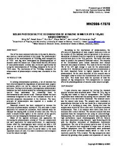
Proc. Natl. Acad. Sci. USA 83 (1986) 8.6kb
Xbal
fragmentj
5kb
3.6kb
2.8kb
sizes
5'
XB
S E
HF(X)E
EHHEB
E X
II
I I
I( 1
I
I1f1
I I
(E)HH X BHXBH B
IrIInu1III
E
i 3'
ApoB Gene EcoR 1 fragment
1.2kb 1.7kb
2.1 kb
12.0kb I
sizes
14. 1kb
cDNA Probe AB 1 FIG. 1. Restriction map of the 3' half of the apoB gene. The sizes of Xba I and EcoRI fragments detected by the cDNA probe ABI (11-13) are shown. Solid box indicates the position of the A+T-rich repeat shown in Fig. 3. X, Xba I; B, BamHI; S, Sal I; E, EcoRI; H, HindIII.
b
kb
_m 0
^w
a
sew
4-
-- -14 -12
kb
__n.
a*
40 *t *IN
4f
-58.6 c
_i
-
5.0
-2.1 A_
__
of
kb 2.8 -;;- ~~2.6
-1.7 :.. ':
*nQiNS 4 El a0
m- 2.8
--
-1.2
---
lqqwl.l
F, --Mdmhm wqw"m;wm4w
INIEW
MI&
\ 2.2
FIG. 2. Autoradiographs of Southern blots. (a) RFLP of the restriction endonuclease Xba I showing homozygotes for 8.6-kb and 5.0-kb fragments and heterozygotes. (b) RFLP of the restriction endonuclease EcoRI showing homozygotes for 14.1-kb and 12.0 plus 2.1-kb fragments and heterozygotes. (c) RFLPs generated by cleavage with the restriction endonuclease Msp I on either side of the A+T-rich repeat shown in Fig. 3. The sizes of the allele fragments are 2.6, 2.4, and 2.2 kb.
R
EM
I
II
H
H
X
I
I
B
M I
H II
R
A
TTTTATAATTA AAATATTTATAATT
0
A
ATA]20
1KB
FIG. 3. A+T-rich repeat in 3' flanking sequence of apoB gene. Solid box represents 20 copies of the 30-base-pair repeat. Solid triangle indicates the position of the polyadenylylation signal (AATAAA) commonly used in apoB mRNAs. R, EcoRV; E, EcoRI; M, Msp I; H, HindIII; X, Xba I.
Proc. Natl. Acad. Sci. USA 83 (1986)
Genetics: Berg et al.
7369
Table 1. Lod scores between immunogenetic Ag polymorphism in LDL and RFLPs at the apoB locus Segregation from Males
Females
Family no. 29185 45107 46147 33036 45166
Scored as ZI, 2:0 Z1, 2:0 ZI, 2:0 ZI, 4:0 Z1, 3:0 Total ZI, 3:0 Z1, 3:0 Z1, 2:0 Z1, 3:0 Total
29076 40262 37042 45297
Both
0.00 0.301 0.301 0.301 0.903 0.602 2.408 0.602 0.602 0.301 0.602 2.107 4.515
0.05 0.258 0.258 0.258 0.814 0.533 2.121 0.533 0.533
0.258 0.533 1.857 3.978
RESULTS DNA Polymorphisms. A restriction map of the 3' half of the human apoB gene is shown in Fig. 1. The map was constructed from restriction digests of human genomic DNA and from overlapping X bacteriophage that span the apoB gene. Polymorphic Xba I and EcoRI sites are shown. The DNA restriction pattern produced by Xba I consisted of an invariant 2.8-kilobase (kb) fragment and either 8.6-kb (genotype X2X2) or 5.0-kb (genotype XX,) fragments in homozygous individuals or both 8.6- and 5.0-kb fragments in heterozygotes (genotype XX2) (Fig. 2a). The pattern for human genomic DNA digested with EcoRI consists of invariant 1.7-kb and 1.2-kb fragments and either a 14.1-kb (genotype R2R2) or a 12- and a 2.1-kb (genotype RR1) fragment in homozygotes or 14.1-, 12-, and 2.1-kb fragments in heterozygotes (genotype RR2) (Fig. 2b). DNA sequencing and restriction mapping of the 3' flanking sequence of the apoB gene has identified a simple repetitive element composed of a 30-base-pair A+T-rich sequence (Fig. 3). The number of copies of the repeat varies between individuals giving rise to RFLPs, which can be generated by restriction enzymes such as Msp I, BamHI, and EcoRV that cleave the genomic DNA on either side of the repeat. The pattern of this repeat after cleavage with Msp I is shown in Fig. 2c and consists of an invariant 2.8-kb fragment and various combinations of 2.6-kb, 2.4-kb, and 2.2-kb allele fragments. This suggests a three-allele system due to variation in copy number of the 30-base-pair repeat. Similar repeats have been reported in the flanking sequence of the insulin, c-Ha-rasl, type II collagen, and globin genes (21-25). Genetic Analysis. Nine two-generation families with a total of 24 children were informative for the linkage relationship between Ag(x) and apoB. Four of the families were scored with the Msp I polymorphism and five were scored with the Xba I polymorphism. Of the four families analyzed with Msp I, two were also informative for the EcoRI polymorphism. There was no recombination between the three DNA polymorphisms. Table 2. Association tests between the Ag(x) polymorphism of
allotypic LDL variants and apoB DNA polymorphisms detectable with endonucleases Xba I and EcoRI, respectively DNA
No. of individuals Ag(x+) Total 22 25 3 X2 = 7.3 40 16 24 P = 003 10 25 21 48 27 4 22 18 P 0 2 JP00 2
polymorphism Genotype Ag(x-) Xba I
XIX,
X1X2
X2X2 EcoRI
RR, RR2
R2R2
2'= =50.06
Recombination fraction 0.10 0.20 0.215 0.134 0.215 0.134 0.215 0.134 0.517 0.720 0.465 0.318 1.830 1.237 0.465 0.318 0.465 0.318 0.215 0.134 0.465 0.318 1.610 1.088 3.440 2.325
Enzyme used 0.30 0.064 0.064 0.064 0.298 0.170 0.660 0.170 0.170 0.064 0.170 0.574 1.234
0.40 0.017 0.017 0.017 0.094 0.049 0.194 0.049 0.049 0.017 0.049 0.164 0.358
variation Xba I Xba I Xba I Msp I Msp I, EcoRI Msp I, EcoRI Msp I Xba I Xba I
Table 1 shows the lod scores at different values of the recombination fraction for Ag (determined by immunological tests) and the apoB DNA polymorphisms for males, females, and both sexes combined. No evidence for recombination was found and the total lod score at recombination fraction zero is 4.5. This proves genetic linkage between Ag(x) and apoB by conventional criteria. Association Tests. Seventy-five unrelated persons were used to test for association between Ag(x) and the RFLPs. A strong association was found between Ag(x) and the 8.6-kb allele in the polymorphism detected with Xba I (Table 2). However, the Xba I polymorphism cannot reflect the Ag(x) variation itself, since Ag(x+) as well as Ag(x-) persons were found in both categories of Xba I homozygotes. An association of borderline significance was found between Ag(x) and the 12/2.1-kb allele detected with EcoRI (Table 2). No association was found between Ag and the polymorphism detected with Msp I (results not shown). The Xba I and EcoRI polymorphisms exhibited very strong population association (x2 = 40.4).
DISCUSSION This study shows that alleles of the Ag system of immunogenetic variants are very closely linked to the human apoB gene. This is demonstrated by cosegregation of RFLPs at the apoB locus with Ag(x) alleles (lod score 4.5 at zero recombination fraction). Thus, the Ag system almost certainly reflects protein polymorphism of apoB, and not primary genetic differences in the posttranslational processing of apoB or in the amount of lipid bound to apoB. From these results, we can therefore assign Ag to human chromosome 2 in the p23-p24 region to which the apoB gene has been assigned (10, 26). Strong association has been found between Ag(x) and a polymorphic site generated by the restriction endonuclease Xba I. The polymorphic Xba I site is in the coding sequence of the apoB mRNA, the entire 14.1-kb sequence of which we have recently determined. However, loss of the Xba I site is caused by a silent cytosine to thymine mutation in the third base of the threonine codon at residue 2488 in the mature protein and cannot represent the Ag(x) determinant (unpublished results). The Ag(x) determinant must represent a distinct protein sequence change that is in strong linkage disequilibrium with the polymorphic Xba I site. To establish the precise nature of the mutation that underlies the Ag(x) determinant, expressed fragments of cDNA representing the two Xba I alleles are being studied. Since the Ag(x) determinant is associated with altered plasma lipid levels, its identification may focus attention on functionally important lipid binding domains of apoB (7). It is possible, as suggested
7370
Genetics: Berg et al.
by Fisher et al., that the Ag system affects the size of the lipoprotein particles (27). The EcoRI site polymorphism alters the apoB protein sequence by substituting lysine (AAA) for glutamic acid (GAA) at residue 4154 in the mature protein. However, the association between the EcoRI site polymorphism and Ag(x) is weak compared to the association between Ag(x) and the Xba I polymorphism, and the EcoRI polymorphism does not affect lipid levels (unpublished results). No association has been found betwen Ag(x) and the alleles generated by the A+T-rich repeat in the 3' flanking sequence of the gene. The lack of association between Ag(x) and the alleles generated by the 3' repeats, the strong association between Ag(x) and the Xba I polymorphism, and the borderline association between Ag(x) and the EcoRI polymorphism suggest that Ag is 5' to the polymorphic EcoRI site (Fig. 1). The allelic association discovered suggests that it will be valuable to study the effect of specific haplotypes of Ag and various RFLPs on lipid and lipoprotein variation. This is particularly interesting because of the association between Ag types and lipid levels (7), since it is possible that some haplotypes are associated with a greater part of the effect on lipid level apparently caused by the Ag variation. This view is supported by our recent finding of strong association between the Xba I polymorphism and altered plasma cholesterol and triglyceride levels (28). The skillful assistance of Elisabeth M0kleby, Helen Brunt, and Linda Priestley is gratefully acknowledged. This work was supported by the Norwegian Research Council for Science and the Humanities, the Norwegian Council on Cardiovascular Disease, and Anders Jahres Foundation for the Promotion of Science. 1. Havel, R. J., Goldstein, J. L. & Brown, M. S. (1980) in Metabolic Control and Disease, eds. Bondy, P. K. & Rosenberg, L. E. (Saunders, Philadelphia), pp. 393-494. 2. Goldstein, J. L. & Brown, M. S. (1983) in The Metabolic Basis of Inherited Disease, eds. Stanbury, J. B., Wyngaarden, J. B., Fredrickson, D. S., Goldstein, J. L. & Brown, M. S. (McGraw-Hill, New York), 5th Ed., pp. 672-712. 3. Kane, J. P. (1983) Annu. Rev. Physiol. 45, 637-650. 4. Allison, A. C. & Blumberg, B. S. (1961) Lancet i, 634-637. 5. Berg, K. & Beam, A. G. (1970) Clin. Genet. 1, 104-120. 6. Berg, K. (1979) in The Biochemistry of Atherosclerosis, eds. Scanu, A. M., Wissler, R. W. & Getz, G. S. (Dekker, New York), pp. 419-490. 7. Berg, K., Hames, C., Dahlen, G., Frick, M. H. & Krishan, I. (1976) Proc. Natl. Acad. Sci. USA 73, 937-940.
Proc. Natl. Acad. Sci. USA 83 (1986) 8. Berg, K. (1983) in Progress in Medical Genetics, eds. Steinberg, A. G., Beam, A. G., Motulsky, A. G. & Childs, B. (Saunders, Philadelphia), Vol. V, pp. 35-90. 9. Rapacz, J., Elson, C. E. & Lalich, J. J. (1977) Exp. Mol. Pathol. 27, 249-261. 10. Knott, T. J., Rall, S. C., Jr., Innerarity, T. L., Jacobson, S. F., Urdea, M. S., Levy-Wilson, B., Powell, L. M., Pease, R. J., Eddy, R., Nakai, H., Byers, M., Priestley, L. M., Robertson, E., Rall, L. B., Betscholtz, C., Shows, T. B., Mahley, R. W. & Scott, J. (1985) Science 230, 37-43. 11. Priestley, L., Knott, T., Wallis, S., Powell, L., Pease, R. & Scott, J. (1985) Nucleic Acids Res. 18, 6790. 12. Priestley, L., Knott, T., Wallis, S., Powell, L., Pease, R. & Scott, J. (1985) Nucleic Acids Res. 18, 6792. 13. Priestley, L., Knott, T., Wallis, S., Powell, L., Pease, R. & Scott, J. (1985) Nucleic Acids Res. 18, 6793. 14. Deeb, S. S., Motulsky, A. G. & Albers, J. J. (1985) Proc. Natl. Acad. Sci. USA 82, 4983-4986. 15. Huang, L. S., Bock, S. C., Feinstein, S. I. & Breslow, J. L. (1985) Proc. NatI. Acad. Sci. USA 82, 6825-6829. 16. Lusis, A. J., West, R., Mehrabian, M., Reuben, M. A., LeBoeuf, R. C., Kaptein, J. S., Johnson, D. F., Schumaker, V. N., Yuhasz, M. P., Schotz, M. C. & Elovson, J. (1985) Proc. Natl. Acad. Sci. USA 82, 4597-4601. 17. Protter, A. A., Handman, D. A., Schilling, J. W., Miller, J., Appleby, V., Chen, G. C., Kirsher, S. W., McEnroe, G. & Kane, J. P. (1986) Proc. Natl. Acad. Sci. USA 83, 1467-1471. 18. Berg, K. (1981) Twin Research 3: Epidemiological and Clinical Studies (Liss, New York), pp. 117-130. 19. Humphries, S. E., Berg, K., Gill, L., Cumming, A. M., Robertson, F. W., Stalenhoef, A. F. H., Williamson, R. & B0rresen, A.-L. (1984) Clin. Genet. 26, 389-396. 20. Maynard-Smith, S., Penrose, L. S. & Smith, C. A. B. (1961) Mathematical Tables for Research Workers in Human Genetics (Churchill, London). 21. Spritz, R. A. (1981) Nucleic Acids Res. 9, 5037-5047. 22. Bell, G. I., Selby, M. J. & Rutter, W. J. (1982) Nature (London) 299, 31-35. 23. Proudfoot, N. J., Gil, A. & Maniatis, T. (1982) Cell 31, 553-563. 24. Capon, D. J., Chen, E. Y., Levinson, A. D., Seeburg, P. H. & Goeddel, D. V. (1983) Nature (London) 302, 33-37. 25. Stoker, N. G., Cheah, K. S. E., Griffin, J. R., Pope, F. M. & Solomon, E. (1985) Nucleic Acids Res. 13, 4613-4622. 26. Huang, L.-S., Miller, D. A., Bruns, G. A. P. & Breslow, J. L. (1986) Proc. Natl. Acad. Sci. USA 83, 644-648. 27. Fisher, W. R., Hammond, M. G., Mengel, M. C. & Warmke, G. L. (1975) Proc. Natl. Acad. Sci. USA 72, 2347-2351. 28. Law, A., Wallis, S. C., Powell, L. M., Pease, R., Brunt, H., Priestley, L. M., Knott, T. J., Scott, J., Altman, D. G., Miller, G. J., Rajput, J. & Miller, N. E. (1986) Lancet i, 1301-1303.