© 2002 Oxford University Press
Human Molecular Genetics, 2002, Vol. 11, No. 4
397–409
Aire deficient mice develop multiple features of APECED phenotype and show altered immune response Chris Ramsey1, Ola Winqvist2, Lea Puhakka1, Maria Halonen3,4, Aune Moro1, Olle Kämpe2, Petra Eskelin3, Markku Pelto-Huikko5 and Leena Peltonen1,3,6,* 1Department
of Human Genetics, UCLA School of Medicine, Gonda Center, University of California Los Angeles, Los Angeles, CA, USA, 2Department of Internal Medicine, University Hospital of Uppsala, Uppsala, Sweden, 3Department of Human Molecular Genetics, National Public Health Institute, Helsinki, Finland, 4Hospital for Children and Adolescence, Helsinki University Hospital, Helsinki, Finland, 5Department of Developmental Biology and Pathology, Medical School, Tampere University and Tampere University Hospital, Tampere, Finland and 6Department of Medical Genetics, University of Helsinki, Helsinki, Finland Received October 5, 2001; Revised and Accepted December 5, 2001
Autoimmune polyendocrinopathy-candidiasis-ectodermal dystrophy (APECED) is a monogenic autosomal recessive disease caused by mutations in the AIRE gene. Here we have produced knock-out mice for the Aire gene. The Aire–/– mice develop normally; however, autoimmune features of APECED in Aire–/– mice are evident, including multiorgan lymphocytic infiltration, circulating autoantibodies and infertility. The distribution of B and T cells and thymic maturation as well as activation of T cells appear normal, while the TCR-Vβ repertoire is altered in peripheral T cells of Aire–/– mice. When mice are challenged with immunization, the peripheral T cells of Aire–/– mice have a 3–5-fold increased proliferation. These findings suggest that the Aire gene is not necessary for normal T cell education and development, while a defect in immune response detected in challenged Aire–/– mice underlines the crucial role of AIRE/Aire in maintaining homeostatic regulation in the immune system.
INTRODUCTION Autoimmune polyendocrinopathy-candidiasis-ectodermal dystrophy (APECED; OMIM 240300), also known as autoimmune polyglandular syndrome type I (APS 1), is caused by mutations in the AIRE gene on chromosome 21 (1,2). APECED is an autoimmune disorder mapped outside the major histocompatibility complex (MHC) (3,4) region with a higher prevalence in genetically isolated populations, such as Finns, Sardinians and Iranian Jewish (5–7). Clinically, APECED patients suffer from the autoimmune destruction of different endocrine and non-endocrine target organs giving rise to hypoparathyroidism, adrenocortical failure, insulin-dependent diabetes mellitus (IDDM), gonadal failure, pernicious anemia, hypothyroidism and hepatitis. In addition, they suffer from chronic mucocutaneous candidiasis and ectodermal dystrophies (8,9). Immunologically, the APECED syndrome is characterized by the presence of multiple organ-specific autoantibodies against several defined antigens (2). Furthermore, the presence of chronic Candida infections suggests a defect in the T cell response to pathogens and earlier reports indicated an altered control of T cell proliferation in patients (10). The gene for APECED was identified by positional cloning (11,12). It encodes a predicted protein of 545 amino acids, named AIRE, with a hitherto unknown function. The predicted
amino acid sequence includes a nuclear localization signal (NLS), a putative HSR dimerization domain, a SAND domain, four LXXLL motifs, two PHD zinc-finger motifs and a proline-rich region, which suggests a potential transcriptional regulatory activity, also supported by in vitro reporter assays (13–17). It has been demonstrated that AIRE protein is capable of inducing transcription in vitro (18). AIRE is expressed in rare cells of the thymic medulla, spleen, bone marrow and lymph nodes but low expression is also observed in a wide variety of cells and tissues in both human and mouse (19). We produced a targeted disruption of the mouse Aire gene, normally encoding 552 amino acid polypeptide that shares 71% homology with human AIRE (20), in order to obtain a rodent model for APECED. Here we report the characterization of the phenotype and immunological defects in these AIRE-deficient mice. RESULTS Targeted disruption of the Aire gene We designed a targeting construct of the Aire gene to mimic the effect of the most common human APECED mutation, a termination codon in exon 6 (2). The disruption of the mouse Aire gene by homologous recombination resulted in the insertion
*To whom correspondence should be addressed at: Department of Human Genetics, UCLA School of Medicine, Gonda Center, 695 Charles E. Young Drive South, Los Angeles, CA 90095-7088, USA. Tel: +1 310 794 5631; Fax: +1 310 794 5446; Email:
[email protected] The authors wish it to be known that, in their opinion, the first two authors should be regarded as joint First Authors
398
Human Molecular Genetics, 2002, Vol. 11, No. 4
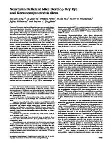
Figure 1. Targeted disruption of the mouse Aire gene and analysis of mRNA species. (A) The exons of the Aire gene are presented as black boxes. The neo and HSVtk genes are marked as a dotted box and a striped box, respectively. The targeted Aire gene after homologous recombination is shown at the bottom. (B) The homologous recombination shown in genomic DNA of Aire–/– mice. The expected 750 bp DNA fragment was detected in the Aire+/+ mice, the 750 and 2000 bp DNA fragments in Aire+/– mice and the 2000 bp DNA fragment represents the allele from Aire–/– mice. (C) RT–PCR was performed with exon 3 (a1) and exon 9 (a4) specific primers from the Aire+/+, Aire+/– and Aire–/– mice using poly(A) mRNA from the thymus as a template. The fragments obtained represent (703 bp) W and targeted (473, 554 and 1465 bp) b, a, c transcripts. (D) Sequence of the RT–PCR product from Aire–/– mice was amplified with Aire exon 3 (a1) and exon 9 (a4) specific primers, representing the (b) 473 bp, (a) 554 bp and (c) 1465 bp. All three Aire–/– transcripts contained a stop codon truncating the Aire polypeptide chain.
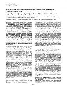
of a Neo-cassette in the beginning of exon 6 (Fig. 1A and B). We verified the targeted disruption by RT–PCR (Fig. 1C) and by sequencing the transcript variants (Fig. 1D). The data confirmed the expected interrupted mRNA species and the predicted early termination of all synthesized polypeptides. To verify the absence of Aire polypeptides, we analyzed sections from thymus, liver and brain of the Aire–/– mice by immunohistochemistry using an Aire-specific antibody. In the wild-type Aire+/+ mice, the thymus shows a strong nuclear Aire-immunoreactivity (IR) in a population of medullary epithelial cells, and characteristically, we observed IR in some epithelial cells in the Hassal’s corpuscles (Fig. 2A and C). Furthermore, the nuclei of liver hepatocytes from Aire+/+ mice were Aire-IR (Fig. 2E). Similarly, in brain sections, Purkinje cells and a large number of the granular neurons of the cerebellar cortex displayed a strong Aire-IR in Aire+/+ mice
(Fig. 2G), but we could not detect reactivity in any of the Aire–/– mice tissues examined (Fig. 2B, D, F and H). Infertility and tissue pathology in Aire-deficient mice The Aire–/– mice did not significantly differ in weight, size or maturation from their Aire+/+ and Aire+/– littermates and the distribution of the genotypes of the progeny abided by the Hardy–Weinberg equilibrium. However, 85% of male or female Aire–/– mice (n = 20) failed to produce any litters when crossed with Aire–/– mice. To confirm sterility, we crossed both male and female non-reproducing Aire–/– mice with C57BL/6 mice and again these mice did not reproduce. Surprisingly, subsequent crosses of Aire+/– with Aire+/– breeding sets (n = 39) also exhibited a 44% reduction in fertile mice relative to control breeding sets (n = 42). The compromised reproductive
Human Molecular Genetics, 2002, Vol. 11, No. 4
399
Figure 2. Immunocytochemical demonstration of Aire-immunoreactivity. (A) In Aire+/+ thymus several Aire-immunoreactive epithelial cells (arrowheads) can be seen in the medulla (me); cortex (co). (C) At higher magnification labeled epithelial cells in Hassal’s corpucles (Hc) are evident and also large number of medullary cells (arrows) are Aire-immunoreactive. (B and D) No labeling can be observed in Aire–/– thymus. (E) Most of the hepatocyte nuclei (arrowheads) are stained in Aire+/+ liver. (F) Liver hepatocytes from Aire–/– mice lack IR. (G) Strongly labeled Purkinje neurons (arrowheads) and granular neurons (arrows) are seen in cortex of the cerebellum (ml, molecular layer; gcl, granular cell layer). (H) No labeled neurons are present in Aire–/– cerebellum. Bar: 100 µm (A, B, E–H), 50 µm (C and D).
success is consistent with the infertility manifested in APECED patients; this occurrence has been associated with testicular or ovarian atrophy (21). We systematically looked at tissue architecture and histology in 2–3-month-old Aire–/– and their Aire+/+ littermates with special attention paid to the thymus, adrenal glands, spleen, liver, testis and ovaries. None of the Aire–/– mice showed observable anatomical or physiological abnormalities.
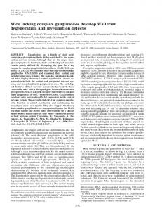
Out of 14 Aire–/– mice examined, the majority of the thymus and spleen tissues showed no altered morphology (Fig. 3A– D) with the exception of two Aire–/– mice displaying thymic atrophy. We did not find any consistent decrease in the cortex to medulla ratio in all of the Aire-deficient mice that were examined. However, five out of 10 Aire–/– mice acquired periportal accumulation of lymphocytes in the liver sections (Fig. 3F), suggestive of autoimmune hepatitis found in 13% of
400
Human Molecular Genetics, 2002, Vol. 11, No. 4
Figure 3. Tissue sections from Aire+/+ and Aire–/– mice stained with H&E. The architecture and cellular organization of the cortex (c) and medulla (m) appear normal in the thymus when comparing Aire+/+ (A) and Aire–/– (B) specimens. The spleen sections from Aire+/+ (C) and Aire–/– (D) appeared normal with white/red pulp and periarteriolar lymphoid sheath (PALS) (p). Liver sections from Aire+/+ mice (E) show no signs of infiltrates but in several of the Aire–/– mice (F) a periportal lymphocytic infiltrate was observed. Ovary from an Aire+/+ (G) as compared to an atrophic ovary lacking follicles from an Aire–/– mouse (H). Normal adrenals from Aire+/+ (I) and Aire–/– mice (J) with intact cortex (c) and medulla (m) were observed. Magnification: 10× (A–D and G–J), 40× (E–F).
the APECED patients (21). Only one mouse lacked ovarian follicles (Fig. 3H). Interestingly, we observed a lymphocytic infiltrate in the margin of this atrophied ovary, suggesting a history of autoimmune-based gonadal atrophy, comparable to APECED patients. We were unable to detect the adrenal
glands in four Aire–/– mice by careful dissection, suggesting dramatic atrophy of these organs. Likewise, adrenal glands observed in Addison’s disease patients were also very small and difficult to locate at autopsy (22). Thyroid glands (n = 3) and pancreas (n = 10) of Aire–/– mice appeared normal in all the
Human Molecular Genetics, 2002, Vol. 11, No. 4
Table 1. Presence of autoantibodies in sera from Aire+/+ and Aire–/– mice Liver
Testis
Pancreas
Adrenal
Aire+/+
0/14
0/14
0/14
0/14
Aire–/–
3/15
8/15
7/15
3/15
mice examined. In the remaining Aire–/– and in all Aire+/+ (n = 14) mice, the adrenals were readily identified and tissue histology appeared normal (Fig. 3J). Taken together, 42% of the Aire–/– mice examined revealed atrophy of thymus, adrenals or ovaries and 50% showed periportal accumulation of lymphocytes in the liver sections. Aire-deficient mice produce circulating autoantibodies Characteristically, sera of APECED patients contain specific autoantibodies as a sign of an ongoing autoimmune response against target organs. Sera from 14 Aire+/+ and 15 Aire–/– mice were analyzed regarding immunoglobulin (Ig) levels and autoantibodies. We found no quantitative difference in total Ig or Ig subclass levels comparing the Aire–/– with Aire+/+ mice using an Ig-specific ELISA (23). We analyzed the presence of autoantibodies in the Aire–/– mice sera using frozen organ sections from healthy mice by indirect immunofluorescence (Table 1) (24). No in situ antibodies of Ig subclass in target organs were detected in 10 Aire-deficient mice using immunofluorescence of frozen sections. We found 20% (n = 3) of Aire–/– sera staining the cytoplasm of hepatocytes in mouse liver sections (Fig. 4A). Interestingly, two of the Aire–/– mice harboring autoantibodies against hepatocytes also displayed a periportal lymphocytic infiltrate when stained with haematoxylin and eosin (H&E) (Fig. 3F). Fifty-three percent of Aire–/– sera stained the spermatogonia and/or spermatids (Fig. 4E), but we did not observe positive staining of the steroid producing Leydig cells. To our surprise, 47% of sera from Aire–/– mice contained autoantibodies against the exocrine pancreas (Fig. 4C). In addition, only one serum of the Aire–/– mouse displayed reactivity against the insulin producing cells in the islet of Langerhans. In contrast, the majority of the APECED patients develop autoreactivity against the islet of Langerhans (25,26) recognizing the major autoantigen in diabetes, the glutamic acid decarboxylase (GAD) and the aromatic L-amino acid decarboxylase (AADC). We also identified adrenocortical autoantibodies in 20% (n = 3) of sera from Aire–/– mice (Fig. 4G). None of the three sera displayed reactivity against the steroid producing Leydig cells of the testis nor any of the sera from Aire+/+ mice displayed autoreactivity against any of the organs tested (Fig. 4B, D, F and H). Overall, sera from 11 out of 15 (73%) Aire–/– mice contained autoantibodies against at least one of the four organs tested and, furthermore, six out of 15 (40%) of the Aire–/– mice sera displayed autoreactivity against two or more of the four target organs (liver, testis, pancreas and adrenal glands), thus mimicking the autoreactivity against multiple organs seen in sera from patients suffering from the APECED (21).
401
Characterization of lymphocytes and immunological responses to challenge To gain insight into the immunological responses in APECED, we characterized the distribution and responsiveness of lymphocytes in both the Aire–/– and control mice. Flow cytometry studies in fractions of CD4+ and CD8+ T cells of the thymus, spleen (Fig. 5A), lymph node and blood (data not shown) showed no significant differences in the distribution of B and T lymphocytes and, furthermore, the ratio of B to T cells were similar in all of the tissues studied between the Aire+/+ and Aire–/– mice as judged by triple staining the cells with anti-CD4, anti-CD8 and anti-B220 antibodies (group size of 10). Since the Aire protein is expressed in medullary epithelial cells of the thymus (Fig. 2A) and has been implicated to participate in negative selection of thymocytes (19,27,28), we triple stained thymocytes with antibodies against CD4 and CD8 combined with an antibody against TCRβ chain, heat stable antigen (CD24), CD43 or CD69. We did not find any differences in the distribution of these surface markers in the populations of single positive, double positive or double negative thymocytes, indicating that no major defect is present in the thymic maturation of the T cells. In addition, no significant difference in apoptotic thymocytes was apparent as judged by annexin-V staining (data not shown), indicating that thymocytes undergoing the selection process were not altered in the Aire–/– mice. The Aire–/– lymphocytes displayed a naive phenotype with CD62Lhi and CD44lo cells that did not differ from the levels expressed on the Aire+/+ cells (Fig. 5B) using antibodies against CD62 ligand, which is downregulated during activation, and CD44, which is highly expressed on the surface of effector and memory type T cells (29,30). Intraperitoneal injection of mice with bromodeoxyuridine (BrdU), which is incorporated into activated cells undergoing proliferation, showed only a low level of in vivo BrdU incorporation into CD4+, CD8+ and B220+ cells from lymph nodes and spleen, indicating naive non-activated cell populations from both the Aire–/– and Aire+/+ mice (Fig. 5C). Furthermore, when we investigated the in vivo labeled thymi, the incorporation of BrdU did not differ between the Aire–/– and Aire+/+ mice, suggesting normal turnover of thymocytes during the thymic education and maturation process. We next stimulated T cells in vitro using an antibody against CD3 that activates and mimics T cell receptor signaling (31). Activation of total lymph node cells from Aire–/– and Aire+/+ mice in vitro in a time course study, measuring the upregulation of the early activation marker CD69 as well as the interleukin-2 receptor (CD25), showed no difference in either CD69 (Fig. 5D) or CD25 (data not shown) surface expressions on either CD4+ or CD8+ T cells between Aire–/– and Aire+/+ mice (n = 4). The combined findings suggest that the T cell receptor signaling pathway is not altered in the absence of Aire expression, at least with respect to the expression of CD69 and CD25 surface markers. Clinical studies show that patients with APECED disease develop organ-specific autoreactivity against self-specific antigens (24,26,32) and since the Aire protein is expressed in the thymic medulla (19,27), mutations in the Aire gene may compromise the fidelity of central tolerance. To test this hypothesis, we purified responder CD4+ T cells and incubated them with T depleted spleen cells as antigen presenting cells (APCs) in a mixed lymphocyte reaction (33). CD4+ responder
402
Human Molecular Genetics, 2002, Vol. 11, No. 4
Figure 4. Immunofluorescence analysis of healthy mouse organs using sera from Aire–/– and Aire+/+ mice. The presence of autoantibodies in circulating blood was demonstrated by staining the healthy mouse frozen organs with sera derived from Aire–/– and Aire+/+ mice using indirect immunofluorescence. Autoantibodies were detected only from the sera of Aire–/– mice reactive to (A) liver, (B) pancreas, (C) testis and (D) adrenal, but not in Aire+/+ mice (E, F, G and H, respectively).
cells from Aire–/– mice failed to respond to their own APCs and likewise, no proliferation was elicited by Aire+/+ APCs (data not shown). Further, there was no responsiveness among CD4+ Aire+/+ cells against self or Aire–/– APCs. These findings are consistent with normal self-tolerance and with normal antigen processing with respect to MHC class II presentation by Aire–/– APCs.
Based on the surface expression levels of CD44 and CD62L, combined with the low BrdU incorporation, we recognized that the Aire–/– immune cells were never challenged in our sterile mouse facility. To monitor the antigen response, we activated lymphocytes in vivo by immunization with hen egg lysozyme (HEL) in complete Freund’s adjuvant. Restimulation of the draining lymph node cells in vitro (34) 8 days post-immunization
Human Molecular Genetics, 2002, Vol. 11, No. 4
403
Figure 5. (Above and overleaf) Flow cytometry and spectratype characterization of T cells from Aire+/+ and Aire–/– mice. (A) Thymus or spleen cells were stained with antibodies reactive to CD4 and CD8 surface antigens. (B) Analysis of lymph node cells for activation markers using antibodies against CD62 ligand and CD44. (C) In vivo BrdU incorporation was measured in proliferating lymph nodes or thymocytes by FACS. Isolated cells from BrdU injected mice were triple stained with anti-CD4, anti-CD8 and anti-BrdU antibodies. Percent incorporation of BrdU is indicated. (D) Levels of CD69 surface expression were measured on CD4+ T cells after in vitro activation with an anti-CD3 antibody for 0, 2, 6 and 16 h by flow cytometry. (E) Proliferative responses from HEL immunized Aire–/– and Aire+/+ mice. Draining lymph node cells were collected 8 days after in vivo immunization and restimulated in vitro with 30 µg/ml of HEL. Each well was pulsed with 1 µCi of [3H]-thymidine for 18 h before harvesting at days 3, 4 and 5 followed by scintillation counts, beta counts per minute (c.p.m.). (F) Concentration dependency was measured at day 5 with cells restimulated with 0, 10, 30 and 100 µg/ml of HEL. T cell receptor Vβ spectratype profile of mouse thymus and spleen. (G) A representative of four spectratype of thymic MuVβ18, MuVβ19 and MuVβ20 Vβ families. (H) A representative of four spectratype of the MuVβ18, MuVβ19 and MuVβ20 families in the spleen cells: x-axis, lengths in nucleotides from the run-off reactions which includes the CDR3 region; y-axis, fluorescence intensities, reflecting the number of clones using each Vβ/CDR3 length combination. Each plot is normalized by taking the ratio of each peak to the highest peak.
displayed 3–5-fold proliferative response in 90% of the Aire–/– mice (n = 10) when compared with relatively weak or no response in Aire+/+ mice (Fig. 5E and F). One Aire–/– mouse did
not show hyperproliferation, most likely due to improper immunization technique (Fig. 5E and F). When we analyzed the lymph node cells with surface markers (CD4, CD8, CD69,
404
Human Molecular Genetics, 2002, Vol. 11, No. 4
Human Molecular Genetics, 2002, Vol. 11, No. 4
CD44, CD62L, CD25 and CD11b) after immunization, we found no expression or distribution differences in any of the markers above; however, the lymph nodes of the Aire–/– mice contained twice as many T cells than the Aire+/+ mice, while the number of B cells and CD11b+ cells were relatively similar (n = 3). It is possible that the exaggerated proliferating response is caused by over-representation of certain T cells that normally are destined to negative selection. To monitor for alterations in the TCR repertoire we analyzed the variations in both length and distribution of the most variable region of the TCR Vβ-chain, the complementarity determining region 3 (CDR3) (35). Using spectratype analysis, we found no apparent difference comparing thymic RNA of Aire–/– to Aire+/+ mice (Fig. 5G) in both length and distribution of TCR-Vβ families (n = 4).
405
However, in the spleen cells, three out of 24 Vβ families (MuBV18, MuBV19 and MuBV20) exhibited a clear alteration in their TCR repertoire (Fig. 5H) and the remaining 19 TCR-Vβ subfamilies were normal. The cause of TCR differences is unclear; however, we consider the clonal expansion unlikely since 6-month-old mice also displayed alterations in the same subset of TCR-Vβ repertoire as 2-month-old mice. DISCUSSION One of the hallmarks of APECED is the presence of multiple serological markers, such as autoreactive antibodies against defined organ-specific antigens (21). Serological characterization of Aire–/– mice showed that 73% of the Aire deficient mice
406
Human Molecular Genetics, 2002, Vol. 11, No. 4
generated at least one circulating autoantibody and >40% of the sera contained two or more autoantibodies reactive to liver, adrenal cortex, pancreas or testis. In conjunction with organspecific autoantibodies, tissues of several organs showed lymphocytic infiltration that was absent from a list of age and sex matched Aire+/+ control mice. Tissue pathology reports confirmed that 50% of Aire–/– mice displayed liver inflammation. Interestingly, 60% of sera that stained liver samples displayed periportal lymphocytic infiltration. This infiltration is most likely related to the absence of Aire protein since knock-out mice deficient in the RelB protein also lacked Aire expression, and these mice were reported to develop inflammatory liver infiltrates (36) similar to those seen in our Aire–/– mice. We did not observe striking destruction of endocrine tissues beyond some ovarial and adrenal changes. However, it is possible that the mice were not challenged by exogenous pathogens in sterile facility. Furthermore, the age of the mice should be considered in the potential development of autoimmunity. Indeed, G protein-coupled receptor, G2A, knock-out mice did not acquire clear signs of autoimmunity for ∼1 year (37) and development of spontaneous autoimmunity in transgenic mice that express a myelin basic protein-specific T cell receptor was restricted to mice in a non-sterile facility, whereas mice that were kept in sterile facility did not develop autoimmunity (38). We are currently aging our mouse population as well as challenging them with a non-sterile environment. No quantitative or qualitative differences of B and T cells in naive Aire–/– mice were observed. Overall, we could not identify any major defects in either spontaneous or in in vitro stimulated activation of T cells. In addition, no major disturbances were evident to cause a difference in thymic negative selection as determined by mixed lymphocyte reactions. The negative results from our immunological studies suggest that Aire does not significantly affect the fidelity of the central tolerance; however, loss of Aire function may potentially influence a small population of thymocytes that escaped our detection method. To convincingly determine the fate of Aire in the central tolerance, we are currently crossing the Aire–/– mice with several different TCR transgenic mice. In an attempt to characterize the TCR repertoire, we identified differences in three of the 24 Vβ subfamilies in the spleen, but not in the thymus. We speculate that over-representation of certain T cells can potentially lead to autoimmunity if not deleted (39). In addition, our suggestive evidence for altered peripheral, but not central T cell repertoire, would indicate a potential defect in peripheral T cell tolerance (40). However, we recognize that differences in a small subpopulation of thymus may be hidden and, further, cells undergoing apoptosis due to negative selection may still contribute to the total RNA pool. Although stimulation of lymphocytes with anti CD3 antibodies did not show altered surface expression of CD69 and CD25 markers, immunization with HEL antigen resulted in a 4–5-fold increase in proliferating response in Aire–/– mice. Indeed, deficiencies in the homeostatic regulation of expanded or autoreactive lymphocytes lead to lymphoproliferative disorders such as autoimmunity (41). The cause of hyperproliferation is not clear. Although there are twice as many T cells in the lymph nodes of immunized Aire–/– mice compared to Aire+/+, we are not certain if the loss of proliferation control is T cell autonomous or APC-dependent.
Besides the causative gene responsible for APECED, individual variations in the genetic background can be an important factor in the overall clinical variability in APECED observed even within a family. We observed similar phenotypic diversity in the Aire–/– mice that also retained heterogeneous genetic background (J129 and C57BL/6). Most probably the variations in genetic background are not solely responsible for the phenotypic variability, either in Aire–/– mice or APECED patients. Challenging the Aire–/– mice with immunization leads to an abnormal T cell proliferation. This would suggest that environmental stimulus that initiates an immune response, such as acquirement of pathogens, is a crucial factor that contributes to the APECED phenotype. Environmental stimuli and heterogeneity in the genetic background would explain the phenotypic variability and the unpredictable age of onset found among APECED patients and Aire–/– mice. We are currently back-crossing our Aire–/– mice to produce a congenic strain to address these issues in more detail. In conclusion, APECED in humans is a monogenic disease model for progressive non-HLA linked autoimmunity leading to multiple organ specific immune destruction. Just like APECED patients, Aire–/– mice seem to express any combination of endocrine and non-endocrine autoimmune components to a various extent and with a variable age of onset. Although preliminary, our findings suggest that when environmental influence, such as acquirement of pathogens, triggers an immune response, mice produce an exaggerated response in the absence of Aire protein. We are here presenting a comprehensive description of the Aire–/– phenotype closely resembling the human APECED disease, thus providing direct evidence that the absence of Aire results in a disturbance of the homeostasis in the immune system. Further analysis of these mice should help to dissect the molecular details of this disturbance. MATERIALS AND METHODS Targeting the mouse Aire gene Targeting sequence of the mouse Aire gene was cloned from a bacteriophage mouse genomic library by using the AIRE human cDNA as a probe. The targeting construct was produced by inserting the 5.3 and 1.8 kb fragments from the mouse Aire gene into the XbaI and the XhoI sites of pPNT, respectively. The fragments were amplified by long range PCR (Expand Long Template) (Roche, Indianapolis, IN) according to the manufacturer’s instructions using oligonucleotides which include the XbaI and the XhoI restriction sites. The 5.3 kb genomic fragment, covering intron 6 flanking sequences (16 bp at the 3′ end) and exons 7–12 (5270 bp), was inserted into a XbaI site of pPNT so that the thymidine kinaseresistant (tkr) gene was located 3′ of the Aire 5.3 kb fragment. The 1.8 kb fragment, covering exons 4–5 (588 bp) and intron 5 flanking sequences (1177 bp), was inserted into an XhoI site of pPNT, thus placing the neomycin-resistant (neor) gene adjacent to interrupted exon 6. Orientation was verified by DNA sequencing. The NotI linearized targeting construct (40 µg) was introduced into the J129 embryonic stem (ES) cells by electroporation. ES cells were maintained in ES cell medium (Dulbecco’s modified Eagle’s medium) with 20% ES cells Qualified Fetal Serum, glutamine, non-essential amino acids, penicillin streptomycin, 2-mercaptoethanol on mitotically
Human Molecular Genetics, 2002, Vol. 11, No. 4
inactivated MEF feeder layers in the presence of LIF-conditioned medium. ES cells were selected in G418 (Invitrogen, San Diego, CA) 350 µg/ml and 2 µM Ganciclovir. After 10 days, colonies were picked, expanded and isolated. Homologous recombination was identified by PCR and positive ES cells were injected into the C57BL/6 blastocysts using standard procedures. Genotyping Genomic DNA was prepared from the tail samples using DNeasy Tissue Kit (Qiagen, Valencia, CA). For analysis by PCR, 10 ng of DNA were amplified on a thermal cycler using the intron 5 (a2) and the exon 7 (a3) primer pair. Both a wild-type and mutated alleles were amplified by these mouse specific primers. PCR products were isolated by agarose gel electrophoresis and visualized by ethidium bromide staining. Poly(A) mRNA was isolated from mouse thymus using Direct mRNA Mini Kit (Qiagen). cDNA first strand was synthesized from 1 µg poly(A) mRNA with oligo(dT)23 primer and SuperScript II H-Reverse Transcriptase (Invitrogen) according to the manufacturer’s protocol. RT–PCR was amplified using the RT-products (1/20 volume of reaction) as a template, 2.5 U Taq polymerase (Amersham Pharmacia, Piscataway, NJ) and the mouse Aire exon 3 (a1) and exon 9 (a4) primers with 1.4 M betaine (N,N,N-trimethylglycine monohydrate; Sigma, St Louis, MO) in the reaction buffer. Amplification products were analyzed on an agarose gel followed by ethidium bromide staining.
407
by two independent observers unaware of the origin of the sera used. Histology For histological studies, 2–3-month-old organs were fixed by immersion in 10% formaldehyde overnight, rinsed in PBS, dehydrated through increasing graded strengths of ethanol, cleared and embedded in paraffin, and cut into 6 µm sections on slides. The serial sections of mouse thymus, liver, spleen, ovary, kidney and adrenal were stained with H&E. Flow cytometry characterization Lymph nodes, spleens or thymus organs of 2–3-month-old mice were grounded in a loose fit glass homogenizer in DMEM containing 2.5% horse serum to obtain cell suspensions for flow cytometry. Cells were washed and 1 × 10⁶ cells were resuspended in FACS buffer containing PBS (Invitrogen), 2% fetal calf serum and 0.02% NaN3. Double or triple staining of cells were performed with antibodies (BD Pharmingen, San Diego, CA) conjugated with fluorescein isothiocyanate (FITC); anti-CD4, anti-H2-Db, anti-BrdU, annexin-V, phycoerythrin (PE); antiTCRβ, anti-CD8, anti-CD4, anti-B220, anti-CD25, anti-CD69, anti-CD62L, anti-CD24, anti-CD43, anti-CD44 (IM7) or allyphycocyanin (APC); anti-CD8. Flow cytometry was performed on a FACScan and analyzed using the Cell Quest software (Becton Dickinson, Franklin Lakes, NJ).
Antibodies and immunohistochemistry
In vivo labeling of proliferating cells
To obtain a specific antibody against the Aire protein, rabbits were immunized by subcutaneous injections with a soluble synthetic peptide of 17 amino acids from the mouse Aire protein (amino acids 160–176) in Freund’s complete adjuvant (19). The primary injection with 50 µg of the peptide was followed by three additional boosters of 100 µg after 10 days, 6 and 10 weeks, respectively. The blood was collected 1 week after the last immunization and the antibodies directed against the Aire polypeptide were affinity purified using nitrocellulosebound CnBr-coupled peptide. The antibody titers and specificity were determined by immunofluorescence and western blotting. For immunohistochemistry, 2–3-month-old adult wild-type and knock-out mice were anesthetized with pentobarbital and perfused transcardially with physiological saline followed with 4% paraformaldehyde (PFA) in phosphate buffered saline (PBS) for 4 min. Subsequently the tissues were excised and immersed in the same fixative for an additional 60 min. The samples were cryoprotected with 15% sucrose in PBS, frozen with dry ice, and 10 µm sections were cut with Micron HM500 cryostat. The sections were incubated with the purified antibody against mouse Aire protein (dilution 1:100–200) at 4°C overnight in a buffer containing 1% bovine serum albumin and 0.3% Triton X-100. Subsequently, the sections were incubated with biotinylated goat anti-rabbit IgG (diluted 1:300) and ABC complex using the Vectastain Elite Kit (Vector Laboratories, Burlingame, CA) for 30 min each. Immunoreaction was visualized with nickel-intensified DAB as a chromogen. The sections were then dehydrated and embedded in Entellan. Frozen sections (6 µm) of organs from healthy wild-type mice were stained with sera from Aire+/+ and Aire–/– mice using indirect immunofluorescence as described (25). Sections were evaluated
Mice were injected intraperitoneally with 1 mg of BrdU twice within 16 h and sacrificed 4 h after the last injection. Cell suspensions from lymph nodes, spleen and thymus were surface stained with anti-CD4 FITC and anti-CD8 APC followed by fixation and permeabilized according to the BrdU flow kit instructions (Becton Dickinson). After deoxyribonuclease treatment anti-BrdU-FITC conjugated antibody was applied as described to reveal analog incorporation into proliferating cells using flow cytometry. In vitro activation assay Cell suspensions from lymph nodes containing 1 × 10⁶ cells were incubated at 37°C in RPMI medium containing 10% fetal calf serum, 5% NCTC, 1% glutamine, 1% penicillin-streptomycin and 5 × 10⁻⁵ M 2-mercaptoethanol in the presence of 0.5 µg of activating anti-CD3 antibody (BD Pharmingen). Cells were pelleted after 0, 2, 6 and 16 h of incubation and triple stained with anti-CD4 FITC, anti-CD8 APC and anti-CD69 PE or antiCD25 PE antibodies, washed, followed by fixation for 10 min in fixation solution (Becton Dickinson). Cells were again washed and resuspended in FACS buffer and kept at 4°C in the dark before FACS analyses. Immunization and proliferation assay Mice were injected in hind footpads and at the tail base with a total of 100 µg of HEL (Sigma) diluted 1:1 with complete Freund’s adjuvant (Invitrogen). Cells from the popliteal and inguinal draining lymph nodes were collected on day 8, pooled together from each individual mouse and used in a proliferation assay at 3 × 10⁵ cells per well in triplicate. The cells were
408
Human Molecular Genetics, 2002, Vol. 11, No. 4
restimulated in vitro with 0, 10, 30 or 100 µg/ml of HEL, pulsed for 18 h with 1 µCi of [3H]-thymidine before harvesting at days 3, 4 and 5 followed by scintillation counting. TCR spectratype analysis Total thymus and spleen RNA from 2-month-old Aire–/– and Aire+/+ mice were extracted using the Qiagen RNeasy kit (Qiagen). Ten micrograms of total RNA was used to make cDNA from each of the four Aire–/– and four Aire+/+ mice using the cDNA synthesis kit (Invitrogen). The samples were measured using a spectrophotometer and diluted to a 20 ng/ml final concentration. PCR reactions with the specific primers for the 24 Vβ-gene families (35) and amplifications were performed using 20 ng of cDNA from each tissue, 2 U Taq polymerase (Amersham Pharmacia), 200 µM dNTP mix, 1× supplier’s buffer, 1 µM 5′ and 3′ primer in a 50 µl final reaction. The amplification started with a denaturation step of 10 min at 95°C, followed by 35 cycles of 2 min at 95°C, 1 min at 60°C, and 2 min at 72°C followed by a 10 min extension at 72°C. Three microliter aliquots of the 24 amplification products were used in the Run-Off Reaction (Primer Extension) primed with Fam-labeled Cβ nested primer. Two microliters of the fluorescent run-off products were mixed with 0.5 µl of ABI TAMARA 500 size standard, 0.5 µl of loading buffer and 2 µl of formamide, and run on 6% polyacrylamide denaturing gels containing 8 M urea in an ABI 377 automated sequencer (Applied Biosystems, Foster City, CA). Fragment sizes were determined automatically using GeneScan 3.0 software (Applied Biosystems). ACKNOWLEDGEMENTS We thank Dr Anu Jalanko for the help in the design of the targeting construct, Dr Nora Rozengurt for analysis of histology sections, Alden Cilindro for performing blastocyst injections, Dr Janna Saarela for valuable discussions, Kristina Dorenbos for preparation of tissue samples, and Kathrin Oelgeschlaeger and Katrin Österlund for excellent technical assistance. We also like to thank Dr Owen Witte for his expert advice and Dr Linda Baum for donating anti-CD43 antibody. We wish to thank the animal husbandry and support team. This study was supported by the Academy of Finland, the Swedish cancer foundation and the Torsten and Ragnar Soderberg foundation. REFERENCES 1. Aaltonen,J., Bjorses,P., Sandkuijl,L., Perheentupa,J. and Peltonen,L. (1994) An autosomal locus causing autoimmune disease: autoimmune polyglandular disease type I assigned to chromosome 21. Nat. Genet., 8, 83–87. 2. Bjorses,P., Aaltonen,J., Horelli-Kuitunen,N., Yaspo,M. and Peltonen,L. (1998) Gene defect behind APECED: a new clue to autoimmunity. Hum. Mol. Genet., 7, 1547–1553. 3. Eisenbarth,G., Wilson,P., Ward,F. and Lebovitz,H. (1978) HLA type and occurrence of disease in familial polyglandular failure. New Engl. J. Med., 298, 92–94. 4. Ahonen,P., Koskimies,S., Lokki,M., Tiilikainen,A. and Perheentupa,J. (1988) The expression of autoimmune polyglandular disease type I appears associated with several HLA-A antigens but not with HLA-DR. J. Clin. Endocrinol. Metab., 66, 1152–1157. 5. Rosatelli,M.C., Meloni,A., Meloni,A., Devoto,M., Cao,A., Scott,H.S., Peterson,P., Heino,M., Krohn,K.J., Nagamine,K. et al. (1998) A common mutation in Sardinian autoimmune polyendocrinopathy-candidiasis-ectodermal dystrophy patients. Hum. Genet., 103, 428–434.
6. Bjorses,P., Aaltonen,J., Vikman,A., Perheentupa,J., Ben-Zion,G., Chiumello,G., Dahl,N., Heideman,P., Hoorweg-Nijman,J.J., Mathivon,L. et al. (1996) Genetic homogeneity of autoimmune polyglandular disease type I. Am. J. Hum. Genet., 59, 879–886. 7. Zlotogora,J. and Shapiro,M. (1992) Polyglandular autoimmune syndrome type I among Iranian Jews. J. Med. Genet., 29, 824–826. 8. Ahonen,P. (1985) Autoimmune polyendocrinopathy–candidosis–ectodermal dystrophy (APECED): autosomal recessive inheritance. Clin. Genet., 27, 535–542. 9. Ahonen,P., Miettinen,A. and Perheentupa,J. (1987) Adrenal and steroidal cell antibodies in patients with autoimmune polyglandular disease type I and risk of adrenocortical and ovarian failure. J. Clin. Endocrinol. Metab., 64, 494–500. 10. Ahonen,P., Myllarniemi,S., Sipila,I. and Perheentupa,J. (1990) Clinical variation of autoimmune polyendocrinopathy-candidiasis-ectodermal dystrophy (APECED) in a series of 68 patients. New Engl. J. Med., 322, 1829–1836. 11. Arulanantham,K., Dwyer,J.M. and Genel,M. (1979) Evidence for defective immunoregulation in the syndrome of familial candidiasis endocrinopathy. New Engl. J. Med., 300, 164–168. 12. The Finnish–German APECED Consortium (1997) An autoimmune disease, APECED, caused by mutations in a novel gene featuring two PHD-type zinc-finger domains. Autoimmune PolyendocrinopathyCandidiasis-Ectodermal Dystrophy. Nat. Genet., 17, 399–403. 13. Nagamine,K., Peterson,P., Scott,H.S., Kudoh,J., Minoshima,S., Heino,M., Krohn,K.J., Lalioti,M.D., Mullis,P.E., Antonarakis,S.E. et al. (1997) Positional cloning of the APECED gene. Nat. Genet., 17, 393–398. 14. Pitkanen,J., Doucas,V., Sternsdorf,T., Nakajima,T., Aratani,S., Jensen,K., Will,H., Vahamurto,P., Ollila,J., Vihinen,M. et al. (2000) The autoimmune regulator protein has transcriptional transactivating properties and interacts with the common coactivator CREB-binding protein. J. Biol. Chem., 275, 16802–16809. 15. Aasland,R., Gibson,T. and Stewart,A. (1995) The PHD finger: implications for chromatin-mediated transcriptional regulation. Trends Biochem. Sci., 20, 56–59. 16. Gibson,T., Ramu,C., Gemund,C. and Aasland,R. (1998) The APECED polyglandular autoimmune syndrome protein, AIRE-1, contains the SAND domain and is probably a transcription factor. Trends Biochem. Sci., 23, 242–244. 17. Heery,D., Kalkhoven,E., Hoare,S. and Parker,M. (1997) A signature motif in transcriptional co-activators mediates binding to nuclear receptors. Nature, 387, 654–655. 18. Bjorses,P., Halonen,M., Palvimo,J.J., Kolmer,M., Aaltonen,J., Ellonen,P., Perheentupa,J., Ulmanen,I. and Peltonen,L. (2000) Mutations in the AIRE gene: effects on subcellular location and transactivation function of the autoimmune polyendocrinopathy-candidiasis-ectodermal dystrophy protein. Am. J. Hum. Genet., 66, 378–392. 19. Heino,M., Peterson,P., Kudoh,J., Nagamine,K., Lagerstedt,A., Ovod,V., Ranki,A., Rantala,I., Nieminen,M., Tuukkanen,J. et al. (1999) Autoimmune regulator is expressed in the cells regulating immune tolerance in thymus medulla. Biochem. Biophys. Res. Commun., 257, 821–825. 20. Halonen,M., Pelto-Huikko,M., Eskelin,P., Peltonen,L., Ulmanen,I. and Kolmer,M. (2001) Subcellular location and expression pattern of autoimmune regulator (Aire), the mouse orthologue for human gene defective in autoimmune polyendocrinopathy candidiasis ectodermal dystrophy (APECED). J Histochem. Cytochem., 49, 197–208. 21. Wang,C., Shi,J., Davoodi-Semiromi,A. and She,J. (1999) Cloning of Aire, the mouse homologue of the autoimmune regulator (AIRE) gene responsible for autoimmune polyglandular syndrome type 1 (ASP1). Genomics, 55, 322–326. 22. Nerup,J. (1974) Addison’s disease – a review of some clinical, pathological and immunological features. Dan. Med. Bull., 21, 201–217. 23. Liljedahl,M., Winqvist,O., Surh,C.D., Wong,P., Ngo,K., Teyton,L., Peterson,P.A., Brunmark,A., Rudensky,A.Y., Fung-Leung,W.P. et al. (1998) Altered antigen presentation in mice lacking H2-O. Immunity, 8, 233–243. 24. Winqvist,O., Karlsson,F. and Kämpe,O. (1992) 21-hydroxylase, a major autoantigen in Addison’s disease. Lancet, 339, 1559–1662. 25. Velloso,L., Winqvist,O., Gustafsson,J., Kampe,O. and Karlsson,F. (1994) Autoantibodies against a novel 51 kDa islet antigen and glutamate decarboxylase isoforms in autoimmune polyendocrine syndrome type I. Diabetologia, 37, 61–69. 26. Rorsman,F., Husebye,E.S., Winqvist,O., Bjork,E., Karlsson,F.A. and Kampe,O. (1995) Aromatic-L-amino-acid decarboxylase, a pyridoxal
Human Molecular Genetics, 2002, Vol. 11, No. 4
27.
28.
29. 30.
31.
32.
33.
phosphate-dependent enzyme, is a beta-cell autoantigen. Proc. Natl Acad. Sci. USA, 92, 8626–8629. Zuklys,S., Balciunaite,G., Agarwal,A., Fasler-Kan,E., Palmer,E. and Hollander,G.A. (2000) Normal thymic architecture and negative selection are associated with Aire expression, the gene defective in the autoimmune-polyendocrinopathy-candidiasis-ectodermal dystrophy (APECED). J. Immunol., 165, 1976–1983. Peterson,P., Nagamine,K., Scott,H., Heino,M., Kudoh,J., Shimizu,N., Antonarakis,S.E. and Krohn,K.J. (1998) APECED: a monogenic autoimmune disease providing new clues to self-tolerance. Immunol. Today, 19, 384–386. Sprent,J. and Tough,D. (1994) Lymphocyte life-span and memory. Science, 265, 1395–1400. Budd,R.C., Cerottini,J.C., Horvath,C., Bron,C., Pedrazzini,T. and Howe,R.C. (1987) MacDonald HR. Distinction of virgin and memory T lymphocytes. Stable acquisition of the Pgp-1 glycoprotein concomitant with antigenic stimulation. J. Immunol., 138, 3120–3129. Ernst,D.N., Weigle,W.O., McQuitty,D.N., Rothermel,A.L. and Hobbs,M.V. (1989) Stimulation of murine T cell subsets with anti-CD3 antibody. Age-related defects in the expression of early activation molecules. J. Immunol., 142, 1413–1421. Winqvist,O., Gustafsson,J., Rorsman,F., Karlsson,F.A. and Kampe,O. (1993) Two different cytochrome P450 enzymes are the adrenal antigens in autoimmune polyendocrine syndrome type I and Addison’s disease. J. Clin. Invest., 92, 2377–2385. Luksch,C.R., Winqvist,O., Ozaki,M.E., Karlsson,L., Jackson,M.R., Peterson,P.A. and Webb,S.R. (1999) Intercellular adhesion molecule-1
34. 35.
36.
37.
38. 39. 40. 41.
409
inhibits interleukin 4 production by naive T cells. Proc. Natl Acad. Sci. USA, 96, 3023–3028. Ron,Y. and Sprent,J. (1987) T cell priming in vivo: a major role for B cells in presenting antigen to T cells in lymph nodes. J. Immunol., 138, 2848–2856. Pannetier,C., Cochet,M., Darche,S., Casrouge,A., Zoller,M. and Kourilsky,P. (1993) The sizes of the CDR3 hypervariable regions of the murine T-cell receptor beta chains vary as a function of the recombined germ-line segments. Proc. Natl Acad. Sci. USA, 90, 4319–4323. Weih,F., Carrasco,D., Durham,S.K., Barton,D.S., Rizzo,C.A., Ryseck,R.P., Lira,S.A. and Bravo,R. (1995) Multiorgan inflammation and hematopoietic abnormalities in mice with a targeted disruption of RelB, a member of the NF-kappa B/Rel family. Cell, 80, 331–340. Le,L.Q., Kabarowski,J.H., Weng,Z., Satterthwaite,A.B., Harvill,E.T., Jensen,E.R., Miller,J.F. and Witte,O.N. (2001) Mice lacking the orphan G protein-coupled receptor G2A develop a late-onset autoimmune syndrome. Immunity, 14, 561–571. Goverman,J., Woods,A., Larson,L., Weiner,L.P., Hood,L. and Zaller,D.M. (1993) Transgenic mice that express a myelin basic protein-specific T cell receptor develop spontaneous autoimmunity. Cell, 72, 551–560. Pender,M.P. (1999) Activation-induced apoptosis of autoreactive and alloreactive T lymphocytes in the target organ as a major mechanism of tolerance. Immunol. Cell. Biol., 77, 216–223. Miller,J.F. and Morahan,G. (1992) Peripheral T cell tolerance. Annu. Rev. Immunol., 10, 51–69. Lenardo,M., Chan,K.M., Hornung,F., McFarland,H., Siegel,R., Wang,J. and Zheng,L. (1999) Mature T lymphocyte apoptosis–immune regulation in a dynamic and unpredictable antigenic environment. Annu. Rev. Immunol., 17, 221–253.
410
Human Molecular Genetics, 2002, Vol. 11, No. 4