YEXNR-11611; No. of pages: 11; 4C: Experimental Neurology xxx (2013) xxx–xxx
Contents lists available at ScienceDirect
Experimental Neurology journal homepage: www.elsevier.com/locate/yexnr
4 5 6 7 8 9 10 11 12 13 14
F
Marta Perez-Alcazar a,b,1, Jonny Daborg b,1, Anna Stokowska a, Pontus Wasling b, Andreas Björefeldt b, Marie Kalm a, Henrik Zetterberg c,d, Karl E. Carlström e, Klas Blomgren a,f,g, Christine T. Ekdahl e,h, Eric Hanse b,⁎, Marcela Pekna a,i,⁎⁎
O
3Q1
R O
2
Altered cognitive performance and synaptic function in the hippocampus of mice lacking C3☆
a
Institute of Neuroscience and Physiology, Department of Clinical Neuroscience and Rehabilitation, The Sahlgrenska Academy at the University of Gothenburg, S-405 30 Gothenburg, Sweden Institute of Neuroscience and Physiology, Department of Physiology, The Sahlgrenska Academy at the University of Gothenburg, S-405 30 Gothenburg, Sweden c Institute of Neuroscience and Physiology, Department of Psychiatry and Neurochemistry, The Sahlgrenska Academy at the University of Gothenburg, S-431 80 Mölndal, Sweden d UCL Institute of Neurology, Queen Square, London WC1N 3BG, UK e Inflammation and Stem Cell Therapy Group, Division of Clinical Neurophysiology, Lund University and Skåne University Hospital, SE-221 84 Lund, Sweden f Department of Pediatrics Oncology, University of Gothenburg, The Queen Silvia Children's Hospital, 413 45 Gothenburg, Sweden g Karolinska Institute, Department of Women's and Children's Health, Karolinska University Hospital Q2:07, 171 76 Stockholm, Sweden h Lund Epilepsy Center, Dept Clinical Sciences, Lund University, Sweden i The Florey Institute of Neuroscience and Mental Health, Parkville, Victoria 3052, Australia b
P
1
i n f o
Article history: Received 29 October 2013 Revised 4 December 2013 Accepted 18 December 2013 Available online xxxx
51
T
C
E
R
R N C O
49 50
Introduction
Complement is part of the innate immune system and plays a major role in the elimination of pathogens and inflammatory response to injury. There is, however, an accumulating body of evidence in support of a range of non-immunological functions of complement, in particular in the CNS. The third complement component (C3)-derived peptide C3a Abbreviations: C3, the third complement component; C1q, the first complement component subunit. ☆ This is an open-access article distributed under the terms of the Creative Commons Attribution-NonCommercial-No Derivative Works License, which permits non-commercial use, distribution, and reproduction in any medium, provided the original author and source are credited. ⁎ Correspondence to: E. Hanse, Institute of Neuroscience and Physiology, Dept. of Physiology; The Sahlgrenska Academy at University of Gothenburg; Box 430, SE-405 30 Gothenburg, Sweden. ⁎⁎ Correspondence to: M. Pekna, Institute of Neuroscience and Physiology, Dept. of Clinical Neuroscience and Rehabilitation; The Sahlgrenska Academy at University of Gothenburg; Box 440, SE-405 30 Gothenburg, Sweden. E-mail addresses:
[email protected] (E. Hanse),
[email protected] (M. Pekna). 1 These authors contributed equally.
U
47 48
Previous work implicated the complement system in adult neurogenesis as well as elimination of synapses in the developing and injured central nervous system. In the present study, we used mice lacking the third complement component (C3) to elucidate the role the complement system plays in hippocampus-dependent learning and synaptic function. We found that the constitutive absence of C3 is associated with enhanced place and reversal learning in adult mice. Our findings of lower release probability at CA3–CA1 glutamatergic synapses in combination with unaltered overall efficacy of these synapses in C3 deficient mice implicate C3 as a negative regulator of the number of functional glutamatergic synapses in the hippocampus. The C3 deficient mice showed no signs of spontaneous epileptiform activity in the hippocampus. We conclude that C3 plays a role in the regulation of the number and function of glutamatergic synapses in the hippocampus and exerts negative effects on hippocampus-dependent cognitive performance. © 2013 The Authors. Published by Elsevier Inc. All rights reserved.
Keywords: Complement cascade Development Epilepsy Hippocampus Synapse elimination
45 44 46
a b s t r a c t
E
a r t i c l e 16 17 18 19 20 21 23 22 24 25 26 27 28 29 30
D
15
31 32 33 34 35 36 37 38 39 40 41 43 42
induces intracellular [Ca2>/>+] elevation and neutrophin expression in cultured microglia (Heese et al., 1998; Möller et al., 1997), and regulates migration and neuronal differentiation of neuronal progenitor cells as well as neurite outgrowth in vitro (Shinjyo et al., 2009). In vivo studies demonstrated the involvement of C3a in neuronal differentiation during the development of rat cerebellum (Bénard et al., 2008) and signaling through C3a receptor stimulates neurogenesis in the adult hippocampus (Rahpeymai et al., 2006). In the immature brain, C3a is protective against hypoxic-ischemic injury and C3a treatment ameliorates hypoxia–ischemia-induced cognitive impairment (Järlestedt et al., 2013). The complement system has also been implicated in developmental elimination of synapse in the thalamus (Stevens et al., 2007) and the sensorimotor cortex (Chu et al., 2010). Microglia play an important role in this process, since inhibition of microglial motility resulted in delayed synapse elimination in the hippocampus (Paolicelli et al., 2011) and disrupting microglia-specific complement receptor 3 (CR3)/C3 signaling led to sustained deficits in synaptic connectivity in the retinogeniculate system (Schafer et al., 2012). In the adult brain, the hippocampus plays a crucial role in the formation of certain types of memory, such as episodic memory and spatial memory (Kesner et al., 2000; Squire, 1992), and hippocampal lesions
0014-4886/$ – see front matter © 2013 The Authors. Published by Elsevier Inc. All rights reserved. http://dx.doi.org/10.1016/j.expneurol.2013.12.013
Please cite this article as: Perez-Alcazar, M., et al., Altered cognitive performance and synaptic function in the hippocampus of mice lacking C3, Exp. Neurol. (2013), http://dx.doi.org/10.1016/j.expneurol.2013.12.013
52 53 54 55 56 57 58 59 60 61 62 63 64 65 66 67 68 69 70 71 72
91
Animals
92 93
C3-deficient (C3 KO) mice (Pekna et al., 1998) were backcrossed onto the C57BL/6 background for 13 generations. Homozygous C3 KO
Animals Male C3 KO mice and WT controls were 2.5–3 months old (first experiment, Fig. 1A) and 2 months old (second experiment, Fig. 1B–D), respectively when behavioral testing began. Before weaning, all mice were anesthetized with isoflurane (Abbott Laboratories, North Chicago, IL, USA) and implanted subcutaneously with microtransponders (DATAMARS, PetLink, Youngstown, OH, USA) to allow to individual animal identification in the IntelliCages. After weaning, the mice were, kept in groups of up to 10 and separated by genotype.
102
Testing of place and reversal learning using IntelliCage® The IntelliCage platform (New Behavior, Zurich, Switzerland) for unbiased monitoring of mouse behavior in a home cage setting has been described elsewhere (Barlind et al., 2010; Galsworthy et al., 2005;
111 112
E T C E R R
86 87
101
O
84 85
Behavioral testing
C
82 83
N
80 81
U
79
F
Methods
77 78
94 95
O
90
75 76
and wild type (WT) control mice were housed in standard cages with a 12-hour light/dark cycle and free access to food and water. All experiments were performed in accordance with the guidelines of the local ethical committee for animal research at the University of Gothenburg or the Malmö-Lund Ethical Committee for the use of laboratory animals and were conducted in accordance with European Union directives on animal rights.
D
88 89
are associated with impaired spatial learning and spatial pattern separation (Gallagher and Holland, 1992; Gilbert et al., 1998; Morris et al., 1982); in particular when the environment complexity is high (Moses et al., 2007). Both synaptic plasticity and generation of new neurons have been implicated in hippocampal function (Deng et al., 2010; Dragoi et al., 2003; Morris et al., 1986; Nakazawa et al., 2003). As C3 seems to regulate both the number of synapses and hippocampal neurogenesis, in the present study, we sought to determine the effects of constitutive genetic ablation of C3 on the functions of an unchallenged adult hippocampus by assessing place and reversal learning ability and synaptic function in C3 deficient (C3 KO) mice. Our results show that C3 KO mice have an enhanced hippocampus dependent learning and provide evidence that supports the involvement of the complement system in excitatory synapse elimination in the hippocampus. Further, we report that the C3 deficiency does not result in spontaneous epileptiform activity in the hippocampus as the increased number of functional synapses is compensated by reduced presynaptic glutamate release probability.
R O
73 74
M. Perez-Alcazar et al. / Experimental Neurology xxx (2013) xxx–xxx
P
2
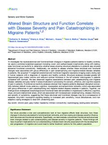
Fig. 1. C3 KO mice show enhanced place and reversal learning compared to WT. (A) Exploratory behavior, expressed as number of nose pokes, in the 5 first days declined over time in both groups of mice but the C3 KO mice (n = 10) were less active compared to WT mice (n = 15) (B) In a separate experiment, the exploratory activity of C3 KO mice (n = 9) was lower compared to WT mice (n = 10) during the place learning period (corner 1), and place learning (corner 1) and reversal learning (corner 2) showed significantly lower incorrect visit ratio in C3 KO compared to WT mice (C). (D) The C3 KO mice made less incorrect nose pokes over the 5 days of both learning periods (corner 1 and 2). * = time × genotype, ¤ = time, # = genotype, P b 0.05*, #, P b 0.01**, P b 0.001¤¤¤.
Please cite this article as: Perez-Alcazar, M., et al., Altered cognitive performance and synaptic function in the hippocampus of mice lacking C3, Exp. Neurol. (2013), http://dx.doi.org/10.1016/j.expneurol.2013.12.013
96 97 98 99 100
103 104 105 106 107 108 109 110
113 114
M. Perez-Alcazar et al. / Experimental Neurology xxx (2013) xxx–xxx
126 127 128 129 130 131 132 133 134 135 136 137 138 139 140 141
Electrophysiology
143 144
For electrophysiological recordings, hippocampal slices from 17–30day-old male WT and C3 KO mice were used as follows: In the pairedpulse ratio (PPr) experiment, WT n = 5 and C3 KO n = 7; in the MK801 experiment, WT n = 10 and C3 KO n = 10; in the 10-stimulus train experiment WT n = 5, C3 KO n = 6; in the patch clamp experiment WT n = 7, C3 KO n = 9. Multiple slices were used per animal, as described in the following section.
154 155 156 157 158 159 160 161 162 163 164 165 166 167 168 169 170 171 172 173 174 175 176
C
E
R
152 153
Slice preparation Mice were anesthetized with isoflurane and decapitated. Brains were rapidly removed and placed in ice-cold (4 ºC) preparation solution: 140 mM choline Cl, 2.5 mM KCl, 0.5 mM CaCl2, 7 mM MgCl2, 25 mM NaHCO 3 , 1.25 mM NaH2PO4 , 1.3 mM ascorbic acid, and 7 mM dextrose. Transverse hippocampal slices (400 μm thick) were cut with a vibratome (HM 650 V Microm, Germany) in the same ice-cold solution. Slices were subsequently stored in artificial cerebrospinal fluid (ACSF) containing: 129 mM NaCl, 3 mM KCl, 2 mM CaCl2 , 4 mM MgCl2, 20 mM NaHCO 3, 1.24 mM NaH2PO4 , 0.5 mM ascorbic acid, 3 mM myo-inositol, 4 mM D,L-lactic acid, and 10 mM D -glucose at 25 °C. After a minimum of 1 h, a single slice was transferred to a recording chamber where it was kept submerged in a constant flow (around 2 ml/min) at 25–28 °C. The perfusion solution consisted of 129 mM NaCl, 20 mM NaHCO 3 , 1.24 mM NaH2 PO4 , 3 mM KCl, and 10 mM D -glucose. CaCl2 and MgCl 2 content varied between the different experimental paradigms; in the single and paired pulse experiments, the solution contained 2 mM each of CaCl2 and MgCl 2; in the MK-801 experiments, the solution contained 3 mM CaCl2 and no MgCl2 to enable NMDA receptor opening; in the whole-cell patch-clamp recordings and the burst experiments, the solution contained 4 mM each of CaCl2 and MgCl2 to suppress the population spike. Picrotoxin (100 μM; Sigma-Aldrich, Stockholm, Sweden) was always present in the perfusion solution to block GABA receptor-mediated activity. All solutions were continuously bubbled with 95% O 2 and 5% CO 2 (pH 7.4).
R
150 151
N C O
149
U
147 148
T
142
145 146
Drugs In the burst experiments, the selective NMDA receptor antagonist AP5 (50 μM; Accent Scientific, Bristol, UK) was used to block NMDA receptors in order to hinder long-term potentiation. MK-801 (Dizocilpine, 20 μM, Tocris Cookson, Bristol, UK), a non-competitive NMDA receptor antagonist, was used in one set of experiments to block NMDA receptor signaling such that after each electrical stimulation, a proportion of NMDA receptors dependent on the synaptic release probability was blocked. During these experiments, NBQX (10 μM; Tocris Cookson) was added to block AMPA receptors. Picrotoxin (100 μM) was included to block GABAA receptors in all experiments.
230
F
124 125
178 179
O
122 123
For field excitatory postsynaptic potential (fEPSP) recordings, electrical stimulation and recordings of synaptic responses were carried out in the CA1 region of the hippocampus. A glass pipette (filled with 1 M NaCl, resistance 1.5–3 MΩ) was used for field recordings. fEPSPs were recorded at a sampling frequency of 10 kHz and filtered at 1 kHz, using a MultiClamp 700B amplifier and a Digidata 1440A data acquisition system (Molecular Devices, Sunnyvale, CA, USA). Evoked responses were analyzed off-line in a blinded manner using custommade IGOR-Pro software (WaveMetrics, Lake Oswego, OR, USA). fEPSP magnitude was evaluated as the initial (first 0.6–0.8 ms) slope of the fEPSP. The stimulation and recording electrode were positioned in the stratum radiatum at a fixed distance from the pyramidal cell layer. Stimuli consisted of biphasic constant current pulses (0.2 ms, 12–34 μA, STG 1002 Multi-Channel systems MCS GmbH, Reutlingen, Germany) delivered through tungsten wires (resistance 0.1 MΩ). The stimulation intensity was set such that it would not evoke firing in the postsynaptic neurons, as evidenced by the absence of a population spike distorting the fEPSP. Signs of population spike activity could, however, occur on facilitated fEPSPs during high-frequency burst stimulation, or paired-pulse stimulation. The early measurement of the fEPSP initial slope (cf. above) minimizes the risk that such population spike activity would influence the evaluation of the fEPSP magnitude. Stimuli were delivered at 0.2 Hz in the input/output experiments (Fig. 2) and in the paired-pulse (50 ms inter-stimulus interval) experiments (Fig. 3). In the MK-801 experiments (Fig. 4), stimuli were delivered at 0.5 Hz. In the burst experiments (Fig. 6), a 10 impulse, 20 Hz train was delivered every 30 s and a total of 30 train stimulations were elicited. Whole-cell patch-clamp recordings were performed on visually identified pyramidal cells, using infrared differential interference contrast video microscopy mounted on an Olympus BX51WI microscope. The pipette solution contained (in mM) 130 Cs-methanesulfonate, 2 NaCl, 20 HEPES, 0.6 EGTA, 5 QX-314, 4 Mg-ATP, 0.4 GTP (pH ~>/>7.3 and osmolality 280–300 mOsm) and 0,5% neurobiotin. Liquid junction potential was measured to be about 8 mV and was not corrected for. Patch pipette resistance was 3–6 MΩ. mEPSCs were recorded at a sampling frequency of 10 kHz and filtered at 1 kHz, using an EPC-9 amplifier (HEKA Elektronik, Lambrecht, Germany). mEPSC analyses were based on voltage clamp recordings at −>/>70 mV for 2–5 min and were analyzed in a blinded manner using Mini-analysis program (version 5.1.4; Synaptosoft, Decatur, GA, USA). Tissue slices from patch-clamp recordings and concomitant neuron loading with neurobiotin were post-fixed in 4% paraformaldehyde in PBS at 4 °C overnight. After wash with PBS, slices were permeabilized with 0.01% Triton-X in PBS for 1 h at room temperature, followed by 3 washes in PBS. Slices were incubated with streptavidin conjugated Alexa Fluor 488 (1:1000, Invitrogen, Eugene, OR, USA) in PBS for 3 h at room temperature, washed 5 times in PBS and mounted with Prolong Gold mounting medium (Invitrogen). Coverslips were sealed to prevent dehydration of tissue. Slices were stored at −>/>20 °C.
R O
121
177
P
119 120
Recordings and analysis
D
117 118
Knapska et al., 2006; Onishchenko et al., 2007). The animals were housed in the IntelliCages in groups of up to 10 animals per cage. The C3 KO (n = 10) and WT (n = 15) mice that were tested for exploratory behavior were housed in the cages only during that period. In the second experiment, the C3 KO (n = 9) and WT (n = 10) mice had an introduction period of 5 days in the IntelliCages during which the animals were acclimatized to performing nose pokes to gain access to the water bottles. The introduction period was followed by a place learning period for which each animal was randomized to one corner (the most visited corner during the introduction was excluded). This allocated corner was programmed as the correct corner while the other three corners were programmed as incorrect. The mice were only allowed to drink from the water bottles in the correct corner, where a nose poke would open a door and give them access to the two water bottles. In incorrect corners, the doors to the water bottles could not be opened by performing nose pokes. After 5 days, the animals were randomized to a new corner (the previous corner excluded) for the reversal learning. Animals that failed to drink in the IntelliCages or were not registered as visiting any corners at all (indicating misplaced or missing microtransponders) were removed from the IntelliCages. Food was provided ad libitum during the experiments in the IntelliCage and red plastic houses were provided as shelters. Data from the IntelliCages were analyzed using the IntelliCage software (IntelliCage Plus, 2.4, NewBehavior AG, Zurich, Switzerland) followed by statistical analysis. Only the active (dark) period was analyzed and visits lasting longer than 180 s were excluded from the analysis.
E
115 116
3
Please cite this article as: Perez-Alcazar, M., et al., Altered cognitive performance and synaptic function in the hippocampus of mice lacking C3, Exp. Neurol. (2013), http://dx.doi.org/10.1016/j.expneurol.2013.12.013
180 181 182 183 184 185 186 187 188 189 190 191 192 193 194 195 196 197 198 199 200 201 202 203 204 205 206 207 208 209 210 211 212 213 214 215 216 217 218 219 220 221 222 223 224 225 226 227 228 229
231 232 233 234 235 236 237 238 239 240
M. Perez-Alcazar et al. / Experimental Neurology xxx (2013) xxx–xxx
T
E
D
P
R O
O
F
4
R
E
C
Fig. 2. Synaptic responses relative to the number of axons in hippocampus slices do not differ between WT and C3 KO mice. (A) Schematic drawing of a hippocampus slice, showing placement of the stimulation and recording electrodes. (B) Example sweeps from two individual experiments using 5 different stimulus intensities. Each trace is an average of 10 recordings at the same stimulus intensity. Pulses were delivered at 0.2 Hz. (C) Diagram showing the slope of the fEPSP plotted against the slope of the fiber volley from the sweeps depicted in Fig. 2B. The dashed lines show a linear regression line through the data points and origo. (D) Bar graphs showing the average slope coefficient of the regression lines (see Fig. 2C) from all n = 22 WT slices and n = 18 C3 KO slices.
Neurobiotin visualization of neurons, assessment of spine density
242
Neuron images were obtained on Zeiss 700 inverted confocal microscope (Carl Zeiss, Germany) at 63× magnification and image resolution
N
C
O
of 1028 × 1028 pixels. Z-stacks of dendritic segments were taken at 0.5 μm steps. Dendritic spine density measurements were performed on apical secondary and tertiary dendrites of hippocampal CA1 pyramidal neurons. Dendritic spines were counted manually with the help of
U
243
R
241
Fig. 3. C3 KO mice have a higher paired-pulse ratio than WT mice. (A) Representative sweeps averaged from 20 paired-pulse recordings (50 ms inter-stimulus interval) recorded at 0.2 Hz. (B) Bar graphs showing the average paired pulse ratio from n = 22 WT slices and n = 22 C3 KO slices. * P b 0.05.
Please cite this article as: Perez-Alcazar, M., et al., Altered cognitive performance and synaptic function in the hippocampus of mice lacking C3, Exp. Neurol. (2013), http://dx.doi.org/10.1016/j.expneurol.2013.12.013
244 245 246 247
5
R O
O
F
M. Perez-Alcazar et al. / Experimental Neurology xxx (2013) xxx–xxx
P
Fig. 4. C3 KO mice show a slower blockage rate of NMDAR responses than WT mice in slices treated with the irreversible NMDAR antagonist MK-801. (A) The diagram shows the decay of the NMDA fEPSPs (measured as the EPSP area and plotted against the number of stimulations [delivered at 0.5 Hz]). Circles represent the average response (n = 14 and n = 15 slices for C3 KO and WT mice, respectively) and lines show the corresponding best-fit curves for the two strains. Resulting time constants are τWT = 65.25 and τC3 KO = 82.22 (Note that these values differ from the ones in the text and the bar graph in B as those are based on averaging τ values obtained from best fits from each individual experiment/animal). Inserts depict an average of the first 10 (a) and the last 10 (b) NMDA fEPSPs from a representative experiment on a C3 KO slice. (B) Bar graphs showing average time constants (tau) of individual exponential fits. * P b 0.05.
ImageJ software (NIH) and in a blind manner with regard to the experimental groups. Spine densities were calculated as the number of spines divided by the length of the segment.
251
EEG recordings in vivo
252 253
For in vivo EEG recording, individually housed, three months old male mice (C3 KO, n = 3 and WT, n = 4) were anesthetized with isoflurane (1%–2%) and implanted unilaterally with a bipolar stainless steel recording electrode (Plastics One, Roanoke, VA, USA) in the ventral hippocampal CA3-CA1 region (coordinates: 2.9 mm caudal and 3.0 mm lateral to bregma, 3.0 mm ventral from dura, tooth bar set at −3.3 mm) as previously described (Mohapel et al., 2004). EEG activity was recorded 10 days later during the light phase of the light/dark cycle for two 12-hour sessions with LabChart software (AD Instruments, Bella Vista, NSW, Australia). An investigator blind to genotype evaluated the occurrence of seizures (abnormal high-frequency epileptiform activity) and focal interictal spike activity.
259 260 261 262 263
T
C
E
R
258
R
256 257
N C O
254 255
Iba1 immunohistochemistry
265
Immunohistochemistry Six months old male C3 KO (n = 14) and WT (n = 14) mice were deeply anesthetized with 0.3 mL 2.5% thiopental i.p. and transcardially perfused with 0.9% saline followed by 4% paraformaldehyde. Brains were removed and post-fixed in 4% paraformaldehyde for 24 h at 4 °C, then switched to 30% sucrose. Brains were sliced to 30 μm coronal sections using a sliding microtome. Free-floating sections containing the hippocampus were selected for immunohistochemistry, and blocked with 5% normal goat serum and 0.25% Triton X-100 in PBS, shaking at room temperature for 1 h and then incubated with the primary antibody, polyclonal rabbit anti-Iba1 (1:1000, Wako Chemicals, Neuss, Germany) overnight, shaking at 4 °C. Sections were then incubated with the secondary antibody, Alexa Fluor 488 goat anti-rabbit IgG (1:200, Invitrogen) shaking at room temperature for 2 h. Sections were mounted on slides with Pertex mounting medium (HistoLab, Västra Frölunda, Sweden), dried at room temperature overnight, and then stored at −20 °C.
268 269 270 271 272 273 274 275 276 277 278 279 280 281
U
264
266 267
Imaging and data analysis Cell counts were performed bilateral in six–eight hippocampal sections per animal located 3.2–5.2 mm posterior to bregma by an observer blind to genetic background. The Iba1-positive cells were counted using an Olympus BX61 epifluorescence microscope in the dentate hilus and the granule cell layer (GCL) and the subgranular zone (SGZ), the latter defined as two cell body diameters below the GCL. Data are presented as number of cells per section.
282 283
Statistics
290
Statistical significance was assessed by the use of Student's t-test unless otherwise specified (see below). Significance level was set at P b 0.05. All data are presented as mean ± SEM. For the behavioral analysis generalized estimating equations (GEE) were used to estimate the average response of the populations for the different parameters measured in the IntelliCage (Karlsson et al., 2011). Data were first analyzed for a Time × Treatment effect and if no significant effect was detected, time and treatment were analyzed separately. All the analyses were performed using SigmaStat 2.0 (SPSS, Chicago, IL, USA). For the statistical analyses of all electrophysiology experiments, brain slices were treated as replicates, i.e., reported n equals n slices analyzed. Measurement values identified as outliers according to the Extreme Studentized Deviate test as well as values that resulted from poor quality recordings were excluded from the analysis. In the MK experiment, release probability was modeled by a single exponential decay function using values for all the animals (all slices) representing each strain. Resulting best-fit curves for C3 KO and WT animals were compared globally with the F-test, i.e., comparing the sum of squares of fits of two separate curves for each data set versus fitting one common curve for both strains. Additionally, the most meaningful estimated parameter of the curves, the time constant (tau), was analyzed with Student's t-test after adjusting for the correct df number (used in the curve estimation). Modeling and model comparison was performed in GraphPad Prism software version 5.0a (GraphPad Software, San Diego, CA, USA (www.graphpad.com)) using built-in functions.
291
E
249 250
D
248
Please cite this article as: Perez-Alcazar, M., et al., Altered cognitive performance and synaptic function in the hippocampus of mice lacking C3, Exp. Neurol. (2013), http://dx.doi.org/10.1016/j.expneurol.2013.12.013
284 285 286 287 288 289
292 293 294 295 296 297 298 299 300 301 302 303 304 305 306 307 308 309 310 311 312 313 314 315 316 317 318
6
M. Perez-Alcazar et al. / Experimental Neurology xxx (2013) xxx–xxx
319
Results
320
Spatial learning is enhanced in C3 KO mice
321 322
369
To investigate possible functional effects caused by the absence of C3, the IntelliCage platform was used to assess activity and learning at the age of 2–3 months. The IntelliCage platform enables unbiased analysis of learning over time in a social context and a home cage environment and has been used to evaluate hippocampal function (Berry et al., 2012) and detect spatial learning deficits in models of disrupted neurogenesis and irradiation-induced hippocampal injury (Barlind et al., 2010; Huo et al., 2012; Kalm et al., 2013; Karlsson et al., 2011; Zhu et al., 2010). First, the exploratory behavior was assessed by measuring the total number of nose pokes made during the initial five days on the IntelliCage. In both groups, activity expressed as the number of nose pokes declined over time, indicating habituation to the cages (P = 0.00014). However, the C3 KO mice showed a lower activity compared to the WT mice during the introduction period (P = 0.016; Fig. 1A). In a separate experiment and using different mice, we measured activity during the place learning period (corner 1). During this period, activity did not change over time, but the number of nose pokes made by C3 KO mice was significantly lower compared to WT mice (P = 0.014) which indicates that the C3 KO mice were less active regardless of the novelty of the environment or habituation (Fig. 1B). Next, we assessed the hippocampus-dependent cognitive function by measuring place and reversal learning. In the IntelliCage system, the learning task is more difficult for every corner period, since it requires the mice first to learn where to drink (place learning, corner 1); then in the subsequent phase (reversal learning, corner 2) mice have to unlearn what they learned previously and learn to find the new allocated corner. The incorrect visit ratio is the number of visits in incorrect corners out of the total number of visits for a specific day. This measure therefore takes into consideration the degree of exploratory behavior/ activity and is not affected by the overall activity levels. We observed a significant difference in the incorrect visit ratio between the C3 KO and WT mice over the five days of corner 1 (P = 0.009) and corner 2 period (P = 0.039, treatment x time, Fig. 1C). The C3 KO mice displayed a 23.2% and 25.3% improvement in odds ratio per day during corner 1 and 2 period, respectively, compared to 11% and 14.4% improvement in odds ratio per day during corner 1 and 2, respectively, for WT mice. The incorrect nose poke ratio (the ratio of attempts to open the door in non-allocated, incorrect corners) was used as a measure of the animal's ability to learn that it cannot open the door in an incorrect corner, i.e. there was an inverse relationship between memory retention and the ratio of nose pokes in non-allocated corners. Also with this measure, we found a significant difference between the C3 KO and WT mice over the 5 days of corner 1 (P = 0.004) and corner 2 period (P = 0.0063, treatment x time, Fig. 1D). The C3 KO mice improved their odds ratio with 33.3% per day during corner 1 and 37% per day during corner 2 period, whereas the WT mice displayed an 11.6% improvement in odds ratio per day in corner 1 and 14.3% during corner 2 period. Together, these results indicate enhanced place and reversal learning in C3 KO compared to WT mice.
370
No net change in synaptic efficacy in the hippocampus of C3 KO mice
371
To assess the efficacy of hippocampal synapses, we first recorded evoked fEPSPs in CA1 in acute hippocampal slices from C3 KO mice and WT controls (Fig. 2A). To estimate the relative number of functional synapses, we calculated a ratio between the fEPSP and the fiber volley at five different stimulation intensities for each slice (Fig. 2B). When plotted, these ratios showed a linear relationship (Fig. 2C), and therefore the linear regression was calculated. The slope coefficients of the linear fits were taken as a measure of functional synapse density, as they represent the average synapse efficacy per activated axon. In C3 KO mice, the average slope coefficient was 0.556 ± 0.052 (n = 18 slices, in 4
344 345 346 347 348 349 350 351 352 353 354 355 356 357 358 359 360 361 362 363 364 365 366 367 368
372 373 374 375 376 377 378 379 380
Unaltered quantal amplitude in C3 KO pyramidal cells
412
The findings of reduced release probability (p) but unchanged synaptic efficacy suggest that the number of synapses (n) or quantal size (q) is increased in C3 KO mice, according to the formula for synaptic efficacy n*p*q. To determine if the average quantal size (q) is altered at CA3-CA1 synapses in C3 KO mice, we recorded miniature EPSCs (mEPSCs) in WT (n = 22) and C3 KO (n = 17) mice (mean age: WT, P19.8 ± 0.323; C3 KO, 20.4 ± 0.648 P = 0.438). The average amplitude of the mEPSCs, recorded in the presence of the sodium channel blocker tetrodotoxin (TTX; 500 nM), was not significantly different in C3 KO mice as compared to WT mice (mean mEPSC amplitude: C3 KO, 12.5 ± 1.20 pA, n = 17; WT, 11.1 ± 0.943 pA, n = 22; P = 0.359; Fig. 5B). We did not find any significant difference in the frequency of mEPSCs between C3 KO and WT mice (mean mEPSC frequency: C3 KO, 1.38 ± 0.307 Hz, n = 17; WT, 1.42 ± 0.322 Hz, n = 22; P = 0.794; Fig. 5C). As the mEPSC frequency reflects the total number of functional glutamate synapses onto the postsynaptic CA1 neuron and their release probabilities, our results suggest that the combined effect of these parameters is not altered in the C3 KO mice. Therefore, together with the field recording results showing reduced release probability, these findings point to an increased number of functional CA3–CA1 synapses in the C3 KO hippocampus.
413 414
Normal dendritic spine density in CA1 pyramidal cells in C3 KO mice
434
As excitatory synapses are preferentially located on dendritic spines, we next calculated the density of spines on secondary and tertiary apical dendrites of CA1 pyramidal cells in C3 KO and WT male mice at age of P17-P20. We did not find any difference in spine density between WT and C3 KO dendrites (0.942 ± 0.067 spines/μm, n = 12 dendrites
435 436
P
R O
O
F
387 388
D
342 343
383 384
Next, we performed paired-pulse recordings to estimate the release probability of CA3–CA1 synapses in C3 KO and WT mice (Fig. 3A). The C3 KO mice exhibited significantly higher paired-pulse ratio (PPr) than WT mice (1.43 ± 0.052, n = 22 vs. 1.28 ± 0.035, n = 22; P = 0.026) (Fig. 3B). This finding indicates decreased release probability in C3 KO mice. To confirm these results, we carried out an independent set of experiments measuring release probability using the NMDA receptor (NMDAR) antagonist MK-801, an irreversible open-channel blocker. In these experiments, the rate of decay of the NMDAR EPSP magnitude is directly dependent on the presynaptic release probability (Hessler et al., 1993; Wasling et al., 2004). When a synaptic vesicle containing glutamate is released in the presence of MK-801, the opposing NMDARs open and become irreversibly blocked, which means that the synaptic NMDARs are turned off in a manner dependent on the release probability. To test whether release probability differed between C3 KO and WT mice, we modeled decay of NMDAR EPSPs by exponential functions and compared the resulting curves. Model comparison revealed a significant difference between genotypes (F-test, F(3,8894) = 60.52, P b 0.0001). In line with the global comparison results, MK-curves from recordings in C3 KO mice showed a slower decay compared to WT mice (Fig. 4A). Time constant (tau) values were significantly greater in C3 KO mice (tauC3 KO = 82.3 ± 8.48, n = 14; tauWT = 61.1 ± 4.76, n = 15; P = 0.030; Fig. 4B), supporting the conclusion that release probability is decreased in C3 KO mice.
E
340 341
381 382
Hippocampal CA3–CA1 synapses have lower release probability in C3 KO 385 mice 386
T
338 339
C
336 337
E
334 335
R
332 333
R
330 331
O
328 329
C
326 327
N
324 325
U
323
out of the 22 slices used, the data obtained was too limited to allow the construction of a slope coefficient) compared to 0.493 ± 0.047 (n = 22 slices) in WT mice (P = 0.374, Fig. 2D) indicating no difference in the average synaptic efficacy between C3 KO and WT mice.
Please cite this article as: Perez-Alcazar, M., et al., Altered cognitive performance and synaptic function in the hippocampus of mice lacking C3, Exp. Neurol. (2013), http://dx.doi.org/10.1016/j.expneurol.2013.12.013
389 390 391 392 393 394 395 396 397 398 399 400 401 402 403 404 405 406 407 408 409 410 411
415 416 417 418 419 420 421 422 423 424 425 426 427 428 429 430 431 432 433
437 438 439
7
R
R
E
C
T
E
D
P
R O
O
F
M. Perez-Alcazar et al. / Experimental Neurology xxx (2013) xxx–xxx
440 441
N C O
Fig. 5. WT and C3 KO mice do not differ in the amplitude or frequency of miniature AMPAR-mediated EPSCs, and dendritic spine density. Representative recording traces of AMPAR mEPSCs of the WT and C3 KO mice in the presence of 500 nM TTX (A). No differences were detected in mEPSC amplitudes (B) or frequency (C) in C3 KO (n = 17) compared to WT (n = 22) mice. Scale bars represent 10 pA and 100 ms. (D) Representative confocal microscopy images of neurobiotin filled apical dendrites of hippocampal CA1 neurons in C3 KO and WT mice. Dendritic spine density in C3 KO and WT mice (n = 12 dendrites from 4 mice per genotype). Scale bars, 5 μm.
from four mice vs. 0.994 ± 0.082 spines/μm, n = 12 dendrites from four mice, P = 0.626; Fig. 5D).
Facilitated synaptic burst response in C3 KO mice
443 444
The above results indicate that C3 KO mice have decreased release probability but a normal overall synaptic efficacy. The tested synaptic efficacy was, however, measured as response to single action potentials evoked at low frequency. A physiologically more relevant measure of synaptic efficacy is a response to a high-frequency burst of action potentials. We therefore compared the magnitude of the burst responses in C3 KO and WT mouse slices (Fig. 6A). When normalized to the magnitude of the first EPSP in the burst response, the magnitude of the overall burst response was significantly larger in C3 KO mice compared with WT mice (9.11 ± 0.558, n = 12 vs. 7.52 ± 0.241, n = 8, P = 0.020) (Fig. 6B and C). These findings indicate that although the synaptic efficacy in response to single action potentials is well compensated, the synaptic efficacy measured as a burst response is larger in C3 KO mice.
445 446 447 448 449 450 451 452 453 454 455 456
U
442
In vivo EEG Recordings do not show spontaneous seizures in C3 KO mice
457
In order to detect possible spontaneous epileptiform activity caused by alternation in excitatory signal transmission, C3 KO and WT mice were implanted with a recording electrode in the hippocampal CA3–CA1 region and EEG activity was recorded twice for 12 h. The recordings showed no spontaneous seizures or interictal focal spike activity in C3 KO or WT mice in vivo (Fig. 7A) during this recording period, suggesting low risk of spontaneous epileptiform activity.
458
Microglia numbers in C3 KO dentate gyrus are not altered
465
As the microglial cells in the dentate gyrus become activated and their numbers increase in response to seizures (Bonde et al., 2006; Ekdahl et al., 2003), an increased number of microglia in the dentate gyrus of C3 KO mice would be suggestive of spontaneous epileptiform activity. However, the quantification of Iba1-positive cells showed comparable numbers of microglia in the subgranular zone/granular cell layer (49.4 ± 2.55, n = 14 vs. 49.7 ± 2.27, n = 14, P = 0.932) and the hilus (34.7 ± 1.77, n = 14 vs. 30.7 ± 2.22, n = 14, P = 0.170) in
466
Please cite this article as: Perez-Alcazar, M., et al., Altered cognitive performance and synaptic function in the hippocampus of mice lacking C3, Exp. Neurol. (2013), http://dx.doi.org/10.1016/j.expneurol.2013.12.013
459 460 461 462 463 464
467 468 469 470 471 472 473
M. Perez-Alcazar et al. / Experimental Neurology xxx (2013) xxx–xxx
R O
O
F
8
In the present study, we show that deletion of the gene encoding the complement protein C3 is associated with enhanced hippocampusdependent spatial learning. The C3 deficiency did not affect glutamatergic synaptic efficacy in the hippocampus when evaluated both as the
481 482
O
R
R
E
C
T
E
480
C
478 479
Discussion
N
476 477
C3 KO mice compared with WT. (Fig. 7B–C). In addition, the vast majority of Iba1-positive cells in both C3 KO and WT mice exhibited a surveying ramified morphology with small soma and long thin processes (Thored et al., 2009). These findings, together with the in vivo EEG recordings support the conclusion that C3 KO mice have low risk of spontaneous epileptiform activity in the hippocampus.
U
474 475
D
P
Fig. 6. C3 KO mice show an increased burst response. (A) Representative sweeps from 20 Hz burst stimulation in WT and C3 KO mice. Traces show the average response from 6 recordings at the same stimulus intensity. (B) Diagram showing the average burst response from 8 WT and 12 C3 KO slices, measured as the initial slope of the fEPSP, normalized to the first fEPSP in the burst. (C) Bar graphs show the sum of the slopes in the bursts from Fig. 5B. * P b 0.05.
Fig. 7. C3 KO mice do not show spontaneous epileptiform activity in the hippocampus. (A) Representative 200 s long EEG recordings in vivo from the hippocampus of C3 KO (n = 3) and WT (n = 4) mice. Two 12-hour recordings during daylight did not show any signs of epileptiform activity. (B) The morphology and (C) numbers of microglial cells in the hilus and subgranular zone/granular cell layer (SGZ/GCL) of the dentate gyrus did not differ between C3 KO and WT mice. The broken line in B indicates the border between GCL and SGZ. Scale bars (A) horizontal 2 s, vertical 2 mV, (B) 100 μm.
Please cite this article as: Perez-Alcazar, M., et al., Altered cognitive performance and synaptic function in the hippocampus of mice lacking C3, Exp. Neurol. (2013), http://dx.doi.org/10.1016/j.expneurol.2013.12.013
483 484
M. Perez-Alcazar et al. / Experimental Neurology xxx (2013) xxx–xxx
506 507 508 509 510 511 512 513 514 515 516 517 518 519 520 521 522 523 524 525 526 527 528 529 530 531 532 533 534 535 536 537 538 539 540 541 542 543 544 545 546 547 548 549 550
F
O
R O
504 505
P
502 503
D
500 501
response to chronic activity blockage (Vitureira et al., 2012). Decrease in presynaptic transmitter release was described as a result of increased muscle innervation in Drosophila (Davis and Goodman, 1998) and agerelated reduction in presynaptic release combined with reduced mESPC frequency and amplitude at the auditory nerve endbulb synapse were associated with hearing loss in mice (Wang and Manis, 2005). Our results provide evidence for compensatory decrease in release probability in the rodent hippocampal CA1. In the CX3CR1 deficient mice, in which elimination of synapses is delayed due to impaired microglia migration, both mEPSC amplitude and frequency were increased in CA1 pyramidal neurons at P13–P16 (Paolicelli et al., 2011). Although the authors did not assess presynaptic release, it is tempting to speculate that the reduced transmitter release as a compensatory homeostatic synaptic plasticity for excessive number of functional synapses develops at a later maturation stage. An intriguing finding from our study is the enhanced spatial learning of adult C3 KO mice, not least in light of our previous report showing that C3 plays a positive role in adult neurogenesis and that the C3 KO mice have a lower number of newly formed and surviving neurons in the hippocampal dentate gyrus (Rahpeymai et al., 2006). Both synaptic plasticity and generation of new neurons have been implicated in hippocampal function (Deng et al., 2010; Dragoi et al., 2003; Morris et al., 1986; Nakazawa et al., 2003), and the enhanced place and reversal learning of C3 KO mice implies that neural plasticity at the level of synaptic function is a more important factor in determining hippocampusdependent learning than the rate of generation of new neurons in the dentate gyrus, at least in the context of C3 deficiency. The role of C3 in the elimination of synapses in the context of a neurodegenerative pathology such as Alzheimer's disease and the possible benefit for cognitive performance of synapse sparing by C3 inhibition warrants further studies on the role complement in the aging or diseased hippocampus. In contrast to Chu et al, who reported spontaneous cortical seizures in C1q deficient mice (Chu et al., 2010), our EEG recordings and morphological findings did not show any evidence for spontaneous epileptiform activity in the hippocampus of C3 KO mice. Thus, whereas the increased number of excitatory synapses is associated with spontaneous epileptiform activity in the neocortex, homeostatic plasticity mechanisms seem to prevent excessive excitatory synapses from causing epileptiform activity in the hippocampus. In view of the fact that the C1q and C3 KO mice show the same phenotype with regard to the defective elimination of the retinogeniculate synapses (Stevens et al., 2007), and that the classical complement pathway deficiency is associated with increased numbers of excitatory synapses also in the neocortex (Chu et al., 2010) and hippocampus (present study), it seems unlikely that the mechanisms underlying synapse elimination would differ between the different brain regions. Although we cannot exclude that the animal age plays an important role in the occurrence of spontaneous epileptiform activity (1 month in the study by Chu et al. (2010), 3 months in our study), it is also possible that the different regions of the brain have different capacities to employ homeostatic plasticity to compensate for these defects. Notably, a recent study showed that adult C3 KO mice have reduced activity-dependent synaptic potentiation of perforant path to dentate gyrus synapses, a phenotype inverse to adult C1q-deficient mice. These findings suggest that C3 and C1q have individual and conceivably classical complement activation independent functions in the CNS (Stephan et al., 2013). The down-regulation of release probability found in the present study only partially compensates for the proposed increased number of glutamate synapses. When activated with a single stimulation, the overall synaptic efficacy was well compensated in C3 KO mice. However, when activated with a high-frequency burst, the burst response was larger in C3 KO mice, indicating that short-term synaptic plasticity during the burst activation overrides the compensatory decrease in release probability. The delayed elimination of synapses in CX3CR1 deficient mice was associated with lower susceptibility to pentylene tetrazole induced seizures during the early postnatal period but not in adulthood, a
E
498 499
T
496 497
C
494 495
E
492 493
R
491
R
489 490
N C O
487 488
ratio between a single fEPSP and its preceding fiber volley, and as amplitude and frequency of mEPSCs. However, the overall efficacy of a high frequency burst response was higher in C3 KO mice and paired-pulse and MK-801 experiments indicated that the synaptic release probability in response to single stimulations was lower in the C3 KO hippocampus. Although the effect of C3 deficiency on each of these parameters per se is relatively small, these three independent lines of evidence for reduced release probability in combination with unaltered average quantal size (mEPSC amplitude) and overall synaptic efficacy strongly indicate that constitutive absence of C3 results in more glutamate synapses, consistent with defective synapse elimination. The combination of more synapses with lower release probability points to compensatory homeostatic regulations of overall synaptic efficacy of CA3–CA1 synapses. Previous studies have shown that the classical complement pathway is involved in the elimination of retinogeniculate connections and synapses in the sensorimotor cortex (Chu et al., 2010; Stevens et al., 2007) in a microglia and C3 and neural activity-dependent manner (Schafer et al., 2012). Our results in the hippocampus add further support to the notion that the complement cascade is an important mediator of excitatory synapse elimination in the central nervous system. In the sensorimotor cortex, Chu et al. found that the frequency, but not the amplitude, of mEPSCs recorded from layer V pyramidal neurons was increased in mice lacking complement component C1q (Chu et al., 2010). Together with our findings that the mEPSC amplitude among hippocampal synapses is not altered in C3 KO mice, these results indicate that overall quantal size (q) is unaltered in mice unable to activate the classical complement pathway. As we did not find any evidence for an overall change in synaptic efficacy but instead our data indicate a reduction in release probability (p), we conclude that the number of functional synapses, or release sites (n) is increased in the C3 KO mice. It is important to note that our finding of an unchanged mEPSC frequency is fully consistent with this conclusion as mEPSC frequency reflects the overall quantal content, i.e. the product of n and p. Impaired synapse elimination does not have to result in a very large increase in the absolute number of synapses, especially considering the space limitations in the brain. In our study, we used three independent methods to estimate release probability. The paired-pulse response was increased by 12% in the C3 KO mice, the burst response by 21% and the decay time constant of the MK-curve by 34%. A change in paired-pulse is usually less than inversely related to a change in release probability (e.g. Wasling et al., 2004), but an overall conservative estimate of the change in release probability would be 20%. Since we did not detect any change in the quantal size, or in overall synaptic efficacy, we conclude that C3 KO mice have about 20% more functional CA3–CA1 glutamate synapses compared to WT mice when they are near puberty (postnatal day 25–30). This number is close to the estimated number of supernumerary glutamate synapses in the sensorimotor cortex of the C1q KO mice, based on mEPSC frequency and axonal bouton density (Chu et al., 2010). The delayed synaptic elimination due to genetic deletion of the microglial fractalkine receptor CX3CR1 was associated with about 20% increase in spine density in the hippocampal CA1 region in the pre-pubertal period (Paolicelli et al., 2011). In contrast, we did not find any differences in dendritic spine density between C3 KO and WT mice. The discrepancy between the electrophysiology-based data and the morphological findings in C3 deficient CA1 could be due the detection limits of the method used for spine visualization. Alternatively, the supernumerary CA3-CA1 synapses in the C3KO mice are shaft synapses that terminate directly on the dendrites and the clarification of this issue merits further investigation. Another conclusion of our study is that the neurons seem to compensate for the increased number of synapses by decreasing synaptic release probability. This compensatory change in synaptic efficacy could be regarded as a form of homeostatic plasticity, the most recognized form of which is a change in the number, or density, of synaptic AMPA receptors (Turrigiano, 2008). There are numerous examples of increased presynaptic release probability as a homeostatic plasticity
U
485 486
9
Please cite this article as: Perez-Alcazar, M., et al., Altered cognitive performance and synaptic function in the hippocampus of mice lacking C3, Exp. Neurol. (2013), http://dx.doi.org/10.1016/j.expneurol.2013.12.013
551 552 553 554 555 556 557 558 559 560 561 562 563 564 565 566 567 568 569 570 571 572 573 574 575 576 577 578 579 580 581 582 583 584 585 586 587 588 589 590 591 592 593 594 595 596 597 598 599 600 601 602 603 604 605 606 607 608 609 610 611 612 613 614 615 616
M. Perez-Alcazar et al. / Experimental Neurology xxx (2013) xxx–xxx
660
We acknowledge the Centre for Cellular Imaging at the Sahlgrenska Academy, University of Gothenburg for the use of imaging equipment and for the support from the staff and James Wood for the technical assistance with the microglia quantification. We are grateful for the skillful technical assistance of Rita Grandér. This study was supported by grants from The Swedish Research Council (grant no 20116 to M. Pekna, 12600 to E. Hanse, 14250 to K Blomgren, and 21562 to H. Zetterberg), ALF Göteborg (grant 142821 to M. Pekna, 13699 to E. Hanse and 144341 to H. Zetterberg), ALF Lund (C. Ekdahl), Hjärnfonden, The Göteborg Foundation for Neurological Research, U. and R. Amlöv's Foundation, E. Jacobson's Donation Fund, Sten A. Olsson Foundation for Research and Culture, W. and M. Lundgren's Foundation, Alzheimer's Foundation, The Swedish Childhood Cancer Foundation (Barncancerfonden), the King Gustav V Jubilee Clinic Cancer Research Foundation (JK-fonden), The Frimurare Barnhus Foundation, The Gothenburg Medical Society, Sahlgrenska Foundations and The Swedish Society of Medicine.
661
References
662 663 664 665 666 667 668 669 670 671 672 673 674 675 676 677 678 679 680 681
Barlind, A., Karlsson, N., Björk-Eriksson, T., Isgaard, J., Blomgren, K., 2010. Decreased cytogenesis in the granule cell layer of the hippocampus and impaired place learning after irradiation of the young mouse brain evaluated using the IntelliCage platform. Exp. Brain Res. 201, 781–787. Bénard, M., Raoult, E., Vaudry, D., Leprince, J., Falluel-Morel, A., Gonzalez, B.J., Galas, L., Vaudry, H., Fontaine, M., 2008. Role of complement anaphylatoxin receptors (C3aR, C5aR) in the development of the rat cerebellum. Mol. Immunol. 45, 3767–3774. Berg, A., Zelano, J., Stephan, A., Thams, S., Barres, B.A., Pekny, M., Pekna, M., Cullheim, S., 2012. Reduced removal of synaptic terminals from axotomized spinal motoneurons in the absence of complement C3. Exp. Neurol. 237, 8–17. Berry, A., Amrein, I., Nötzli, S., Lazic, S.E., Bellisario, V., Giorgio, M., Pelicci, P.G., Alleva, E., Lipp, H.-P., Cirulli, F., 2012. Sustained hippocampal neurogenesis in females is amplified in P66Shc −/− mice: an animal model of healthy aging. Hippocampus 22, 2249–2259. Bonde, S., Ekdahl, C.T., Lindvall, O., 2006. Long-term neuronal replacement in adult rat hippocampus after status epilepticus despite chronic inflammation. Eur. J. Neurosci. 23, 965–974. Chen, Jerry L., Villa, Katherine L., Cha, Jae W., So, Peter T.C., Kubota, Y., Nedivi, E., 2012. Clustered Dynamics of inhibitory synapses and dendritic spines in the adult neocortex. Neuron 74, 361–373.
646 647 648 649 650 651 652 653 654 655 656 657 658 659
E
T
640 641
C
638 639
E
636 637
R
634 635
R
632 633
O
630 631
C
628 629
N
626 627
U
624 625
F
644 645
623
O
Acknowledgments
621 622
R O
643
619 620
Chu, Y., Jin, X., Parada, I., Pesic, A., Stevens, B., Barres, B., Prince, D.A., 2010. Enhanced synaptic connectivity and epilepsy in C1q knockout mice. Proc. Natl. Acad. Sci. U. S. A. 107, 7975–7980. Davis, G.W., Goodman, C.S., 1998. Synapse-specific control of synaptic efficacy at the terminals of a single neuron. Nature 392, 82–86. Deng, W., Aimone, J.B., Gage, F.H., 2010. New neurons and new memories: how does adult hippocampal neurogenesis affect learning and memory? Nat. Rev. Neurosci. 11, 339–350. Dragoi, G., Harris, K.D., Buzsáki, G., 2003. Place representation within hippocampal networks is modified by long-term potentiation. Neuron 39, 843–853. Ekdahl, C.T., Claasen, J.-H., Bonde, S., Kokaia, Z., Lindvall, O., 2003. Inflammation is detrimental for neurogenesis in adult brain. Proc. Natl. Acad. Sci. U. S. A. 100, 13632–13637. Gallagher, M., Holland, P.C., 1992. Preserved configural learning and spatial learning impairment in rats with hippocampal damage. Hippocampus 2, 81–88. Galsworthy, M.J., Amrein, I., Kuptsov, P.A., Poletaeva, I.I., Zinn, P., Rau, A., Vyssotski, A., Lipp, H.-P., 2005. A comparison of wild-caught wood mice and bank voles in the Intellicage: assessing exploration, daily activity patterns and place learning paradigms. Behav. Brain Res. 157, 211–217. Gilbert, P.E., Kesner, R.P., DeCoteau, W.E., 1998. Memory for spatial location: role of the hippocampus in mediating spatial pattern separation. J. Neurosci. 18, 804–810. Heese, K., Hock, C., Otten, U., 1998. Inflammatory signals induce neurotrophin expression in human microglial cells. J. Neurochem. 70, 699–707. Hessler, N.A., Shirke, A.M., Malinow, R., 1993. The probability of transmitter release at a mammalian central synapse. Nature 366, 569–572. Huo, K., Sun, Y., Li, H., Du, X., Wang, X., Karlsson, N., Zhu, C., Blomgren, K., 2012. Lithium reduced neural progenitor apoptosis in the hippocampus and ameliorated functional deficits after irradiation to the immature mouse brain. Mol. Cell. Neurosci. 51, 32–42. Järlestedt, K., Rousset, C.I., Ståhlberg, A., Sourkova, H., Atkins, A.L., Thornton, C., Barnum, S.R., Wetsel, R.A., Dragunow, M., Pekny, M., Mallard, C., Hagberg, H., Pekna, M., 2013. Receptor for complement peptide C3a: a therapeutic target for neonatal hypoxic–ischemic brain injury. FASEB J. 27, 3797–3804. Kalm, M., Karlsson, N., Nilsson, M.K.L., Blomgren, K., 2013. Loss of hippocampal neurogenesis, increased novelty-induced activity, decreased home cage activity, and impaired reversal learning one year after irradiation of the young mouse brain. Exp. Neurol. (in press). Karlsson, N., Kalm, M., Nilsson, M.K., Mallard, C., Bjork-Eriksson, T., Blomgren, K., 2011. Learning and activity after irradiation of the young mouse brain analyzed in adulthood using unbiased monitoring in a home cage environment. Radiat. Res. 175, 336–346. Kesner, R.P., Gilbert, P.E., Wallenstein, G.V., 2000. Testing neural network models of memory with behavioral experiments. Curr. Opin. Neurobiol. 10, 260–265. Knapska, E., Walasek, G., Nikolaev, E., Neuhäusser-Wespy, F., Lipp, H.-P., Kaczmarek, L., Werka, T., 2006. Differential involvement of the central amygdala in appetitive versus aversive learning. Learn. Mem. 13, 192–200. Mohapel, P., Ekdahl, C.T., Lindvall, O., 2004. Status epilepticus severity influences the longterm outcome of neurogenesis in the adult dentate gyrus. Neurobiol. Dis. 15, 196–205. Möller, T., Nolte, C., Burger, R., Verkhratsky, A., Kettenmann, H., 1997. Mechanisms of C5a and C3a complement fragment-induced [Ca2 +]i signaling in mouse microglia. J. Neurosci. 17, 615–624. Morris, R.G., Garrud, P., Rawlins, J.N., O'Keefe, J., 1982. Place navigation impaired in rats with hippocampal lesions. Nature 297, 681–683. Morris, R.G., Anderson, E., Lynch, G.S., Baudry, M., 1986. Selective impairment of learning and blockade of long-term potentiation by an N-methyl-D-aspartate receptor antagonist, AP5. Nature 319, 774–776. Moses, S.N., Winocur, G., Ryan, J.D., Moscovitch, M., 2007. Environmental complexity affects contextual fear conditioning following hippocampal lesions in rats. Hippocampus 17, 333–337. Nakazawa, K., Sun, L.D., Quirk, M.C., Rondi-Reig, L., Wilson, M.A., Tonegawa, S., 2003. Hippocampal CA3 NMDA receptors are crucial for memory acquisition of one-time experience. Neuron 38, 305–315. Onishchenko, N., Tamm, C., Vahter, M., Hökfelt, T., Johnson, J.A., Johnson, D.A., Ceccatelli, S., 2007. Developmental exposure to methylmercury alters learning and induces depression-like behavior in male mice. Toxicol. Sci. 97, 428–437. Paolicelli, R.C., Bolasco, G., Pagani, F., Maggi, L., Scianni, M., Panzanelli, P., Giustetto, M., Ferreira, T.A., Guiducci, E., Dumas, L., Ragozzino, D., Gross, C.T., 2011. Synaptic pruning by microglia is necessary for normal brain development. Science 333, 1456–1458. Pekna, M., Hietala, M.A., Rosklint, T., Betsholtz, C., Pekny, M., 1998. Targeted disruption of the murine gene coding for the third complement component (C3). Scand. J. Immunol. 47, 25–29. Rahpeymai, Y., Hietala, M.A., Wilhelmsson, U., Fotheringham, A., Davies, I., Nilsson, A.-K., Zwirner, J., Wetsel, R.A., Gerard, C., Pekny, M., Pekna, M., 2006. Complement: a novel factor in basal and ischemia-induced neurogenesis. EMBO J. 25, 1364–1374. Schafer, Dorothy P., Lehrman, Emily K., Kautzman, Amanda G., Koyama, R., Mardinly, Alan R., Yamasaki, R., Ransohoff, Richard M., Greenberg, Michael E., Barres, Ben A., Stevens, B., 2012. Microglia sculpt postnatal neural circuits in an activity and complementdependent manner. Neuron 74, 691–705. Shinjyo, N., Ståhlberg, A., Dragunow, M., Pekny, M., Pekna, M., 2009. Complement-derived anaphylatoxin C3a regulates in vitro differentiation and migration of neural progenitor cells. Stem Cells 27, 2824–2832. Squire, L.R., 1992. Memory and the hippocampus: a synthesis from findings with rats, monkeys, and humans. Psychol. Rev. 99, 195–231. Stephan, A.H., Madison, D.V., Mateos, J.M., Fraser, D.A., Lovelett, E.A., Coutellier, L., Kim, L., Tsai, H.-H., Huang, E.J., Rowitch, D.H., Berns, D.S., Tenner, A.J., Shamloo, M., Barres,
D
642
finding consistent with a delay in brain circuit development (Paolicelli et al., 2011). Therefore, the possibility that the deficiency of C3 is associated with altered seizure susceptibility and epileptogenesis warrants further investigation. Intriguingly, recent reports (Chen et al., 2012; van Versendaal et al., 2012) demonstrate that also inhibitory synapses are removed in response to changes in sensory input and that the remodeling of inhibitory synapses onto pyramidal cell dendrites plays a role in visual cortex plasticity. Whether such inhibitory synapse turnover is part of plasticity changes in other brain regions remains to be studied. However, in a model of axotomy-induced synapse elimination, we have recently shown that inhibitory synapses are removed from the axotomized spinal cord motoneurons in a manner that is dependent on C3 but not C1q (Berg et al., 2012). Thus, the complement system mediates also the elimination of inhibitory synapses, at least in the context of injury, and the mechanisms involved could be different from those governing the removal of excitatory synapses during development. In conclusion, our study shows that in the constitutive absence of C3, the function of hippocampal CA3–CA1 synapses is altered and spatial learning is enhanced. Our findings point to the role of the complement system in the regulation of the number of functional glutamatergic synapses in the hippocampus. Further, we propose that the increased neuronal activity, an expected consequence of excessive number of glutamatergic synapses, is partially counteracted by reduced release probability and these homeostatic plasticity changes prevent the occurrence of spontaneous epileptiform activity in the hippocampus of adult C3 KO mice.
617 618
P
10
Please cite this article as: Perez-Alcazar, M., et al., Altered cognitive performance and synaptic function in the hippocampus of mice lacking C3, Exp. Neurol. (2013), http://dx.doi.org/10.1016/j.expneurol.2013.12.013
682 683 684 685 686 687 688 689 690 691 692 693 694 695 696 697 698 699 700 701 702 703 704 705 706 707 708 709 710 711 712 713 714 715 716 717 718 719 720 721 722 723 724 725 726 727 728 729 730 731 732 733 734 735 736 737 738 739 740 741 742 743 744 745 746 747 748 749 750 751 752 753 754 755 756 757 758 759 760 761 762 763 764 765 766 767
M. Perez-Alcazar et al. / Experimental Neurology xxx (2013) xxx–xxx
768 769 770 771 772 773 774 775 776 777 778 779 780 781
B.A., 2013. A Dramatic increase of C1q protein in the CNS during normal aging. J. Neurosci. 33, 13460–13474. Stevens, B., Allen, N.J., Vazquez, L.E., Howell, G.R., Christopherson, K.S., Nouri, N., Micheva, K.D., Mehalow, A.K., Huberman, A.D., Stafford, B., Sher, A., Litke, A.M., Lambris, J.D., Smith, S.J., John, S.W., Barres, B.A., 2007. The classical complement cascade mediates CNS synapse elimination. Cell 131, 1164–1178. Thored, P., Heldmann, U., Gomes-Leal, W., Gisler, R., Darsalia, V., Taneera, J., Nygren, J.M., Jacobsen, S.E.W., Ekdahl, C.T., Kokaia, Z., Lindvall, O., 2009. Long-term accumulation of microglia with proneurogenic phenotype concomitant with persistent neurogenesis in adult subventricular zone after stroke. Glia 57, 835–849. Turrigiano, G.G., 2008. The self-tuning neuron: synaptic scaling of excitatory synapses. Cell 135, 422–435. van Versendaal, D., Rajendran, R., Saiepour, M.H., Klooster, J., Smit-Rigter, L., Sommeijer, J.P., Zeeuw, De, Chris, I., Hofer, Sonja B., Heimel, J.A., Levelt, Christiaan N., 2012. Elimi-
11
nation of inhibitory synapses is a major component of adult ocular dominance plasticity. Neuron 74, 374–383. Vitureira, N., Letellier, M., Goda, Y., 2012. Homeostatic synaptic plasticity: from single synapses to neural circuits. Curr. Opin. Neurobiol. 22, 516–521. Wang, Y., Manis, P.B., 2005. Synaptic transmission at the cochlear nucleus endbulb synapse during age-related hearing loss in mice. J. Neurophysiol. 94, 1814–1824. Wasling, P., Hanse, E., Gustafsson, B., 2004. Developmental changes in release properties of the CA3–CA1 glutamate synapse in rat hippocampus. J. Neurophysiol. 92, 2714–2724. Zhu, C., Gao, J., Karlsson, N., Li, Q., Zhang, Y., Huang, Z., Li, H., Kuhn, H.G., Blomgren, K., 2010. Isoflurane anesthesia induced persistent, progressive memory impairment, caused a loss of neural stem cells, and reduced neurogenesis in young, but not adult, rodents. J. Cereb. Blood Flow Metab. 30, 1017–1030.
782 783 784 785 786 787 788 789 790 791 792 793 794 795 796
U
N C O
R
R
E
C
T
E
D
P
R O
O
F
797
Please cite this article as: Perez-Alcazar, M., et al., Altered cognitive performance and synaptic function in the hippocampus of mice lacking C3, Exp. Neurol. (2013), http://dx.doi.org/10.1016/j.expneurol.2013.12.013