Malcolm A. Moore 1,2, Hiroyuki Tsuda 1, Hans Jorg Hacker 3, Kiyomi Sato 4, Peter Bannasch 3, and Nobuyuki Ito 1, 5. 1 First Department of Pathology, Nagoya ...
V'urJwws.4rddvB
Virchows Arch B (1987) 53:272-278
9 Springer-Verlag 1987
Altered enzyme expression in propylnitrosamine-induced Syrian hamster lung lesions* Malcolm A. Moore 1,2, Hiroyuki Tsuda 1, Hans Jorg Hacker 3, Kiyomi Sato 4, Peter Bannasch and Nobuyuki Ito 1, 5
3,
1 First Department of Pathology, Nagoya City University Medical School, Nagoya 467, Japan 2 Present Address: Carcinogenesis Research Unit, School of Pathology, University of NSW, Kensington 2033, Australia 3 Department of Cytopathology, German Cancer Research Center, Im Neuenheimer Feld, 6900 Heidelberg, Federal Republic of Germany 4 Second Department of Biochemistry, Hirosaki University School of Medicine, Zaifu-cho 5, Hirosaki 036, Japan
Summary. Focal proliferative and neoplastic lung lesions induced in Syrian hamsters by dihydroxy-di-n-propylnitrosamine (DHPN) were investigated using a combined histochemical, autoradiographic and electron microscopic approach. Expression of elevated glucose-6-phosphate dehydrogenase (G6PD) and gammaglutamyl-transpeptidase (GGT) activites and levels of immunohistochemically demonstrable glutathione S-transferase placental form (GST-P) were evident in epithelial cells of focal proliferative populations and bronchioloalveolar neoplasms. Binding for the GST-C form, normally only weak, became very pronounced in the stromal elements of DHPN-induced lesions. Increased labelling with tritiated thymidine was associated with increase in morphological atypia within the tumours. Although the enzyme phenotype findings were equivocal the presence of lameUar bodies in some cells of focal proliferative and neoplastic lesions suggested an origin from alveolar type II cells. The present resuits regarding changed enzyme phenotype in lung lesions suggest important similarities at the biochemical level for the process of neoplasia in the different target organs of DHPN in the hamster and indicate that GST-P may be a useful 'marker' for lung neoplasia. Key words: Propylnitrosamine carcinogenesis Lung - Enzyme phenotype - Proliferation kinetics
* Supported in part by Grants-in-Aid for Cancer Research from the Ministry of Health and Welfare, the Ministry of Education, Science and Culture, Japan and the Society for Promotion of Pathology in Nagoya, Japan Offprint requests to." N. Ito at the above address
Introduction
The propylnitrosamine DHPN is a wide spectrum carcinogen whose target organs in the Syrian hamster includes the pancreas, liver and lung (Pour et al. 1974; Pour and Salmasi 1982). D H P N has also been demonstrated as inducing lung neoplasia in the rat, mouse, rabbit and tree shrew (Konishi et al. 1976; 1978; Kondo etal. 1978; Rao and Reddy 1980), the morphology of the focal proliferative and neoplastic lesions being similar in all species tested. Classified as bronchiolo-alveolar tumours, the cell of origin is considered by some authors to be the Clara cell of the bronchioles (Dermer 1982; Kennedy et al. 1977). However, recent studies utilizing antibodies to surfactant apoprotein and Clara cell antigen have pointed to a histogenesis from the alveolar type II cell in both mouse and rat (Oshima et al. 1985; Ward et al. 1985). The present investigation aimed to supplement the hitherto scant information available as to changes in enzyme phenotype occurring in focal proliferative and neoplastic lung lesions (Kennedy and Little 1977). Attention was concentrated on alteration in the 'marker' enzymes for preneoplastic populations in the rat liver, GGT, G6PD and GST-P (Hanigan and Pitot 1985; Hacker etal. 1982; Sato et al. 1984), since one or more of these enzymes have been demonstrated as increasing in both pancreatic and liver lesions induced by DHPN in the Syrian hamser (Moore et al. 1983; 1985; 1986). Materials and methods Syrian golden hamsters (purchased from Hoechst, Versuchstieranlage, Frankfurt, FRG) aged approximately 6 weeks at the
M.A. Moore et al. : Altered enzyme expression in Syrian hamster lung lesions
273
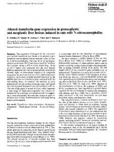
Fig. 1. Adenoma cells demonstrating a homogeneous pattern. H& E a x 120 b Fig. 2. Adenocarcinoma cells characterized by nuclear atypia and cellular heterogeneity (polymorphism). H & E a • 120 b
commencement, were maintained under constant conditions (22~ C, 12 h light/dark cycle) and fed Altromin A (Lage-Lippe) and tap water ad libitum. Lung lesions were induced by a series of weekly 250 mg/kg body weight s.c. injections of DHPN (generous gift of Dr. Wiessler, Institute for Toxicology and Chemotherapy, German Cancer Research Center) for twenty weeks and hamsters were sacrificed under ether anesthesia five weeks later. For investigation of tritiated thymidine incorporation a group of 12 DHPN-treated and 3 control animals received 5 daily 0.8 ~tCi/g body weight i.p. injections of (H3)thymi dine (specific activity 5.0 Ci/mmol, Amersham Buchlcr, Braunschwcig, FRG) in the week before sacrifice. Lung lobes were excised immediately upon sacrifice and either fixed in Carnoy's solution, quick frozen in liquid nitrogen-precooled isopentane at - 140~ C or fixed in 4% glutaraldehyde and post-fixed in osmium tetroxide for subsequent pro-
cessing for electron microscopic examination. 4 ~tm paraffin sections were cut in series and stained with H & E or for binding of antibodies raised against GST-B, C and P forms (Sato et al. 1984; Kitahara et al. 1984) or G6PD (generous gift of Prof. Ichihara, Tokushima University, Japan) (Nakamura et al. 1982) using the avidin-biotin-peroxidase complex (ABC) method (Hsu et al. 1981) (Vectastain Kit purchased from Vector Laboratories, Burlingame, CA, USA). A total of 65 lesions were investigated immunohistochemically in serial sections. For autoradiographic analysis sections were covered with K O D A K AR 10 fine grain stripping film, exposed for 30 days and subsequently fixed and stained with H & E. Assessment of incorporation of label was made at the microscope with the aid of an eye-piece grid and expressed as labelled cells per 1000 cells examined. For histochemical analysis 10 ~m cryostat sections, includ-
274
M.A. Moore et al. : Altered enzyme expression in Syrian hamster lung lesions
Table 1. Lung lesions: Incorporation of H 3 thymidine label Alveolar cells
Control DHPN-Treated
1.5 16
Bronchiolar cells
0.3 7
Focal proliferations
Adenomas
72
Adenocarcinomas
Solid (pale)
Papillary (basophilic)
64
143
295
Results expressed as labelled nuclei/1000 cells counted
Table 2. Phenotype of DHPN-induced lung lesions
Normal
Carcinogen-induced
Alveoli Bronchioli Focal Proliferation Adenoma (Solid) Adenoma (Papillary) Adenocarcinoma
- , Absent; + , present; - / + ,
G6PD
GGT
GST-P
GST-B
GST-C
+ + + + +
- /+ + + + + + +/+
+ + + + +
+ + + +
*+ *+ + /+ *+ *+ + *+ +
/+ + + +/+ + +
weak or absent; + + , prevalent; + + / - ,
ing 37 carcinogen-induced focal lesions were stained by PAStoluidine blue or reacted for demonstration of G6PD (membrane method after Meijer and Vries 1974) and GGT (Lodja et al. 1976) activities. For electron microscopical investigation epon embedded blocks from 7 adenomas and 3 adenocarcinomas diagnosed on the presence of cellular atypia were selected after high resolution light microscopic examination of toluidine blue stained semi-thin sections (0.5 gm). Ultrathin sections contrasted with lead citrate were examined with a Zeiss electron microscope.
Results
At final sacrifice a range of focal lesions were observed in the lungs of hamsters treated with DHPN, these being classified as focal proliferations, adenomas of solid (see Fig. 1) or papillary morphology and adenocarcinomas (see Fig. 2) demonstrating cellular atypia, invasive character and necrotic central regions. In animals administered carcinogen the background level of tritiated thymidine incorporation was elevated in both the alveolar and bronchiolar cell compartments as compared to controls (see Table 1). Papillary tumour formations were associated with a higher rate of labelling than the less basophilic sheets of cells in solid adenomas and most pronounced incorporation of tritiated thymidine was evident in large, atypical cells within carcinomas. Enzyme phenotype (see Table 2) of normal appearing alveoli and bronchioli did not differ between the group given carcinogen and that maintained as untreated control, type I and type II alveolar cells being weakly positive for G6PD, demonstrating moderate binding for GST-B and negative for the other parameters investigated. Bron-
+/+/+ +/+ + +
+/+ /+ /+ +/-
+ + / + , heterogeneous pattern; *, stromal compartment
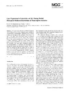
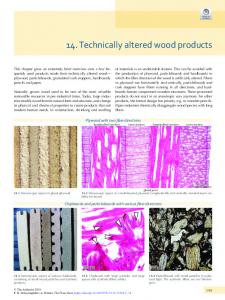
chiolar cells, in contrast, were characterized by a strong G6PD reaction and especially in superficial epithelium, pronounced binding for GST-B and P species. GST-C was weakly positive in the connective tissue elements. As summarized in Table 2, carcinogen-induced focal lesions were characterized by distinctive patterns of expression of enzyme phenotype. GST-P (see Figs. 3-5) served as a good marker for all the carcinogen-induced focal populations, almost invariably observed together with increased GST-C binding in surrounding connective tissue. GST-P staining was particularly remarkable in nucleoplasm. G6PD binding was strong within all proliferating populations although the reaction in more solid adenomas was somewhat weaker than in other lesions (see Fig. 6). GGT activity, not a feature of normal lung bronchiolar epithelia and only slightly positive in alveolar cells, was evident especially in papillary adenomas. GST-B binding differed from that of the other antibodies in the majority of focal proliferative lesions, a reduction also being apparent in the adenomas of more solid pattern. Under the electron microscope the majority of lesions (7 adenomas) were characterized by a homogeneous population of epithelial cells possessing varying numbers of lamellar bodies (see Fig. 7). However, in three cases diagnosed as carcinomas on the basis of cellular atypia, a much more heterogeneous (polymorphic) population was observed, epithelial cells with few if any lamellar bodies and dense accumulations of free ribosomes in the cytoplasm predominating (see Fig. 8).
M.A. Moore et al. : Altered enzyme expression in Syrian hamster lung lesions
275
Fig. 3. Small hyperplastic lesion. Immunohistochemical demonstration of a GST-P b GST-C. x 100 Fig. 4a, b. Solid adenoma. Immunohistochemical demonstration of a GST-P b GST-C. x 40 Fig. 5a, b. Portion of adenocarcinoma. Immunohistochemical demonstration of a GST-P b GST-C. x 80. Note in all three cases the prevalent binding of GST-P antibody in the nuclei of epithelial cells and the localization of GST-C in surrounding stroma
Discussion
The present morphological, autoradiographic and histochemical findings, although not sequentially performed, point to a histogenetic link between small focal proliferations, adenomas and adenocarcinomas. Foci were observed both adjacent to bronchioles and apparently free in the alveolar tissue, and, while it was not possible to preclude
bronchiolar involvement in any particular case, the electron microscopic evidence clearly suggests an origin from alveolar type II cells. Similar ultrastructural findings were reported for butylnitrosamine-induced tumours in European hamsters (Reznik-Schuller and Mohr 1975) and lung lesions arising after diethylnitrosamine treatment of Syrian golden hamsters (Straks and Feron 1973). This conclusion of a histogenesis from alveolar epitheli-
276
M.A. Moore et al. : Altered enzyme expression in Syrian hamster lung lesions
Fig. 6. Histochemical demonstration of enzyme activity in a papillary adenoma a G6PD b GGT. • 80
um is in agreement with that made earlier for rats and mice (Ohshima et al. 1985; Ward et al. 1985) on the basis of immunocytochemical localization of surfactant apoprotein but not Clara cell antigen in similar bronchiolo-alveolar type tumours. The relative lack of lamellar bodies within adenocarcinoma cells (cf Flaks and Flaks 1970) might thus be interpreted as indicating a loss of differentiative features with increasing malignancy, a well known characteristic of neoplastic development. Whether this might partially explain the electron microscopic findings in the tree shrew (Rao and Reddy 1980) and polonium 210-induced lesions in the Syrian hamster lung (Kennedy et al. 1977) or whether two distinct tumour types, of separate histogenesis, may exist (Kauffman et al. 1979) remains unclear. In man ultrastructural and immunohistochemical
evidence has also indicated type II cell-like differentiation in a number of cases of bronchioalveolar tumours (Espinoza et al. 1984) although the presence of Clara cell granules may complicate the question of histogenesis (Espinoza etal. 1984; Ogata and Endo 1984). Further studies of the potential of tumor and normal lung cells for change in differentiation is required before this problem can be resolved. If the conclusion of an origin from type II cells in the present model is correct, the histochemical results are suggestive of a nonrandom switch within carcinogen-induced lesions to an enzyme phenotype more typical of normal bronchiolar epithelium. This would imply an increase of G6PD and GST-P in concert, along with the appearance of GGT activity, all three enzymes sharing a physio-
M.A. Moore et al. : Altered enzyme expression in Syrian hamster lung lesions
277
Fig. 7. Electron micrograph illustrating appearance of adenoma cells. Note homogeneity of cell population and presence of lamellar bodies, x 6000 Fig. 8. Electron micrograph illustrating appearance of adenocarcinoma cells. Note heterogeneity of cell population and absence of lamellar bodies. x 6000
logical link via glutathione metabolism, and might suggest enhanced drug detoxification potential within carcinogen-induced focal lung lesions reflecting some adaptive response (Farber 1984). However, while available data for hepatocellular (Buchmann etal. 1985; Kitahara etal. 1984; Moore et al. 1985), pancreatic (Moore et al., unpublished work) and cholangiocellular (Moore et al. 1986) lesions support a possible role for detoxifying enzymes in their induction, the situation with regard to kidney (Tsuda et al. 1985) and gallbladder (Moore et al. 1986) where a number of the enzymes in question are reduced illustrates the caution which must be used in drawing general conclusions. Nevertheless, the shared features of
focal proliferative lesions induced by propylnitrosamines in their various target organs is impressive. GST-P seems particularly promising as a 'marker' enzyme for preneoplastic foci and nodules. The meaning of the increased GST-C binding evident in the stromal component of DHPN-induced lesions is unclear. Similar increase in GST-C staining was associated with both cholangiocellular and pancreatic lesions induced by propylnitrosamine in the Syrian hamster (Moore et al. 1986, 1987) although the finding of periductal increase also after common duct ligation suggests t h a t it may be a non-specific response. The finding is, however, of interest as pointing to a role for epithe-
278
M.A. Moore et al. : Altered enzyme expression in Syrian hamster lung lesions
lial-endothelial communication in the control of GST enzyme form expression.
References Buchmann A, Kuhlmann W, Schwartz M, Kunz W, Wolf CR, Moll E, Friedberg T, Oesch F (1985) Regulation and expression of four cytochrome P-450 isoenzymes, NADPH-cytochrome P-450 reductase, the glutathione transferases B and C and microsomal epoxide hydrolase in preneoplastic and neoplastic lesions in rat liver. Carcinogenesis 6:513-521 Definer GB (1982) Origin of bronchioloalveolar carcinoma and peripheral bronchial adenocarcinoma. Cancer 49:881-887 Espinoza CG, Balis JU, Saba SR, Paciga JE, Shelley SA (1984) Ultrastructural and immunohistochemical studies of bronchio-alveolar carcinoma. Cancer 54: 2182-2189 Farber E (1984) Pre-cancerous steps in carcinogenesis - their physiological adaptive nature. Biochim Biophys Act 738:171-180 Flaks B, Flaks A (1970) Fine structure of murine pulmonary adenocarcinomata induced by treatment with 20-methylcholanthrene in organ culture. Eur J Cancer 6: 477-482 Hacker H J, Moore MA, Mayer D, Bannasch P (1982) Correlative histochemistry of some enzymes of carbohydrate metabolism in preneoplastic and neoplastic lesions in the rat liver. Carcinogenesis 3:1265-1272 Hanigan MH, Pitot HC (1985) Gamma-glutamyl transpeptidase - its role in hepatocarcinogenesis. Carcinogenesis 6:165-172 Hsu SM, Raine L, Fanger H (1981) Use of avidin-biotin-peroxidase complex (ABC) in immunoperoxidase techniques: a comparison between ABC and unlabelled antibody PAP procedures. J Histochem Cytochem 29:577-580 Kauffman SL, Alexander L, Sass L (1979) Histologic and ultrastructural features of the Clara cell adenoma of the mouse lung. Lab Invest 40:708-716 Kennedy AR, Little JB (1977) Histochemistry of normal lungs and 210 Po induced pulmonary tumors in hamsters. Acta Histochem 58 : 353-359 Kennedy AR, McGandy RB, Little JB (1977) Histochemical, light and electron microscopic study of Polonium 210 induced peripheral tumors in hamster lungs: evidence implicating the Clara cell as the cell of origin. Eur J Cancer 13 : 1325-1340 Kitahara A, Satoh K, Nishimura K, Ishikawa T, Kiuke K, Sato K, Tsuda H, Ito N (1984) Changes in molecular forms of rat hepatic glutathione S-transferase during chemical carcinogenesis. Cancer Res 44: 2698-2703 Kondo H, Ikeda T, Yoshimura Y, Hoshimura H, Konishi Y (1978) Carcinogenic effect of N-nitrosobis(2-hydroxypropyl)amine in rabbits. Cancer Lett 5 : 339-343 Konishi Y, Denda A, Kondo H, Takahashi S (1976) Lung carcinomas induced by oral administration of N-bis(2hydroxypropyl)-nitrosamine in rats. Gann 67:773-780 Konishi Y, Kondo H, Inui S, Denda A, Ikeda T, Kojima K (1978) Organotropic effect of N-bis(2-hydroxypropyl)-nitrosamine: production of lung and liver tumors by its oral administration in mice. Gann 69 : 77-84 Lojda Z,. Gossrau R, Schiebler TH (1976) Enzyme Histochemical Methods. Springer, Berlin Heidelberg New York Meijer AEFH, Vries EP (1974) Semipermeable membranes for improving the histochemical demonstration of enzyme activities in tissue sections. IV. Glucose-6-phosphate dehydrogenase and 6-phosphogluconate dehydrogenase (decarboxylating). Histochemistry 40:349-359 Moore MA, Fukushima S, Ichihara A, Sato K, Ito N (1986) Intestinal metaplasia and altered enzyme expression in pro-
pylnitrosamine-induced Syrian hamster cholangiocellular and gallbladder lesions. Virchows Arch [Cell Pathol] 51 : 29-38 Moore MA, Bannasch P, Sato K, Ito N (1987) Immunohistochemically demonstrated increase in glutathione S-transferase species within propylnitrosamine-induced preneoplastic and neoplastic pancreatic lesions. Virchows Arch [Cell Pathol] 52:479-488 Moore MA, Satoh K, Kitahara A, Sato K, Ito N (1985) A protein cross-reacting immunohistochemically with rat glutathione S-transferase placental form as a marker for preneoplasia in Syrian hamster pancreatic and hepatocarcinogenesis. Jpn J Cancer Res (Gann) 76:1-4 Moore MA, Takahashi M, Ito N, Bannasch P (1983) Early lesions induced in Syrian hamsters by DHPN or DOPN. I. Histologic, histochemical and radioautographic findings. Carcinogenesis 4:431-437 Nakamura T, Yoshimoto K, Aoyama K, Ichihara A (1982) Hormonal regulation of glucose-6-phosphate dehydrogenase and lipogenesis in primary cultures of rat hepatocytes. J Biochem 91:681-693 Ogata T, Endo K (1984) Clara cell granules of peripheral lung cancers. Cancer 54:1635-1644 Ohshima M, Ward JM, Singh G, Katyal SL (1985) Immunocytochemical and morphological evidence for the origin of nitrosomethylurea-induced and naturally occurring primary lung tumours in F344/NCr rats. Cancer Res 45:2785-2792 Pour P, Salmasi SZ (1982) Comparative studies of neoplastic response to a single dose of nitroso compounds. 7. The effect of N-nitrosobis(2-hydroxypropyl)amine in Syrian golden hamsters. J Cancer Res Clin Oncol 102:265-269 Pour P, Kruger FW, Althoff J, Cardesa A, Mohr U (1974) Effect of beta-oxidized nitrosamines on Syrian hamsters: III 2,2'-dihydroxy-di-n-propylnitrosamine. J Natl Cancer Inst 54:141-145 Rao MS, Reddy JK (1980) Carcinogenicity of 2,2'-dihydroxydi-n-propylnitrosamine in the tree shrew (Tupaia glis) : light and electron microscopic features of pulmonary adenomas. J Natl Cancer Inst 65:835-840 Reznik-Schuller H, Mohr U (1975) The ultrastructure of Ndibutylnitrosamine induced pulmonary tumours (adenocarcinomata) in European hamsters. Br J Cancer 32:230-238 Sato K, Kitahara A, Satoh K, Ishikawa T, Tatematsu M, Ito N (1984) The placental form of glutathione S-transferase as a new marker protein for preneoplasia in rat chemical hepatocarcinogenesis. Gann 75 : 19%202 Strake W, Feron VJ (1973) Ultrastructure of pulmonary adenomas induced by intratracheal instillation of diethylnitrosamine in Syrian golden hamsters. Eur J Cancer 9 : 359-362 Toth B, Shimizu H (1974) 1-Carbamyl-2-phenylhydrazine tumorigenesis in Swiss mice. Morphology of lung adenomas. J Natl Cancer Inst 52:241-251 Tsuda H, Moore MA, Asamoto M, Satoh K, Tsuchida S, Sato K, Ichihara A, Ito N (1985) Comparison of the various forms of glutathione S-transferase with glucose-6-phosphate dehydrogenase and gamma glutamyltranspeptidase as markers of preneoplastic and neoplastic lesions in rat kindey induced by N-ethyl-N-hydroxyethylnitrosamine.Jpn J Cancer Res (Gann) 76:919-929 Ward JM, Singh G, Katyal SL, Anderson LM, Kovatch RM (1985) Immunocytochemical localization of the surfactant apoprotein and Clara cell antigen in chemically induced and naturally occurring pulmonary neoplasms of mice. Am J Pathol 118 : 493-499
Received October 11, 1986 / Accepted May 15, 1987