European Heart Journal – Cardiovascular Imaging (2013) 14, 228–234 doi:10.1093/ehjci/jes139
Altered left ventricular longitudinal diastolic function correlates with reduced systolic function immediately after anthracycline chemotherapy Paul W. Stoodley1,2*, David A.B. Richards3,4, Anita Boyd4, Rina Hui5, Paul R. Harnett5, Steven R. Meikle1,2,6, Jillian L. Clarke1,2, and Liza Thomas3,4,7 1 Faculty of Health Science, University of Sydney, Lidcombe, NSW, Australia; 2Westmead Private Cardiology, Suite 1 Westmead Private Hospital, Corner Mons and Darcy Roads, Westmead, NSW 2145, Australia; 3Department of Cardiology, Liverpool Hospital, Liverpool, NSW, Australia; 4Faculty of Medicine, University of New South Wales, Sydney, NSW, Australia; 5Crown Princess Mary Cancer Centre, Westmead, NSW, Australia; 6Brain and Mind Research Institute, University of Sydney, Camperdown, NSW, Australia; and 7Faculty of Medicine, University of Sydney, Sydney, NSW, Australia
Received 25 April 2012; accepted after revision 17 June 2012; online publish-ahead-of-print 10 July 2012
Aims
The benefits from anthracycline chemotherapy are undermined by potentially life-threatening cardiotoxicity. Transthoracic echocardiography is the most commonly used method for monitoring cardiotoxicity, and centres on the measurement of left ventricular systolic function. The aim of this study was to utilize two-dimensional speckle tracking echocardiography (2DSTE) at baseline and immediately after anthracycline chemotherapy to investigate whether patients with significant changes in systolic function after anthracycline therapy would also develop alterations in diastolic parameters. ..................................................................................................................................................................................... Methods Fifty-two women with histologically confirmed breast cancer were prospectively recruited. Echocardiograms were and results performed 1 week prior to and 1 week following chemotherapy (always before adjuvant trastuzumab or thoracic radiotherapy). Conventional Doppler, tissue velocity imaging (TVI), and 2DSTE were used to measure diastolic function. 2DSTE measurements included longitudinal diastolic strain, early (E-Sr), and late (A-Sr) myocardial strain rate. 2DSTE and left ventricular ejection fraction (LVEF) were used to measure longitudinal systolic function. Altered LV diastolic function (including E-Sr) was observed in the entire cohort after chemotherapy, with a differential reduction in participants with a post therapy LVEF ,55%. Pre-chemotherapy systolic strain was found to predict reduced E-Sr post therapy (P ¼ 0.04). Univariate predictors of E-Sr were LVEF post therapy (P ¼ 0.049) and systolic strain posttherapy (P ¼ 0.01). In a multivariate analysis, systolic strain after chemotherapy was the strongest independent predictor (P ¼ 0.001). ..................................................................................................................................................................................... Conclusion Altered LV diastolic function was observed immediately after the administration of therapeutic doses of anthracycline chemotherapy. Furthermore, our analysis indicates that the changes in diastolic function are associated with reduced systolic function.
----------------------------------------------------------------------------------------------------------------------------------------------------------Keywords
Diastolic function † Two-dimensional speckle tracking echocardiography (2DSTE): anthracyclines
Introduction Anthracycline agents are an essential part of breast cancer treatment due to their highly effective antineoplastic properties.1 However, as anthracyclines are potentially cardiotoxic, close monitoring of cardiac function with their administration is mandatory.2,3 Transthoracic echocardiography (TTE) is the most common
method for monitoring cardiotoxicity,4 and in this setting is centred on measuring left ventricular (LV) systolic function. Yet, both components of the cardiac cycle, systole and diastole are essentially linked and together must function normally for optimal cardiac performance.5 Conventional Doppler measurements have demonstrated changes in diastolic function 6 years after childhood anthracycline
* Corresponding author. Tel: +61 02 9687 0866; fax: +61 02 9687 0422. Email:
[email protected] Published on behalf of the European Society of Cardiology. All rights reserved. & The Author 2012. For permissions please email:
[email protected]
Altered left ventricular longitudinal diastolic function
chemotherapy,6 and tissue velocity imaging (TVI) measured 3 months and then 3 years after anthracyclines in a small adult population, revealed persistently reduced E′ velocities.7 Strain imaging may enhance the evaluation of diastolic function;8,9 however, there is a lack of published literature to support this. Previously, we reported reduced LV systolic strain immediately after anthracylines,10 our aim in this study was to investigate LV diastolic function using conventional Doppler, TVI, strain, and strain rate before and immediately after anthracyclines. We hypothesized that those with significant changes in systolic function immediately following therapy would also develop changes in diastolic parameters.
Methods The Sydney West Area Health Service and the University of Sydney Research Ethics Committees approved the study, and written informed consent was obtained from all participants. Fifty-two consecutive anthracycline naı¨ve women with histologically confirmed breast cancer were prospectively recruited. All echocardiograms were performed at a single site, although chemotherapy was administered at one of four sites (depending on proximity to the patient’s residence). The cumulative anthracycline dose was documented. The clinical history, physical examination, and echocardiographic characteristics were used to establish eligibility. Exclusion criteria included LV ejection fraction (LVEF) ,50% prior to chemotherapy, rhythms other than sinus, more than mild valvular stenosis or regurgitation, prosthetic valve, previous ischaemic heart disease, or pacemaker. Height was measured at baseline, while weight and blood pressure were measured at the time of each echocardiogram. A detailed cardiac history, including clinical risk factors for heart disease (hypertension, diabetes, cholesterol, smoking, and family history) was obtained at recruitment. Participants were also evaluated for any cardiac symptoms especially those of heart failure. The initial echocardiogram was performed 1 week prior to the commencement of chemotherapy. The follow-up echocardiogram was performed 1 week following completion of chemotherapy (i.e. 12 or 18 weeks after the first study, depending on whether four or six cycles of chemotherapy were administered) and always performed before the commencement of trastuzumab or thoracic radiotherapy. Echocardiograms were performed by two accredited sonographers using a Vivid 7 digital ultrasound system (GE medical systems, Norway) with a 2.5 MHz variable frequency transducer optimized for image quality. All TTE’s were performed with patients in the left lateral position and images were obtained from the parasternal, apical, and subcostal views. The transmitral early (E) and late (A) peak diastolic filling velocities, E/A ratio, E-wave deceleration time, and A-wave duration were measured using conventional pulsed wave Doppler echocardiography with the sample volume placed at the mitral leaflet tips in the apical four-chamber view. The velocity time integral (VTI) of the A-wave was measured and the atrial emptying fraction estimated as the A-wave VTI divided by the total mitral inflow VTI. Maximum left atrial (LA) volume (just prior to mitral valve opening) and minimum LA volume were measured using the biplane method of disks, according to the European Association of Echocardiography recommendations11 and indexed to the body surface area. Peak velocity and the VTI of the systolic, diastolic, and atrial components of the pulmonary vein flow were measured with pulsed wave Doppler by placing the sample volume in the proximal 1 cm of the right upper pulmonary vein. The systolic fraction was calculated as the systolic wave VTI divided by the total forward pulmonary vein
229 flow VTI. Isovolumic relaxation time was measured from the continuous wave Doppler LV outflow tract signal. Pulsed wave tissue Doppler imaging (TDI) was used to measure the early (E′ ) and late (A′ ) peak velocities from the septal annulus. Two-dimensional speckle tracking echocardiography (2DSTE) was used to measure bi-plane global longitudinal systolic and diastolic strain (DS) from the apical four- and two-chamber views, with observers blinded to LVEF results. The LV regions of interest for strain and strain rate analysis were manually selected by marking the endocardial border in the apical four- and apical two-chamber views. The tracking quality of individual LV segments was identified prior to analysis using the software scoring table on the off-line measuring package. Tracking quality was over-ridden in segments with no more than two rejected segments where the observer deemed tracking quality to be clearly acceptable. Images with persistently unacceptable tracking quality (more than two segments) were excluded from the final analysis. Measurements were made from three consecutive cardiac cycles and then averaged in order to obtain global strain and strain rate results. Systolic strain was measured as the peak negative strain during systole. DS was estimated by measuring the time interval from the ECG R-wave to peak E velocity with conventional transmitral Doppler echocardiography. The same time interval was applied to the 2DTSE trace to measure early global LV longitudinal DS as previously described12 (see Figure 1A and B). The heart rate was largely unchanged during both measurements. E/DS was calculated as a measure
Figure 1 (A) and (B) Diastolic strain (Ds) measurement: calculation is made by measuring the time interval from the ECG R wave to the peak E velocity. This time interval is then applied to the 2DSTE trace to measure early global LV longitudinal diastolic strain (DS) from the dotted white line.
230
P.W. Stoodley et al.
Table 1 Participant demographics, cancer location, and chemotherapy data Number of participants
52
Age (years)
49 + 9
Height (cm) Weight (kg)
161 + 5 76 + 23
Side of breast cancer Right Left
28 24
Anthracycline type
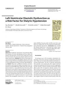
Figure 2 Global 2DSTE diastolic strain rate measurements: the early peak diastolic strain rate (E-Sr) indicated by the white arrow and late peak diastolic strain rate (A-Sr) indicated by the yellow arrow. The coloured lines represent segmental measurements of the strain rate and the dotted line the global average strain rate.
of elevated LV end diastolic pressure.12 2DSTE was also used to measure the biplane longitudinal DS rate. Global peak velocities in early (E-Sr) and late (A-Sr) diastole were measured with reference to the ECG (see Figure 2). Fifteen participants were randomly selected to determine interobserver and intra-observer variability in E-Sr and A-Sr, with the differences reported as mean difference + standard deviation. One observer performed repeated measurements of E-Sr, A-Sr on a different occasion in order to measure intra-observer variability. Inter-observer and intra-observer variability for systolic longitudinal strain has been previously reported.10 All values were expressed as a mean + SD unless otherwise stated. Paired t-tests were used to compare conventional Doppler, TVI, and 2DSTE parameters before and after chemotherapy in the entire cohort (and within subgroups) to compare within patient changes. Linear regression and Chi square analysis were performed to determine univariate determinants of E-Sr as appropriate. Univariate variables with a significant correlation were entered into a stepwise multiple regression analysis to determine independent predictors of E-Sr. Data were analysed using SPSS version 19 (SPSS, Inc., Chicago, IL, USA), and considered significant if P , 0.05.
Results All 52 participants had a baseline TTE before chemotherapy and a follow-up TTE 1 week following completion of chemotherapy. Limited image quality, most often due to left-sided mastectomy and/or breast implant, before and/or after chemotherapy meant that all measurements were not possible in all participants. Three participants (6%) had a history of ischaemic heart disease, 11 (22%) had hypercholesterolaemia, 6 (12%) were smokers, 13 (26%) had hypertension, and 2 (4%) were diabetic. Four (n ¼ 34) or six (n ¼ 18) cycles of anthracycline chemotherapy (doxorubicin or epirubicin) were administered, as determined by the oncology team. No participant reported symptoms or demonstrated signs
Doxorubicin Mean Dox dose (mg/m2)
40 (77%) 236 + 33
Epirubicin
12 (23%)
Mean Epi dose (mg/m2)
408 + 110
of cardiac failure during the follow-up. The maximum doxorubicin dose was 318 mg/m2 and the maximum epirubicin dosage was 581 mg/m2 (see Table 1). Paired measurements of conventional Doppler transmitral diastolic filling velocities were possible in 49 of the 52 participants (94%) and of the pulmonary vein flow in 43 of the 52 participants (83%). Among the transmitral measurements, peak A (P , 0.05) and the atrial fraction (P , 0.001) were significantly increased, whereas the E/A ratio (P , 0.05) was significantly reduced after chemotherapy. Of the pulmonary vein flow measurements, the systolic fraction was significantly increased (P , 0.001) and the diastolic VTI was significantly reduced (P , 0.05) after chemotherapy. LA maximum and minimum volumes and isovolumetric relaxation time did not change after chemotherapy (see Table 2). Paired measurements of TVI diastolic parameters measured at the septal annulus were possible in 46 of the 52 participants (88%). No significant change in E′ velocity, the E/E′ ratio or in A′ velocity was observed after chemotherapy (see Table 2). 2DSTE DS and the early and late DS rate (E-Sr and A-Sr, respectively) measurements were feasible in 45 of the 52 participants (87%). The E-Sr was significantly reduced (P , 0.01) after chemotherapy, with an E-Sr reduction greater than the mean minus 1 SD of the baseline measurement in 13 participants (25%). No significant change was observed in DS or E/DS (see Table 2). Previous analysis of LV systolic function in this cohort revealed significantly reduced global LV longitudinal systolic strain after chemotherapy.10 In order to investigate the relationship between reduced systolic function and diastolic function measurements after chemotherapy, participants were divided into two groups; Group 1 (n ¼ 38) comprised participants with an LVEF after chemotherapy .55% and Group 2 (n ¼ 14) of those with an LVEF of ,55% after chemotherapy. Within Group 1, only the E-Sr was significantly reduced (P ¼ 0.03) after chemotherapy. In contrast, within Group 2, the A velocity and atrial fraction were significantly increased. The pulmonary vein atrial reversal duration minus the transmitral A duration was increased, almost reaching statistical significance (P ¼ 0.05). The E/A ratio was significantly reduced. The diastolic VTI and atrial reversal duration decreased
231
Altered left ventricular longitudinal diastolic function
Table 2 Conventional Doppler, tissue velocity imaging, and 2D speckle tracking echocardiography measurements (2DSTE) of diastolic function in the entire study cohort before and after anthracycline chemotherapy Before
After
Doppler measurements Peak E velocity (m s21)
0.72 + 0.15
0.70 + 0.13
Peak A velocity (m s21)
0.66 + 0.13
0.69 + 0.15*
E/A ratio Deceleration time (m s21)
1.13 + 0.30 220 + 36
1.05 + 0.28* 225 + 38
Atrial fraction
36.0 + 6.9
38.4 + 7.5**
PV systolic VTI (cm s21) PV diastolic VTI (cm s21)
15.8 + 3.5 11.5 + 3.5
16.5 + 4.7 10.5 + 2.5*
................................................................................
Systolic fraction PV atrial reversal duration (ms) Tissue velocity imaging
57.5 + 9.3
62.3 + 8.5**
119 + 19
116 + 15
E′ velocity (cm s21)
8.3 + 2.0
8.2 + 4.7
E/E′ ratio A′ velocity (cm s21)
8.8 + 2.3 9.8 + 4.2
9.2 + 2.6 9.0 + 1.8
Diastolic strain (%) Early strain rate (ESR) (s21)
10.9 + 2.5 1.00 + 0.24
10.4 + 2.0 0.90 + 0.22*
Active strain rate (ASR) (s21)
0.63 + 0.16
0.63 + 0.16
2DSTE measurements
PV, pulmonary vein; VTI, velocity time integral. *P , 0.05 vs. before chemotherapy value. **P , 0.01 vs. before chemotherapy value.
No significant association was present on Chi-square analysis for baseline clinical risk factors (hypertension, diabetes, cholesterol, smoking, and family history) and reduced E-Sr post-therapy. There was no significant difference in blood pressure measurements. As expected, linear regression analysis demonstrated a correlation with age and baseline LVEF and post-therapy E-Sr (r ¼ 20.35, P ¼ 0.04 and r ¼ 20.54, P , 0.001, respectively). Baseline systolic strain was also found to be related to post-chemotherapy E-Sr (r ¼ 20.35, P ¼ 0.04). No association was found between E-Sr post-therapy and baseline LVEF, LV mass, or cumulative anthracycline dose. Univariate predictors of post-chemotherapy biplane E-Sr were LVEF (P ¼ 0.049) and biplane systolic strain (P ¼ 0.01); with age proving to be the strongest predictor (see Figure 3A and B). Multivariate predictors of biplane E-Sr post-therapy were LVEF and biplane systolic strain, with post-therapy biplane systolic strain being the strongest predictor (standardized co-efficient b ¼ 20.48, P ¼ 0.002) (age was excluded from the multivariate analysis based on its established co-dependence with diastolic function). Inter-observer and intra-observer differences in the measurement of the DS rate were calculated. For E-Sr, the mean inter-observer difference was 0.08 + 0.12 s21 and the mean intra-observer difference was 0.01 + 0.05 s21. For A-Sr, the mean inter-observer difference was 0.06 + 0.12 s21 and the mean intra-observer difference 0.01 + 0.08 s21. For global longitudinal systolic strain, the mean inter-observer difference was 21.73 + 1.0 and the mean intra-observer difference was 20.86 + 0.59 (as previously reported).8 Bland-Altman plots and additional correlation analysis figures have been included as supplementary data.
Discussion Table 3 Measurements of systolic and diastolic function with significant variation after chemotherapy in Group 2 (n 5 14) Before
After
218.8 + 2.81
215.6 + 2.47**
................................................................................ Systolic function Bi-plane longitudinal systolic strain (%) Diastolic function Peak A velocity (ms21)
0.64 + 0.14
0.69 + 0.13***
E/A ratio
1.17 + 0.28
1.04 + 0.26***
Atrial fraction Diastolic VTI
35.8 + 5.72 12.76 + 2.89
39.2 + 5.45*** 11.07 + 2.53*
Atrial reversal duration
125 + 37.2
117 + 19.3***
E′ velocity (cm s21) Early strain rate (E-Sr) (s21)
8.24 + 1.57 1.04 + 0.19
7.48 + 1.43*** 0.80 + 0.17*
*P , 0.01 vs. before chemotherapy. **P ¼ 0.01 vs. before chemotherapy. ***P , 0.05 vs. before chemotherapy value.
and the systolic fraction increased significantly after chemotherapy in Group 2. Moreover, E′ and E-Sr were significantly reduced in Group 2 (see Table 3).
The assessment of LV diastolic function is an essential part of a standard TTE examination. However, when monitoring cardiotoxicity, evaluation has typically focused on measuring LV systolic function by the LVEF. We previously reported reduced systolic function using strain imaging immediately after anthracycline chemotherapy;10 in the present study we observed alterations in diastolic function immediately after treatment. Reduced baseline systolic strain was found to be predictive of lower postchemotherapy E-Sr. Moreover, a differential reduction in diastolic function parameters in participants with the LVEF ,55% after chemotherapy was observed. Early identification of patients most likely to develop significant LV dysfunction following anthracyclines is of considerable clinical value, as it will enable targeted monitoring as well as cardioprotective therapy. Anthracycline-related diastolic dysfunction in paediatric6 and adult populations7 in the intermediate and long term has been described. To our knowledge, our study is the first to report altered diastolic 2DSTE strain measurements and its association with systolic dysfunction immediately after anthracycline therapy: thus, our results provide an important insight to early myocardial changes following anthracyclines. Longer-term followup for the development of symptomatic heart failure is required to determine whether the early changes we have demonstrated will help identify patients at risk. For now, identifying altered diastolic function early may assist in the recognition and confirmation of anthracycline-induced systolic dysfunction.
232
P.W. Stoodley et al.
Figure 3 (A) and (B) Pearson’s linear regression analysis revealed a significant correlation between the biplane ESR and LVEF postchemotherapy, P ¼ 0.049 (A) and a significant correlation between biplane ESR and peak longitudinal systolic strain post-chemotherapy, P ¼ 0.01 (B).
Using TVI to measure systolic and diastolic function is now routine in TTE examinations. Reduced TVI E′ velocities 3 months after anthracycline chemotherapy that persisted at 3 years, with
similar reductions in LVEF and S′ ’ velocity have been reported in a study comprising 20 patients.7 However, while the reduction in the LVEF reached statistical significance in that study, it was not
233
Altered left ventricular longitudinal diastolic function
clinically significant (mean LVEF post-therapy 56 + 8%). In contrast, we demonstrate a differential reduction in E′ and E-Sr measurements in patients with the LVEF ,55% post-chemotherapy (Group 2). Ho et al.13 recently reported reduced E velocity, E/A ratio, and E′ velocity in women treated with anthracyclines over a longer duration with subjects having been treated with anthracyclines up to 6 years earlier. They also reported reduced 2DSTE longitudinal and radial systolic strain, but failed to report a link between diastolic and systolic measurements. The E/E′ ratio is an accurate and widely used estimate of LVEDP, although it has been suggested that using 2DSTE to calculate E/DS may provide a more robust measure of LVEDP.12 In the present study, no significant change in E/E′ , DS, E/DS, or A-Sr in the entire study population or in either of the subgroups was observed. The absence of change in E/E′ or E/DS may reflect the fact that these parameters are altered with chronic disease rather than changes that occur acutely (as the follow-up echocardiogram was performed within 1 week of completion of chemotherapy). It is unclear why changes in E-Sr precede changes in DS, but it is likely that the rate of myocardial deformation is altered prior to a reduction in the deformation per se. Isolated reduction in E-Sr with normal A-Sr levels has been reported in hypertension14 and may represent early change in diastolic function. In contrast to systole, diastole comprises two distinct phases. The first phase of diastole, active relaxation, occurs in a series of energy consuming steps that are dependent upon numerous cellular processes for normal function.8 Alterations in myocardial collagen properties, abnormal calcium handling and an increase in ventricular fibrosis are known to result in reduced LV compliance and relaxation, and occur to a greater degree with advancing age.15 – 18 The exact mechanism responsible for myocardial dysfunction as a result of anthracycline administration is unclear; however, the myocardial damage is thought to be mediated by intracellular oxidative stress that results in mitochondrial dysfunction, apoptosis, and myocyte necrosis.19 What is evident from this study is that a decline in early LV relaxation occurs as a result of anthracyclines. This decline can be measured 1 week after the completion of therapy, and results in a compensatory increase in the atrial contribution—similar to alterations in diastolic function that occurs with advancing age.18 – 20 Reports that document an increased risk of morbidity and mortality in association with isolated diastolic dysfunction further validate the importance of our observation21 in a cohort of patients with a mean age of 52 years. Reduced baseline systolic strain was found to be predictive of lower post-chemotherapy E-Sr. Moreover, measurements of early LV relaxation (E′ and E-Sr) were differentially reduced in patients with the LVEF ,55% post-chemotherapy (Group 2), and were strongly associated with reduced LV systolic strain. Our study is limited by the relatively small number of participants, particularly for subgroup analysis; however, within patient differences were still observed. The study reports changes immediately after anthracycline chemotherapy and does not include longer-term follow-up on the clinical implications of early anthracycline-induced changes in myocardial strain. Our study includes TVI E′ measurements sampled only at the septal annulus; an average of septal and lateral E′ velocity is lacking.
The variability in E-Sr in this study although similar to previous reports, is an inherent limitation of strain measurements and therefore clinical decisions cannot be based on this parameter alone. Our study does not include another imaging modality or the serial evaluation of cardiac biomarkers, which may provide additional information, as the performance of these additional tests was beyond the scope of the present study. In the present study, we observed changes in LV diastolic function using 2DSTE immediately after administration of therapeutic doses of anthracycline chemotherapy. Our analysis also indicates that the changes in diastolic function are related to reduced systolic function. Recognition of altered diastolic function could serve as an indicator of altered systolic function, thereby ensuring that patients who require closer monitoring when additional treatment with trastuzumab is required are identified. In the context of the future development of heart failure, our data support the need for longer-term follow-up with 2DSTE in an important clinical setting that requires close collaboration between oncology and cardiology.
Supplementary data Supplementary data are available at European Heart Journal – Cardiovascular Imaging online. Conflict of interest: none declared.
References 1. Gianni L, Herman EH, Lipshultz SE, Minotti G, Sarvazyan N, Sawyer DB. Anthracycline cardiotoxicity: from bench to bedside. J Clin Oncol 2008;26:3777 –84. 2. Bird BR, Swain SM. Cardiac toxicity in breast cancer survivors: review of potential cardiac problems. Clin Cancer Res 2008;14:14–24. 3. Singal PK, Iliskovic N. Doxorubicin-induced cardiomyopathy. N Engl J Med 1998; 339:900–5. 4. Jurcut R, Wildiers H, Ganame J, D’hooge J, De Backer J, Denys H et al. Strain rate imaging detects early cardiac effects of pegylated liposomal Doxorubicin as adjuvant therapy in elderly patients with breast cancer. J Am Soc Echocardiogr 2008;21: 1283 –9. 5. Lester SJ, Tajik AJ, Nishimura RA, Oh JK, Khandheria BK, Seward JB. Unlocking the mysteries of diastolic function: deciphering the Rosetta Stone 10 years later. J Am Coll Cardiol 2008;51:679 –89. 6. Dorup I, Levitt G, Sullivan I, Sorensen K. Prospective longitudinal assessment of late anthracycline cardiotoxicity after childhood cancer: the role of diastolic function. Heart 2004;90:1214 –6. 7. Tassan-Mangina S, Codorean D, Metivier M, Costa B, Himberlin C, Jouannaud C et al. Tissue Doppler imaging and conventional echocardiography after anthracycline treatment in adults: early and late alterations of left ventricular function during a prospective study. Eur J Echocardiogr 2006;7:141 –6. 8. Zile MR, Brutsaert DL. New concepts in diastolic dysfunction and diastolic heart failure: part II: causal mechanisms and treatment. Circulation 2002;105:1503 –8. 9. Nagueh SF, Appleton CP, Gillebert TC, Marino PN, Oh JK, Smiseth OA et al. Recommendations for the evaluation of left ventricular diastolic function by echocardiography. Eur J Echocardiogr 2009;10:165 –93. 10. Stoodley PW, Richards DA, Hui R, Boyd A, Harnett PR, Meikle SR et al. Twodimensional myocardial strain imaging detects changes in left ventricular systolic function immediately after anthracycline chemotherapy. Eur J Echocardiogr 2011; 12:945–52. 11. Lang RM, Bierig M, Devereux RB, Flachskampf FA, Foster E, Pellikka PA et al. Recommendations for chamber quantification. Eur J Echocardiogr 2006;7:79– 108. 12. Dokainish H, Sengupta R, Pillai M, Bobek J, Lakkis N. Usefulness of new diastolic strain and strain rate indexes for the estimation of left ventricular filling pressure. Am J Cardiol 2008;101:1504 – 9. 13. Ho E, Brown A, Barrett P, Morgan RB, King G, Kennedy MJ et al. Subclinical anthracycline- and trastuzumab-induced cardiotoxicity in the long-term follow-up of asymptomatic breast cancer survivors: a speckle tracking echocardiographic study. Heart 2010;96:701–7.
234 14. Saghir M, Areces M, Makan M. Strain rate imaging differentiates hypertensive cardiac hypertrophy from physiologic cardiac hypertrophy (athlete’s heart). J Am Soc Echocardiogr 2007;20:151 –7. 15. Gilbert JC, Glantz SA. Determinants of left ventricular filling and of the diastolic pressure-volume relation. Circ Res 1989;64:827 –52. 16. Martos R, Baugh J, Ledwidge M, O’Loughlin C, Conlon C, Patle A et al. Diastolic heart failure: evidence of increased myocardial collagen turnover linked to diastolic dysfunction. Circulation 2007;115:888 –95. 17. Innelli P, Sanchez R, Marra F, Esposito R, Galderisi M. The impact of aging on left ventricular longitudinal function in healthy subjects: a pulsed tissue Doppler study. Eur J Echocardiogr 2008;9:241 – 9.
P.W. Stoodley et al.
18. Tighe DA, Vinch CS, Hill JC, Meyer TE, Goldberg RJ, Aurigemma GP. Influence of age on assessment of diastolic function by Doppler tissue imaging. Am J Cardiol 2003;91:254 –7. 19. Zuppinger C, Timolati F, Suter TM. Pathophysiology and diagnosis of cancer drug induced cardiomyopathy. Cardiovasc Toxicol 2007;7:61–6. 20. Klein AL, Burstow DJ, Tajik AJ, Zachariah PK, Bailey KR, Seward JB. Effects of age on left ventricular dimensions and filling dynamics in 117 normal persons. Mayo Clin Proc 1994;69:212 –24. 21. Redfield MM, Jacobsen SJ, Burnett JC Jr, Mahoney DW, Bailey KR, Rodeheffer RJ. Burden of systolic and diastolic ventricular dysfunction in the community: appreciating the scope of the heart failure epidemic. JAMA 2003;289:194 –202.
IMAGE FOCUS
doi:10.1093/ehjci/jes187 Online publish-ahead-of-print 14 September 2012
.............................................................................................................................................................................
A very complex congenital heart anomaly: diagnosis through cardiac CT Vassilis I. Barberis1*, Lambros Mitselos1, Ekaterini Lambrinou2, and Christos P. Christou1 1 Department of Cardiology, American Heart Institute, 215, Spyrou Kyprianou Avenue, 2047 Nicosia, PO Box 25610, 1311 Nicosia, Cyprus and 2Nursing Department, Cyprus University of Technology, 15, Vragadinou str, PO Box 12715, 3041 Limassol, Cyprus
* Corresponding author. Tel: +357 22476661; fax: +357 22322770, Email:
[email protected]
A 30-year-old male patient was referred to our institute by a private cardiologist for cardiac computed tomography (CT) due to atypical chest pain and wall motion abnormality on echocardiography. He had a history of hypercholesterolaemia and smoking, whereas his family history was unremarkable. Rest ECG was entirely normal. Multislice cardiac CT was performed using a 64-slice scanner. This illustrated the presence of a complex congenital anomaly of the coronary arteries consisting of: (i) Abnormal origin of the circumflex artery from the right aortic sinus (thin arrow in panels A and B). This vessel was directed anteriorly to follow an interarterial course between the aorta (AO) and the pulmonary artery (PA) trunk (panel B). (ii) Duplicated LAD, with a larger ‘short’ LAD originating from the left aortic sinus and a second thinner ‘long’ LAD originating from the right aortic sinus and coursing in parallel to the ‘short’ LAD supplying the anterior wall and apex (thick arrow in panels A and C ). Moreover, intense trabeculations at the left ventricular apex were revealed, strongly suggesting the presence of non-compacted myocardium (arrows in panel D). Dedicated echocardiographic study confirmed that criteria for non-compaction cardiomyopathy were fulfilled. The patient underwent stress test that was negative for ischaemia and received conservative treatment. At the 2-year follow-up, he remains free of adverse cardiovascular events. This is a unique case of a very complex congenital heart anomaly not described before, with a cluster of abnormalities diagnosed in combination through multislice cardiovascular CT imaging. Published on behalf of the European Society of Cardiology. All rights reserved. & The Author 2012. For permissions please email:
[email protected]