British Journal of Anaesthesia 99 (5): 704–7 (2007)
doi:10.1093/bja/aem241 Advance Access publication on August 27, 2007
Alternative method for predicting optimal insertion depth of the laryngeal tube in children J. T. Kim, S. Y. Jeon, C. S. Kim, S. D. Kim and H. S. Kim* Department of Anesthesiology, Seoul National University College of Medicine, # 28 Yongondong, Jongnogu, Seoul 110-744, Korea *Corresponding author. E-mail:
[email protected] Background. Little information is available about the accuracy of the teeth mark on the laryngeal tube (LT) as a guide to correct placement in children. The aim of this crossover study was to evaluate three methods for optimal insertion depth of the size (#) 2 tube in children weighing 12 – 25 kg. Method. In 24 children, the LT #2 was consecutively inserted by three different methods: (A) until the thick teeth mark on the tube was aligned with the upper incisors, (B) until resistance was felt, and (C) by inserting to a depth, previously measured, of the curved distance between the cricoid cartilage and the upper incisor. In each case, the depth of insertion, the degree of effective ventilation, the presence of leakage, and the fibreoptic view were assessed. Results. Insertion based on the teeth mark led to a shorter insertion depth and a greater incidence of inadequate ventilation compared with the other two methods. There was no difference in the adequacy of ventilation between methods B and C. The vocal cords were more easily identified with methods B (62.5%) and C (75%) than with method A (12.5%). Conclusions. Insertion of the LT #2 aligned with the teeth mark can result in a shallow insertion depth and inadequate ventilation. The measured distance from the cricoid cartilage to the upper incisor offers alternative guidance for correct LT insertion. Br J Anaesth 2007; 99: 704–7 Keywords: airway; children; equipment; laryngeal tube Accepted for publication: June 25, 2007
Laryngeal tube (LT, VBM, Medizintechnik, Sulz, Germany) is a recently developed supraglottic airway device. Appropriate insertion depth is considered to be determined either by the teeth mark on the LT or by feeling the resistance at the oesophageal inlet.1 However, inadequate ventilation frequently occurred when the LT size (#)2 was positioned by the former method.2 3 Further, the resistance at the oesophageal inlet is not easily appreciated in children, especially by novices. Children requiring the LT #2 vary in height and weight, and therefore variability in the teeth to oesophageal inlet distance is to be expected. Although the use of LT in paediatric patients has been reported,2 – 5 there are few data concerning the insertion depth in children, and in particular, the adequacy of the teeth mark for correct positioning. The oesophageal inlet, at which point the distal balloon of the LT should be positioned, is located just behind the lower edge of the cricoid cartilage.6 Therefore, we assumed that the optimal insertion depth of LT could be
measured as the distance from just posterior of the cricoid cartilage, over the tongue base, to the upper incisor. We hypothesized that, in children, this distance might be a better indicator for the optimal insertion depth than the teeth mark. The purposes of this study are to assess this, in addition to evaluating the teeth mark as an indicator of the insertion depth.
Methods After approval from the hospital Ethics committee, and obtaining written informed consent, we studied 22 ASA 1 and 2 ASA 2 children, weight 12– 25 kg, undergoing elective orthopaedic surgery of ,2 h duration under general anaesthesia. Patients with a risk of pulmonary aspiration and any pathology of the upper respiratory or alimentary tract were excluded. The patients were not pre-medicated. In the operating theatre, monitoring consisted of ECG, non-invasive blood
# The Board of Management and Trustees of the British Journal of Anaesthesia 2007. All rights reserved. For Permissions, please e-mail:
[email protected]
Insertion depth of laryngeal tube
pressure, pulse oximetry, capnography, and inspired and expired sevoflurane concentration (Solar 8000M, GE, Milwaukee, WI, USA). An i.v. catheter was placed before operation. After administration of atropine 0.02 mg kg21, anaesthesia was induced with i.v. thiopental sodium 5 mg kg21. Rocuronium 0.6 mg kg21 was injected to facilitate airway manipulation and surgery. The lungs were ventilated with sevoflurane 4–8 vol.% in oxygen 100% via a facemask before the insertion of LT in order to prevent possible hypoxia. The LT was positioned by an anaesthesiologist with experience in LT insertion in .20 patients. Before insertion, the cuffs were deflated and a water-soluble lubricant was applied. The patient’s head was slightly extended on the neck (sniffing position). The tip of the LT was placed against the hard palate, behind the upper incisors, and the device was inserted in the centre of the mouth. The tube was inserted three times in succession on the same patient in the sniffing position by the three different insertion methods, the order of which was randomized by computer. With method A, the LT was inserted until the thick teeth mark on the tube was at the upper incisor. With method B, the LT was inserted until resistance was felt. With method C, it was inserted until the mark, previously made on the LT, was at the upper incisor; this mark was obtained by placing the distal cuff of the LT just behind the cricoid cartilage on the side of the face of the anaesthetized patient, and then marking the point on the LT at the level of the upper incisor. Using previously obtained lateral head and neck X-rays of children, we had postulated the possible path of the LT. Because the insertion pathway is curved along the upper airway, we imagined a curved line from the upper incisor through the mandibular angle to the cricoid cartilage (Fig. 1). The cuffs were inflated using a cuff inflator (VBM, Medizintechnik) until the pressure reached 60– 70 cm H2O. After each insertion, appropriate ventilation was assessed by gently squeezing the reservoir bag at airway pressure 5 cm H2O, and by observing the capnographic waveform and chest movement. The effectiveness of ventilation was scored from 0 to 3 based on three items (no leakage during an airway pressure of 15 cm H2O, bilateral chest excursion with 20 cm H2O of peak inspiratory pressure, and a squarewave capnogram; each item scored 0 or 1 point). Thus, if all three items satisfied the condition, the score was 3. The insertion depth was recorded with reference to the manufacturer’s thick teeth mark. After fixing the LT, its position was assessed using a fibreoptic bronchoscope. During fibreoptic bronchoscopic examination, jaw thrust or further neck extension was done to facilitate the identification of the vocal cord. The airway view was scored according to the structures that could be visualized: 1, vocal cords; 2, arytenoids or posterior part of the laryngeal inlet; 3, epiglottis; and 4, no glottal view or view of epiglottis.5 Data are presented as mean (SD) [range], median [range], or percentage. Since the primary aim of this study
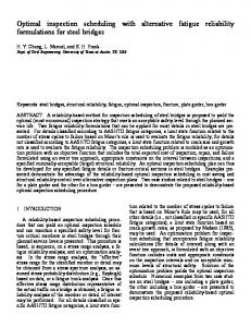
Fig 1 Estimation of the optimal insertion depth of the laryngeal tube. After palpating the cricoid cartilage, the LT was aligned alongside the lateral side of the patient’s head and neck so that the distal balloon of the LT was placed just posterior to the cricoid cartilage. The path of LT was adjusted to pass via the mandible angle, and the anaesthesiologist marked the position of the upper incisor on the tube. The LT was then inserted until the marking point was placed on the upper incisor.
was to compare the adequacy of ventilation between the three insertion methods, on the basis of a pilot study, the sample size of 24 patients was calculated to detect 0.7 of the difference of the effective ventilation score by the Wilcoxon signed rank test, with an a error of 0.05 and a b error of 0.2. The x2 test was used to compare the grade of fibreoptic view of the larynx. A Bonferroni correction was then used to determine the statistical significance.
Results The LT was inserted without any problems in all 24 children, whose characteristics are presented in Table 1. Compared with method A, insertion was 1.8 (0.8) [0 – 3.6] cm deeper with method B and 1.5 (0.9) [0– 3.5] cm deeper with method C (P,0.001); there was no difference between the depth of methods B and C. The median ventilation score was greater with methods B (3 [0– 3]) and C (3 [0 – 3]) than with method A (1.7 [0– 3]) (P,0.001); again there were no differences between methods B and C. A score of three points for effective ventilation was obtained, only 17% with method Table 1 Characteristics of the patients. Values are given as mean (SD) [range] or number Age (yr) Gender (M/F) Height (cm) Weight (kg) ASA class I/II (n)
705
5.2 (1.9) [0.8 –9] 5/19 109.8 (12.3) [79.0 – 125.5] 19.8 (3.5) [12.3 –25.0] 22/2
Kim et al.
Table 2 Fibreoptic examination. Values are given as n (%). E, epiglottis and LI, laryngeal inlet. *P,0.001 vs method A View
Method A (n524)
Method B (n524)
Method C (n524)
3 (12.5)
15 (62.5)*
18 (75)*
0 (0)
1 (4.2)
1 (4.2)
Epiglottis
10 (41.7)
5 (20.8)*
2 (8.3)*
None
11 (45.8)
3 (12.5)*
3 (12.5)*
Vocal cord
Arytenoids or posterior part of the laryngeal inlet
A, vs 83% with methods B and C. Air leaks at an airway pressure ,15 cm H2O were detected more frequently with method A (79%) compared with methods B (17%) and C (17%). In method A, bilateral chest movement was inadequate in five patients (21%) and the capnogram was abnormal in eight patients (33%). Except for one patient, all attempts with methods B and C showed bilateral chest movement and normal capnography. Methods B and C also allowed significantly better views of the vocal cords via fiberoptic bronchoscopy than method A (P,0.001) (Table 2). There was no desaturation below 90%, bleeding, or any injuries to the upper airway during the study.
Discussion The manufacturer’s guideline recommends that the LT should be inserted until the teeth mark is at the level of the upper teeth; this study demonstrates that this leads to unacceptable positioning of the LT #2 in most children. The distance we measured from the cricoid cartilage to the upper teeth and feeling resistance do, however, offer a more reliable guide. The reason for the poor performance of the manufacturer’s recommended method with LT #2 is that it is stated as being suitable for children weighing 12– 25 kg. There is clearly to be expected a great variation in the dimensions of the upper airway within this group. We found that, in the majority of such children, the LT #2 had to be inserted to a greater depth than indicated by the thick teeth mark (1.8 (0.8) [0 – 3.6] cm deeper with method B and 1.5 (0.9) [0 – 3.5] cm deeper with method C). If we consider the distance between the thick teeth mark and thin teeth mark is only 0.9 cm, it is far out of the recommended range. This insufficient depth of insertion of the LT allows the distal cuff to move upwards during inflation, leading to ventilatory obstruction and leakage. For adequate ventilation, optimal cuff size and insertion depth are essential. In all children except one, the lungs
could be successfully ventilated with LT #2 by methods B and C. We assumed that, in the patient whose lungs could not be ventilated (9 yr, 25 kg, 118 cm), the cuff was too small; there was a large leak. Another insertion guideline is the sensation of resistance at the oesophageal inlet. However, the sensation of resistance is not well defined, because the pressure of the upper oesophageal sphincter is reduced during sleep,7 and in infants and the elderly.8 – 10 Thus, this method did not always work in fully relaxed children, or in the hands of novices. Our results offer an alternative guideline as to the optimal depth for LT insertion and positioning. When the anaesthesiologist cannot be sure of the resistance of the oesophageal inlet, method C offers a useful alternative. Previous studies suggested that lifting the chin or head extension improves ventilation.11 – 13 Therefore, we inserted the LT and ventilated the lungs with the patient’s head and neck in the sniffing position. When correctly inserted, the distal tip of the LT is positioned in the hypopharynx and the ventilation holes are opposite the larynx. With the measured length insertion technique, this allowed the vocal cords to be seen in 75% of patients, compared with 0% in Bortone’s study5 and 42% in Cook’s study.9 In conclusion, insertion based on the manufacturer’s teeth mark in paediatric patients can lead to insufficiently deep positioning of the LT and inadequate ventilation. The measured length from the cricoid cartilage to the upper teeth offers a good alternative.
Acknowledgement This study was supported by SNUH fund (0420050380).
References 1 Asai T, Shingu K. The laryngeal tube. Br J Anaesth 2005; 95: 729 –36
706
Insertion depth of laryngeal tube
2 Richebe P, Semjen F, Cros AM, Maurette P. Clinical assessment of the laryngeal tube in pediatric anesthesia. Pediatr Anaesth 2005; 15: 391 – 6 3 Genzwuerker HV, Fritz A, Hinkelbein J, et al. Prospective, randomized comparison of laryngeal tube and laryngeal mask airway in pediatric patients. Pediatr Anaesth 2006; 16: 1251 – 6 4 Genzwuerker HV, Hohl ECh, Rapp HJ. Ventilation with the laryngeal tube in pediatric patients undergoing elective ambulatory surgery. Pediatr Anaesth 2005; 15: 385– 90 5 Bortone L, Ingelmo PM, De Ninno G, et al. Randomized controlled trial comparing the laryngeal tube and the laryngeal mask in pediatric patients. Pediatr Anaesth 2006; 16: 251 – 7 6 Mukherji SK. Pharynx. In: Som PM, Curtin HD, ed. Head and Neck Imaging, 4th Edn. St. Louis: C.V. Mosby, 2003; 1470 – 3 7 Kahrilas PJ, Dodds WJ, Dent J, Haeberle B, Hogan WJ, Arndorfer RC. Effect of sleep, spontaneous gastroesophageal reflux, and a meal on upper esophageal sphincter pressure in normal human volunteers. Gastroenterology 1987; 92: 466 – 71
8 Fulp SR, Dalton CB, Castell JA, Castell DO. Aging-related alterations in human upper esophageal sphincter function. Am J Gastroenterol 1990; 85: 1569 – 72 9 Ribeiro AC, Klingler PJ, Hinder RA, DeVault K. Esophageal manometry: a comparison of findings in younger and older patients. Am J Gastroenterol 1998; 93: 706 – 10 10 Sondheimer JM. Upper esophageal sphincter and pharyngoesophageal motor function in infants with and without gastroesophageal reflux. Gastroenterology 1983; 85: 301 – 5 11 Asai T, Kawashima A, Hidaka I, Kawachi S. The laryngeal tube compared with the laryngeal mask: insertion, gas leak pressure and gastric insufflation. Br J Anaesth 2002; 89: 729 – 32 12 Asai T, Murao K, Shingu K. Efficacy of the laryngeal tube during intermittent positive-pressure ventilation. Anaesthesia 2000; 55: 1099 – 102 13 Cook TM, McKinstry C, Hardy R, Twigg S. Randomized crossover comparison of the ProSeal laryngeal mask airway with the laryngeal tube during anaesthesia with controlled ventilation. Br J Anaesth 2003; 91: 678– 83
707