Microchim Acta (2012) 176:91–99 DOI 10.1007/s00604-011-0698-9
ORIGINAL PAPER
Amperometric sensor for detection of bisphenol A using a pencil graphite electrode modified with polyaniline nanorods and multiwalled carbon nanotubes Sujittra Poorahong & Chongdee Thammakhet & Panote Thavarungkul & Warakorn Limbut & Apon Numnuam & Proespichaya Kanatharana
Received: 30 June 2011 / Accepted: 11 September 2011 / Published online: 22 September 2011 # Springer-Verlag 2011
Abstract We report on a simple and highly sensitive amperometric method for the determination of bisphenol A (BPA) using pencil graphite electrodes modified with polyaniline nanorods and multiwalled carbon nanotubes. The modified electrodes display enhanced electroactivity for the oxidation of BPA compared to the unmodified pencil graphite electrode. Under optimized conditions, the sensor has a linear response to BPA in the 1.0 and 400 μM concentration range, with a limit of detection of 10 nM (at S/N=3). The modified electrode also has a remarkably stable response, and up to 95 injections are possible with a S. Poorahong : C. Thammakhet : P. Thavarungkul : W. Limbut : A. Numnuam : P. Kanatharana (*) Trace Analysis and Biosensor Research Center, Prince of Songkla University, Hat Yai, Songkhla 90112, Thailand e-mail:
[email protected] S. Poorahong : C. Thammakhet : P. Thavarungkul : W. Limbut : A. Numnuam : P. Kanatharana Center of Excellence for Innovation in Chemistry, Faculty of Science, Prince of Songkla University, Hat Yai, Songkhla 90112, Thailand S. Poorahong : C. Thammakhet : A. Numnuam : P. Kanatharana Department of Chemistry, Faculty of Science, Prince of Songkla University, Hat Yai, Songkhla 90112, Thailand P. Thavarungkul Department of Physics, Faculty of Science, Prince of Songkla University, Hat Yai, Songkhla 90112, Thailand W. Limbut Department of Applied Science, Faculty of Science, Prince of Songkla University, Hat Yai, Songkhla 90112, Thailand
relative standard deviation of 4.2% at 100 μM of BPA. Recoveries range from 86 to 102% for boiling water spiked with BPA from four brands of baby bottles. Keywords Bisphenol A . Polyaniline nanorods . Multiwalled carbon nanotubes . Electrochemical sensor
Introduction Endocrine disruptors potentially cause adverse effects on human health and wildlife. Bisphenol A (4,4′-dihydroxy2,2-diphenylpropane, BPA) is one of the many chemicals in this group. BPA is a key monomer in the production of epoxy resins and polycarbonate plastics. These plastics are clear and nearly shatter-proof and are widely used in the chemical industry for the production of bottles used for babies and water containers, food can lining, sports equipment, medical and dental devices, dental fillings and sealants. Upon heating BPA can leach from these containers, such as those used for baby bottles and many beverage containers, due to the breakdown of the polymer [1]. Thus, humans may routinely ingest trace amounts of BPA and its effects on human health have become a growing concern. Moreover, several cases of reproductive abnormalities have also been reported in wildlife exposed to anti estrogenic environmental contaminants. In addition, BPA is postulated to cause a decrease of sperm quality in humans and an increase in various kinds of cancers [2]. To reduce these noxious effects of BPA, an efficient monitoring system is required, so that after detecting exposure from possible sources immediate remediation can occur. The analysis for BPA is most commonly carried out using either high-performance liquid chromatography
92
(HPLC) combined with ultra-violet (UV) detection [3], fluorescence detection [4], electrochemical detection [5] or Gas chromatography-tandem mass spectrometry (GC–MS/ MS) [6]. Although they can provide highly sensitive detection and accurate identification, these methods are quite expensive, time-consuming, need skilled operators and often require sample preparation prior to the analysis [2, 7]. As an alternative, electrochemical sensors are now being continuously developed due to their excellent sensitivity, automation, short analysis time, low power consumption and inexpensive equipment. BPA can be oxidized to exhibit an electrochemical activity since it contains phenolic hydroxyl groups [8, 9]. However, direct determination of BPA using an electrochemical sensor is rare because the response of a traditional electrochemical sensor to BPA is very poor. In addition a relatively high potential is required for the oxidation of phenolic compounds, and this results in an increase of the background current and hence a lowered sensitivity [10]. Moreover, the electrochemical oxidation of BPA can cause inactivation of a glassy carbon or noble metal electrode as the result of the deposition of electropolymerization films [11]. To overcome these drawbacks there is a need for novel sensing materials that can provide high sensitivity, rapid response time with a minimum of fouling effect from the oxidation of BPA. Of the various sensing materials reported so far, nanomaterials have recently found a wide range of applications in the construction of electrochemical sensors. Nanostructure-based electrochemical sensors have demonstrated many remarkable advantages over traditional ones. The large surface-to-volume ratio and the high dispersivity of the nanostructured materials essentially provide a large surface area for the adsorption and thus the enrichment of analytes. Both attributes can lead to an improvement of the sensitivity and selectivity of electrochemical sensors and biosensors [8, 12]. In some cases they also possess an ability to resist electrode fouling. Conducting polymers are an interesting group of substrates for electrode surface modification due to their mechanical flexibilities, high surface areas, chemical specificities, tunable conductivities and easy processing. In recent years, a number of onedimensional (1-D) polymeric nanostructures such as nanowires, nanorods, and nanotubes have been studied with the expectation that these materials will possess the advantages of both low-dimensional systems and organic conductors [13]. Among the conductive polymer materials studied for electrochemical sensors, the most extensively used is polyaniline (PANI), mainly due to its ease of preparation, good biocompatibility and high electrocatalytic activity. For the electrochemical detection of organic compounds, multiwalled carbon nanotubes (MWCNTs) have often been employed due to their strong adsorptive ability, excellent
S. Poorahong et al.
conductivity, high aspect ratio, large specific area and the ability to minimize surface fouling [14]. Thus, the aim of this work was to establish a simple and sensitive electrochemical method for the determination of BPA using the PANI/MWCNTs nanostructured as the sensing material. PANI nanorods were synthesized and applied together with MWCNTs in the chitosan matrix to modify the pencil graphite electrode surface. The PANI nanorods/MWCNTspencil graphite electrode (PGE) offers a significant increase in the anodic current of BPA compared to a bare PGE, giving an enhanced sensitivity for determination of BPA. This method possesses some obvious advantages including high sensitivity, simplicity, rapid response and high stability with minimal surface fouling.
Experimental Apparatus The electrodeposition of PANI nanorods and electrochemical studies were carried out using a μ-Autolab type II (Metrohm Autolab, B.V., The Netherlands, http://www. ecochemie.nl) connected to a personal computer and driven by GPES 4.9 software (Eco Chemie, The Netherlands, http://www.ecochemie.nl). An electrochemical cell composed of a working electrode, 2B PGE, with a diameter of 3.0 mm and a length of 2.0 cm (Staedtler, Nuremberg, Germany, http://www.staedtler.com) modified with PANI nanorods/MWCNTs, a Ag/AgCl (3 M KCl) reference electrode and a stainless steel wire counter electrode. All electrodes were held in a custom-built flow cell (ca. 10 μL). Scanning electron microscope (SEM) images were achieved with the JSM-5200 (JEOL, Japan, http://www. jeol.com) using an acceleration potential of 25 kV. A flow injection system was employed for the analysis of BPA. A peristaltic pump (0.5 mL min−1) (Miniplus 2, Gilson, France, http://www.gilson.com/en) was used to deliver the carrier solution (100 mM glycine-NaOH pH 10.6 containing 0.1 M KCl) while a ten-port valve (Valco, Houston, TX, USA, www.vici.com) was used as the sample injector to control the exact sample volume (300 μL). Materials and chemicals All solutions were prepared with deionized water that was treated by a reverse osmosis system and purified with a Maxima ultrapure water instrument to obtain the resistivity of 18.2 MΩ (ELGA, England, http://www.elgaprocesswater.co. uk). Bisphenol A (BPA) was from Sigma-Aldrich (Seelze, Germany, www.sigmaaldrich.com). Aniline (≥99.5%) was from Aldrich and distilled under reduced pressure prior to use. Multiwalled carbon nanotubes (MWCNTs, 95% purity,
Amperometric sensor for detection of bisphenol A
93
5–15 μm length, 40–60 nm i.d.) were kindly provided by Shenzhen Nanotech Port Co., Ltd. (Nanshan, Shenzhen, China, http://www.nanotubes.com.cn). They were sonicated for 12 h in 2.0 M nitric acid. Nickel Chloride (NiCl2.6H2O) was a CARLO ERBA reagent (Bangkok, Thailand, http:// www.carloerbareagents.com). Boric acid (H3BO3) and nickel sulfate (NiSO4.6H2O) were obtained from Merck (Demstadt, Germany, http://www.merck.com). All other chemicals and reagents were analytical grade. The BPA stock solution was freshly prepared by dissolving 50.00 mM BPA in acetonitrile. The solution was diluted in 100 mM glycine-NaOH pH 10.6 and 10% cetyltrimethylammonium bromide (CTAB) to obtain the desired concentration. All measurements were performed at room temperature in 100 mM glycine-NaOH pH 10.6 containing 0.1 M KCl. Four brands of baby bottles (volume=250 mL) were tested for BPA leaching. Water samples were obtained by adding 100 mL of 100 °C water into the bottles and shaking for 30 min (Wrist action shaker, model 75, Burrell coproration, USA, http://www.burrellsci.com). These water samples were then analyzed with the new system. In the case where BPA was not detectable, an extraction step was applied. Five mL of ethyl acetate (EtOAc) was added into the water sample in the bottle and the shaking was resumed for another 30 min. The 5.00 mL of EtOAc was then removed. Two portions, 2.00 mL each, were separately evaporated. One was rediluted with 1.00 mL of glycineNaOH buffer to be analysed by the amperometric flow injection system at a potential of +0.5 V. The other was rediluted with 1.00 mL of EtOAc for the analysis with GCMS. From this extraction step, the final concentration of the extracted BPA had been pre-concentrated 40 times.
Pittsburgh PA, http://www.fishersci.com). The AAO membrane was then dissolved in 13 M NaOH for 30 min followed by several washing of DI water until the pH of the water reached neutral. The surface of the PGE was mechanically polished with alumina slurries (5, 1 and 0.3 μm) and cleaned by electrochemical etching in 100 mM glycine-NaOH pH 10.6 containing 0.1 M KCl between 0 and +1.0 V vs. Ag/AgCl reference electrode at a scan rate of 50 mV s−1 for 20 scans and dried with nitrogen gas. The electrode surface modification was carried out by mixing 5.0 mg of MWCNTs and 1.0 mg of PANI nanorods in 0.10% w/v chitosan in 0.1 M acetic acid. Chitosan was chosen as the matrix because it can form a stable complex with MWCNTs through non-covalent binding, the stability of MWCNTs in aqueous chitosan solution can result in significant improvements and lead to a uniform distribution of MWCNTs in the chitosan film [15]. Eight μL of the mixture was dropped onto the circular surface (diameter 3.0 mm) of a cleaned PGE and dried at room temperature for 2 h. The procedure was then repeated and the effect of the number of deposition layers was investigated. The number of layers providing the best response was then employed (see 3.2). The PGE modified with only the PANI nanorods or the MWCNTs was also prepared with the same procedures using either 1.0 mg of PANI nanorods or 5.0 mg MWCNTs in 0.10% w/v chitosan in 0.1 M acetic acid. Before use the modified electrode was cleaned in 100 mM glycine-NaOH pH 10.6 containing 0.1 M KCl by cycling the potential from 0 to +1.0 V at 50 mVs−1 for 20 cycles.
Fabrication of the bisphenol A sensor
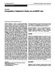
Scanning electron micrographs
An anodic aluminum oxide (AAO) membrane with a thickness of 60 μm and nominal pore diameters of 200 nm (Whatman, England, www.whatman.com) was used as a template for the nanorods. A gold layer was first sputtered on one side of the membrane to serve as the working electrode during the electrodeposition (in connection to an aluminum foil contact). A Ag/AgCl (3 M KCl) and a platinum wire were used as reference and counter electrodes, respectively. Nickel was first deposited from the plating solution, 1.0 M NiSO4.6H2O, 0.2 M NiCl2.6H2O and 0.5 M H3BO3 using the chronocoulometric mode at −1.0 V and a charge of 3.0 C. Following this, PANI was electrodeposited from a solution of 0.1 M aniline and 0.5 M H2SO4 at +1.0 V using a charge of 5.0 C. The magnetic property of Ni made it easier to move the nanorods during the washing steps. After the deposition the sputtered gold was removed from the membrane using fine sand paper along with 0.3 μm alumina powders (Fisher,
Figure 1 shows the scanning electron micrographs of the PGE surfaces. The micrograph of the surface modified with only PANI nanorods (A) shows the nanorods with a 200 nm diameter corresponding to the pore diameter of AAO, and 5 μm in length, uniformly covering the pencil graphite surface. The MWCNTs on the pencil graphite surface is also shown to be uniformly dispersed (B) and this helped to enhance the surface area of the electrode. The morphology of the PANI nanorods/MWCNTs-modified PGE (C) is similar to that of the MWCNTs electrode (B), except for the presence of a few non-uniformly dispersed PANI nanorods covered with MWCNTs.
Results and discussion
Electroactivity of modified electrode towards BPA Cyclic voltammetric (CV) measurements were performed to evaluate the electroactivity of the modified surfaces towards
94
S. Poorahong et al.
Fig. 1 Scanning electron micrographs of the pencil graphite electrode surface modified with PANI nanorods (a) MWCNTs (b) PANI nanorods/ MWCNTs (c) in 0.1% w/v chitosan; at an accelerating voltage: 25 kV
the oxidation of BPA. Figure 2 displays CV at the bare PGE (A), PANI nanorods-modified PGE (B), MWCNTsmodified PGE (C) and PANI nanorods/MWCNTs-modified PGE (D) in the absence and presence of 0.1 mM BPA (dotted and solid lines, respectively). All modified electrodes displayed a defined BPA oxidation peak around +0.5 V. The PANI nanorods electrode (B) provided higher anodic current than bare PGE (A). This may come from the higher surface area of PANI nanorods modified PGE. When compared between PANI nanorods (B) and MWCNTs modified PGE (C), the PANI nanorods provided a broader and lower oxidation current than that obtained from MWCNTs modified PGE which can be attributed to the excellent electrocatalytic activity of MWCNTs [16]. The PANI nanorods/MWCNTs-modified electrode (D) yielded the highest sensitivity over the entire potential range. Such Fig. 2 Cyclic voltammograms recorded in blank (dotted line) and 0.1 mM BPA (solid line) solutions at the (a) bare electrode, (b) PANI nanorods modified graphite electrode, (c) MWCNTs modified graphite electrode and (d) PANI nanorods/MWCNTs modified graphite electrode. Conditions: supporting electrolyte, 100 mM glycine-NaOH pH 10.6 containing 0.10 M KCl; scan rate 50 mVs−1
an improved response is attributed to a higher surface area effect [9]. This is further supported by the background cyclic voltammetric (dotted line) that indicated a change in the background current is stronger due to the modification with the PANI nanorods and MWCNTs [17]. The best number of PANI nanorods/MWCNTs layers (1– 6 layers) was examined by considering the response to 0.1 mM BPA (Fig. 3a). The response increased with the number of modified layer from 1 to 3 layers, then decreased. This is probably due to the limited mass transport of BPA within a thicker film. So the most favorable BPA response was obtained using 3 layers. Figure 3b compared the response of 0.1 mM BPA from 1 to 3 layers. Their peak widths were similar and indicated that the response time is not compromised by the three layers of coating.
Amperometric sensor for detection of bisphenol A
95
Fig. 3 a Effect of the number of layers on the current response for 0.1 mM BPA in 100 mM glycineNaOH (pH 10.6) containing 0.1 M KCl, E=+0.5 V. b The comparison of 0.1 mM BPA in 100 mM glycine-NaOH (pH 10.6) containing 0.1 M KCl, E=+0.5 V response obtained from different modified layers (i) 1 layer (ii) 2 layers (iii) 3 layers
Flow injection analysis of bisphenol A In order to optimize the performance of the flow injection system, the concentration and pH of the carrier solution, detection potential and sample volume were studied by injecting 1.0 mM BPA. Amperometric detection at a constant potential (tested between +0.2 and +0.7 V) of the working electrode was applied to obtain the response of BPA from the PANI nanorods/MWCNTs modified PGE. The response current increased with the applied potential from +0.2 V until +0.5 V and then decreased with a further increase of the potential (Fig. 4a). Thus, +0.5 V was employed for the amperometric detection of BPA. The pH value of the supporting electrolyte is also a key factor to influence the current response and reproducibility. The effect of pH (10.0 to 10.6) and concentration (10– 125 mM) of the glycine-NaOH were investigated together. The amperometric response at +0.5 V was obtained using 1.0 mM BPA in glycine-NaOH at a flow rate of 0.5 mL min−1 and an injection volume (sampling loop) of 300 μL. The highest amperometric response for BPA was obtained at 100 mM of glycine-NaOH pH 10.6 (Fig. 4b). The reason may be because the pKa of BPA is 9.8 and in a highly alkaline medium BPA has a phenolate form which is easier to oxidize than a neutral BPA form [11, 18]. The stability of the bare PGE and PANI nanorods/ MWCNTs-modified PGE was studied by repeatedly injecting 100 μM of BPA in 100 mM glycine-NaOH pH 10.6. Fig. 4 a Effect of the detection potential on the response of 1.0 mM BPA in 100 mM glycine-NaOH pH 10.6 containing 0.1 M KCl. b Effect of the pH and concentration of glycine-NaOH the response of 1.0 mM BPA; E=+0.5 V
The current response obtained from the bare PGE at +0.5 V decreased with the cycle of analysis (Fig. 5a). After 15 analysis cycles the response was only 10% of the original. This is because the electrochemical oxidation of BPA on a carbon electrode resulted in a deposition of an electropolymerization film which is formed when the phenoxy radical attacks an unreacted substrate and this led to the inactivation of the working electrode [11]. In contrast the stability of the PANI nanorods/MWCNTs-modified PGE (Fig. 5b) was greatly improved. Figure 5c shows the percentage responses obtained from PANI nanorods/ MWCNTs-modified PGE. The first 95 analysis cycles yielded a 95±4% response of its original response (relative standard deviation=4.2%). This improvement is due to the ability of the MWCNTs to minimize surface fouling associated with the electrogeneration of phenoxy radicals [2]. Furthermore, the used of the chitosan matrix may also help to improve the stability of the deposit on the electrode due to the forming of a stable complex through non covalent binding between MWCNTs and chitosan. The responses of the PANI nanorods/MWCNTs-modified electrode to various concentrations of BPA are shown in Fig. 6. The response increases linearly with the BPA concentration from 1.0 to 400 μM. The regression line equation between I (the peak height response in μA) and [BPA] (BPA concentration in μM) of Fig. 6 was I=(0.99± 0.02)[BPA]+(11±3); R²=0.995. A very low detection limit of 10.0 nM was also obtained (S/N=3) for the PANI
96
S. Poorahong et al.
Fig. 5 Amperograms obtained by a pencil graphite electrode (a) and a PANI nanorods/ MWCNTs modified pencil graphite electrode (b) for the oxidation of 100 μM BPA in 0.1 M glycine-NaOH (pH 10.6) containing 0.10 M KCl; E= +0.5 V. c The stability of PANI nanorods/MWCNTs modified graphite electrode for the detection of 100 μM BPA in 100 mM glycine-NaOH (pH 10.6) containing 0.10 M KCl, E=+0.5 V
nanorods/MWCNTs electrode. Whereas 100 nM and 70 nM were obtained from the pencil graphite modified with PANI nanorods and MWCNTs, respectively. To test the reproducibility of the electrode responses, three preparations of PANI nanorods/MWCNTs-modified pencil graphite electrodes were used to detect BPA at 10.0, 25.0, 50.0, 75.0 and 100 μM (three replications for each concentration). The regression line equations of the three electrodes were I1 =(1.07±0.04)[BPA]+(5±2); R2 =0.986, I2 =(0.98±0.04)[BPA]+(7±2); R2 =0.987, I3 =(1.11±0.02) [BPA]+(5±3); R2 =0.991. To confirm that the differences between each preparation of the electrode are not significantly different, the slopes of the regression lines obtained from these three electrodes were tested using two-way ANOVA (analysis of variance). The results indicated that each preparation of the modified electrode provided good reproducibility (P>0.05). The average sensitivity (slope of the regression equation) of the three electrodes was 1.05± 0.07 μAμM−1. The sensitivity was 2.5 times higher than
Fig. 6 The amperograms of the various concentrations of BPA in 100 mM glycine-NaOH (pH 10.6) containing 0.10 M KCl, E=+0.5 V, sample loop=300 μL, flow rate=0.5 mLmin−1
the PGE modified with only PANI nanorods (0.42± 0.02 μAμM−1, average of three electrodes) and 1.4 times better than the MWCNTs (0.72±0.01 μAμM−1, average of three electrodes). To evaluate the selectivity of the sensor, some possible interfering substances were examined in 100 mM glycineNaOH solution pH 10.6 containing 20 μM BPA. The results indicated that a 100-fold concentration of phenol, hydroquinone, 2,4 dinitrophenol and 4-chlorophenol had no influence on the signals of BPA with deviations below 5%. Likewise a 500-fold concentration of some ions such as Na+, Ca2+, Mg2+, Fe3+, Ni3+, Cl−, SO42-, No3-, I- and Brhad no influence on the determination of BPA. Analytical applications In order to ascertain the potential application for practical sample analysis, this newly method was applied to detect BPA in water samples from four different brands of baby bottle. The direct analysis of water samples found that BPA was not detectable for any of the brands, i.e., the concentrations in these water samples were less than the detection limit of 10 nM. The extraction method was then carried out as described in the experimental section and the standard addition method was chosen as the analytical method for the analysis of BPA extracted from baby bottles for amperometric detection. The results were also validated by a gas chromatograph coupled with mass spectroscopy. The GC-MS conditions were adopted from GÓmez and coworker [6]. The column was a DB-5 ms capillary column (5% diphenyl 95% dimethylsiloxane), 30 m×0.25 mm i.d., 0.25 μm film thickness. The helium carrier gas flow was set at 1.0 mL min−1. Automatic injections (1.00 μL) were performed in the splitless mode at 280 °C and with the split
Amperometric sensor for detection of bisphenol A Table 1 Comparison of the BPA concentration detected using the flow injection amperometric system and the GC-MS technique extracted from four different brands of baby bottles
Sample
97
Amperometric flow injection system [BPA], μM Added
1 2 3 4
Recovery%
GC-MS RSD%
Found
[BPA], μM Added
Recovery%
RSD%
Found
5.0 10.0 5.0 10.0 5.0 10.0 5.0
5.1±0.3 9.6±0.8 4.4±0.4 9.1±0.6 4.4±0.5 8.6±1.2 4.5±0.5
102±6 96±8 88±7 91±7 89±10 86±12 91±9
5.9 7.9 8.2 7.2 10.6 14.6 10.4
5.0 10.0 5.0 10.0 5.0 10.0 5.0
5.0±0.2 10.1±0.2 4.0±0.1 9.8±0.2 4.7±0.5 8.8±0.3 4.4±0.4
100±4 101±2 80±2 98±2 94±9 88±3 88±8
3.5 1.5 2.5 1.6 9.7 3.7 9.1
10.0
9.7±0.7
97±7
7.0
10.0
9.4±0.4
94±4
3.9
vent opening set at 1.5 min. The transfer line temperature was 290 °C and the ion trap temperature was 200 °C. The oven temperature was programmed as 150 °C for 2 min, and increased to 300 °C at 25 °C min−1 then held for 5 min. The results from both methods again showed no detectable BPA from any of the brands. The method was further evaluated by calculating the percentage recovery of the spiked samples (Table 1). The recoveries of this method were in the range of
86–102% with RSD=5.9–14.6%, indicating that this method has a good accuracy (recommended recoveries; 70–120% by AOAC at analyte concentration levels of ppb and low ppm levels [19]). These were similar to the recoveries obtained from the GC-MS, 80–101% with RSD=1.5–9.7%. That is, this newly developed method for the determination of BPA is effective and feasible. The advantages of the electrochemical method are the short analysis time (3 min for 1 injection as
Table 2 Figures of merits of comparable methods for determination of Bisphenol A Method PANI nanorods/MWCNTs modified PGE GC-MS/MS
Mesoporous silica-based electrochemical sensor Fluorescence sensor
Linear dynamic range
LOD
Applications/Comments
Ref.
1.0–400 μM
10 nM
This work
10–1000 ng L−1
0.5 ng L−1
Applied to extracted water from baby bottles, compared with GC-MS, recovery=86–102% Used solid phase extraction as sample preparation method and applied for hospital effluent samples, long analysis time (25 min) Applied to lake water samples, recovery=91.3– 107.2% Applied for water samples and landfill leachate, recovery=96–106%. LOD higher than recommended safe value for release of BPA Based on inhibitory effect of BPA on the chemiluminescence reaction between luminol and potassium hexacyanoferrate Used Bamboo-activated charcoal for SPE sorbent for extracted BPA from environmental sample, recovery =80.5–119.8% Applied for plastic products, recovery=95.36– 104.39%. High stability of the biosensor (over 30 days) Applied for food package samples, recovery= 98.4–102.8% Applied for water samples, recovery=97–105% Applied for milk samples, recovery=95.3–104%
[16]
0.22–8.8 μM
38 nM
0.079–16.6 μM
70 nM
Flow injection inhibitory chemiluminescence
0.8–12 μM
0.31 μM
LC-ESI-MS/MS
0.1–10 μg L−1
0.02 μg L−1
Tryosinase immobilized on MWCNTs-cobalt phthalocyanine-silk fibroin modified GCE GCE modified carboxylatedMWCNTs PAMAM-AuNPs-SF/GCE Poly(amidoamine) and Fe3O4 magnetic nanoparticles modified GCE MWCNTs-gold nanoparticles modified GCE Cysteamine-coated CdTe quantum dots
0.05–3.0 μM
10–104 nM
30 nM
5 nM
1 nM–1.3 μM 0.01–3.07 μM
0.5 nM 5 nM
0.02–20 μM
7.5 nM
4 nM–0.45 μM
1 nM
Applied to real plastic samples, recovery=93.8– 103.1% Based on fluorescence quenching of BPA, applied for detected in feeding-bottle samples, recovery 95.2–103.7%
[6]
[9] [20]
[21]
[22]
[23]
[24] [25]
[26] [27]
98
opposed to 18 min for the GC-MS method) and is a much cheaper analytical cost than the GC-MS since we used a pencil graphite modified electrode. The recommended safe value for the release of BPA at 50 μgL−1 (22 nM) was adopted by the European Food Safety Authority (EFSA) [1]. This value is higher than our LOD value (10 nM, 2.3 μg L−1), therefore the method could certainly be used to determine whether the BPA release from the baby bottles is above this safety value. Table 2 shows the detailed comparison of the performance of the different methods reported for the determination of BPA. The limit of detection of this work was lower than that was obtained by a fluorescence sensor using pyrene/dimethyl β-cyclodextrin complex [20] and a flow injection inhibitory chemiluminescence [21]. However, this LOD was not as good as those obtained by GC-MS/MS [6] and LC-ESI-MS/MS [22]. Comparing this new method to other electrochemical BPA detectors, the flow injection setup of this electrode provided a wider linear dynamic range (1.0 to 400 μM) than other CNTs composited such as MWCNTs modified GCE [2] or GCE modified tryosinase immobilized on MWCNTs-cobalt phthalocyanine-silk fibroin [23]. Although the limit of detection in this work (10 nM) is higher than or nearly the same as some other works [16, 24–27] (Table 2) it is sufficient to determine the safety value. In the case where a lower concentration is required in a real sample analysis, the sample pre-concentration step could be applied (40 times preconcentrated) that enabled the system to detect as low as 0.25 nM (57 ng L−1).
Conclusions This paper reports a simple and effective preparation of a PANI nanorods/MWCNTs modified PGE. The modified electrode showed a remarkable improvement in the sensor sensitivity compared to PGE. The sensor provides a linear dynamic range for BPA detection from 1.0 μM up to 400 μM, with a detection limit of 10 nM (S/N=3). Different electrodes prepared under the same condition provided the same sensitivity, therefore, recalibration was not required. The sensitivity of the electrode is c.a 2.5 and 1.4 times higher than the electrode modified with only PANI nanorods and MWCNTs, respectively. In addition, this sensor provided high reproducibility, stability (since it can be used up to 95 consecutive times) and low cost, simple and fast. This proposed electrochemical method was successfully demonstrated for the determination of BPA in real water samples obtained from the liquid container bottles. The simple preparation of the modified PGE together with the attractive performance features of this method implied that it would be an effective alternative technique for the rapid and reliable detection of bisphenol A.
S. Poorahong et al. Acknowledgements This project was supported by the National Nanotechnology Center (NANOTEC) Thailand (grant number P-0900501). The Royal Golden Jubilee Ph.D. Program supported by the Thailand Research Fund, Center of Excellence for Innovation in Chemistry (PERCH-CIC), the Commission on Higher Education, Ministry of Education; the National Research University Project of Thailand, Office the Higher Education Commission; Trace Analysis and Biosensor Research Center, Prince of Songkla University; Department of Chemistry, Faculty of Science and Graduate School, Prince of Songkla University, Hat Yai, Thailand are gratefully acknowledged. The authors thank Dr. Brian Hodgson, Prince of Songkla University, Hat Yai, Thailand for assistance with the English.
References 1. Biedermann-Brem S, Grob K (2009) Release of bisphenol A from polycarbonate baby bottles: water hardness as the most relevant factor. Eur Food Res Technol 228:679–684 2. Vega D, Agüí L, González-Cortés A, Yáñez-Sedeño P, Pingarrón JM (2007) Electrochemical detection of phenolic estrogenic compounds at carbon nanotube-modified electrodes. Talanta 71:1031–1038 3. Rezaee M, Yamini Y, Shariati S, Esrafili A, Shamsipur M (2009) Dispersive liquid-liquid microextraction combined with highperformance liquid chromatography-UV detection as a very simple, rapid and sensitive method for the determination of bisphenol A in water samples. J Chromatogr A 1216:1511–1514 4. García-Prieto A, Lunar ML, Rubio S, Pérez-Bendito D (2008) Determination of urinary bisphenol A by coacervative microextraction and liquid chromatography-fluorescence detection. Anal Chim Acta 630:19–27 5. Inoue K, Kato K, Yoshimura Y, Makino T, Nakazawa H (2000) Determination of bisphenol A in human serum by highperformance liquid chromatography with multi-electrode electrochemical detection. J Chromatogr B 749:17–23 6. Gómez MJ, Agüera A, Mezcua M, Hurtado J, Mocholí F, Fernández-Alba AR (2007) Simultaneous analysis of neutral and acidic pharmaceuticals as well as related compounds by gas chromatography-tandem mass spectrometry in wastewater. Talanta 73:314–320 7. Yin HS, Zhou YL, Ai SY (2009) Preparation and characteristic of cobalt phthalocyanine modified carbon paste electrode for bisphenol A detection. J Electroanal Chem 626:80–88 8. Huang W, Yang C (2007) Voltammetric determination of bisphenol A using an acetylene black-dihexadecyl hydrogen phosphate composite film-modified electrode. Anal Lett 40:3280–3289 9. Wang F, Yang J, Wu K (2009) Mesoporous silica-based electrochemical sensor for sensitive determination of environmental hormone bisphenol A. Anal Chim Acta 638:23–28 10. D’Antuono A, Dall’Orto VC, Balbo AL, Sobral S, Rezzano I (2001) Determination of bisphenol A in food-simulating liquids using LCED with a chemically modified electrode. J Agr Food Chem 49:1098–1101 11. Kuramitz H, Nakata Y, Kawasaki M, Tanaka S (2001) Electrochemical oxidation of bisphenol A. Application to the removal of bisphenol A using a carbon fiber electrode. Chemosphere 45:37–43 12. Heli H, Hajjizadeh M, Jabbari A, Moosavi-Movahedi AA (2009) Copper nanoparticles-modified carbon paste transducer as a biosensor for determination of acetylcholine. Biosens Bioelectron 24:2328–2333 13. Huang J, Virji S, Weiller BH, Kaner RB (2002) Polyaniline nanofibers: facile synthesis and chemical sensors. J Am Chem Soc 125:314–315 14. Wang J, Tangkuaram T, Loyprasert S, Vazquez-Alvarez T, Veerasai W, Kanatharana P, Thavarungkul P (2007) Electrocatalytic detection
Amperometric sensor for detection of bisphenol A
15.
16.
17. 18.
19.
20.
of insulin at RuOx/carbon nanotube-modified carbon electrodes. Anal Chim Acta 581:1–6 Babaei A, Afrasiabi M, Babazadeh M (2010) A glassy carbon electrode modified with multiwalled carbon Nanotube/Chitosan composite as a new sensor for simultaneous determination of acetaminophen and mefenamic acid in pharmaceutical preparations and biological samples. Electroanalysis 22:1743–1749 Li J, Kuang D, Feng Y, Zhang F, Liu M (2011) Voltammetric determination of bisphenol A in food package by a glassy carbon electrode modified with carboxylated multi-walled carbon nanotubes. Microchim acta 172:379–386 Wang J, Li M, Shi Z, Li N, Gu Z (2004) Electrochemistry of DNA at single-wall carbon nanotubes. Electroanalysis 16:140–144 Olmo MD, Zafra A, Gonzalez-casado A, Vilchez JL (1998) The use of β-cyclodextrin inclusion complexes for the analysis of bisphenol a residues in water by spectrofluorimetry. Int J Environ An Ch 69:99–110 Bruce P, Minkkinen P, Riekkola ML (1998) Practical method validation: validation sufficient for an analysis method. Microchim Acta 128:93–106 Wang X, Zeng H, Zhao L, Lin JM (2006) Selective determination of bisphenol A (BPA) in water by a reversible fluorescence sensor using pyrene/dimethyl β-cyclodextrin complex. Anal Chim Acta 556:313–318
99 21. Wang S, Wei X, Du L, Zhuang H (2005) Determination of bisphenol A using a flow injection inhibitory Chemiluminescence method. Luminescence 20:46–50 22. Zhao RS, Wang X, Yuan JP (2010) Highly sensitive determination of tetrabromobisphenol A and bisphenol A in environmental water samples by solid-phase extraction and liquid chromatographytandem mass spectrometry. J Sep Sci 33:1652–1657 23. Yin H, Zhou Y, Xu J, Ai S, Cui L, Zhu L (2010) Amperometric biosensor based on tyrosinase immobilized onto multiwalled carbon nanotubes-cobalt phthalocyanine-silk fibroin film and its application to determine bisphenol A. Anal Chim Acta 659:144–150 24. Yin H, Zhou Y, Ai S, Han R, Tang T, Zhu L (2010) Electrochemical behavior of bisphenol A at glassy carbon electrode modified with gold nanoparticles, silk fibroin, and PAMAM dendrimers. Microchim Acta 170:99–105 25. Yin H, Cui L, Chen Q, Shi W, Ai S, Zhu L, Lu L (2011) Amperometric determination of bisphenol A in milk using PAMAM-Fe3O4 modified glassy carbon electrode. Food Chem 125:1097–1103 26. Tu X, Yan L, Luo X, Luo S, Xie Q (2009) Electroanalysis of bisphenol A at a multiwalled carbon nanotubes-gold nanoparticles modified glassy carbon electrode. Electroanalysis 21:2491–2494 27. Kuang R, Kuang X, Pan S, Zheng X, Duan J, Duan Y (2010) Synthesis of cysteamine-coated CdTe quantum dots for the detection of bisphenol A. Microchim acta 169:109–115