J Digit Imaging (2012) 25:179–188 DOI 10.1007/s10278-011-9410-1
An Algorithm for Intelligent Sorting of CT-Related Dose Parameters Tessa S. Cook & Stefan L. Zimmerman & Scott R. Steingall & William W. Boonn & Woojin Kim
Published online: 28 July 2011 # Society for Imaging Informatics in Medicine 2011
Abstract Imaging centers nationwide are seeking innovative means to record and monitor computed tomography (CT)-related radiation dose in light of multiple instances of patient overexposure to medical radiation. As a solution, we have developed RADIANCE, an automated pipeline for extraction, archival, and reporting of CT-related dose parameters. Estimation of whole-body effective dose from CT dose length product (DLP)—an indirect estimate of radiation dose—requires anatomy-specific conversion factors that cannot be applied to total DLP, but instead necessitate individual anatomy-based DLPs. A challenge exists because the total DLP reported on a dose sheet often includes multiple separate examinations (e.g., chest CT followed by abdominopelvic CT). Furthermore, the individual reported series DLPs may not be clearly or consistently labeled. For example, “arterial” could refer to the arterial phase of the triple liver CT or the arterial phase of a CT angiogram. To address this problem, we have designed an intelligent algorithm to parse dose sheets for multi-series CT examinations and correctly separate the total DLP into its anatomic components. The algorithm uses information from the departmental PACS to determine how many distinct CT examinations were concurrently performed. Then, it matches the number of distinct
T. S. Cook (*) : S. R. Steingall : W. W. Boonn : W. Kim Department of Radiology, Hospital of the University of Pennsylvania, 1 Silverstein 3400 Spruce Street, Philadelphia, PA 19104, USA e-mail:
[email protected] S. L. Zimmerman Department of Radiology, Johns Hopkins University, Baltimore, MD, USA
accession numbers to the series that were acquired and anatomically matches individual series DLPs to their appropriate CT examinations. This algorithm allows for more accurate dose analytics, but there remain instances where automatic sorting is not feasible. To ultimately improve radiology patient care, we must standardize series names and exam names to unequivocally sort exams by anatomy and correctly estimate whole-body effective dose. Keywords RADIANCE . Computed tomography . Dose monitoring . CT series separation . Radiation dose . Data extraction . Databases
Introduction As utilization of computed tomography (CT) and the percentage of background radiation attributed to medical sources have increased, so has interest in being able to track radiation doses administered to patients via medical imaging. The number of CTs performed annually has dramatically increased in the last decade as the technology has improved and demand from both patients and physicians has increased [1, 2]. Consequently, the proportion of background radiation in the USA attributed to medical imaging has increased from approximately 15% in 1987 to nearly 50% today [3, 4]. The impact of this imaging boom is uncertain and has been debated in a number of scientific publications [5–8]. It is difficult to quantify the potentially deleterious effects of this increased radiation exposure; many questions exist, and the answers to these questions are not easily obtained. What is clear, though, is that increasing awareness of health care professionals regarding imaging-related radiation dose is integral to improving patient care. The ACR’s
180
white paper on radiation dose states that “…there should be special attention paid to…education for all stakeholders in the principles of radiation safety, the appropriate utilization of imaging…the standardization of radiation dose data to be archived during imaging for its ultimate use in benchmarking, good practice, and finally, the identification and perhaps alternative imaging of patients who may have already reached threshold levels of estimated exposure…” [9]. To this end, there are a number of initiatives underway to standardize the documentation and reporting of radiation dose information. The Digital Imaging and Communications in Medicine Structured Reporting (DICOM SR) standard contains dose objects dedicated to storing CT radiation dose information [10, 11]. Using these radiation dose structured report (RDSR) objects, the Integrating the Healthcare Enterprise initiative has developed a Radiation Exposure Monitoring profile to assist vendors in the implementation of standardized dose reporting by scanner software [12]. The ImageGently and ImageWisely campaigns of the Society of Pediatric Radiology and the ACR, respectively, along with their respective dose registries, are implementing large-scale dose monitoring to enable imaging facilities to identify opportunities for dose reduction [9, 13, 14]. The U.S. Food and Drug Administration recently launched the Initiative to Reduce Unnecessary Radiation Exposure from Medical Imaging [15]. The NIH is also making efforts to track and report radiation dose for all patients imaged at the Institutes [16]. In spite of all these measures, however, multiple challenges remain in the dose monitoring problem. The first is posed by vast repositories of retrospective CT data
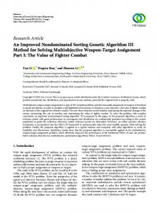
Fig. 1 RADIANCE automated dose extraction pipeline which combines data from the dose sheet, exam header, and radiology information system (RIS) to enable analytics and quality assurance
J Digit Imaging (2012) 25:179–188
that store dose parameters as an image-based dose sheet instead of structured data within the DICOM header. Furthermore, CT scanners currently in use may not have firmware amenable to incorporating radiation dose into image headers. To address this issue, we use RADIANCE (Fig. 1), an automated extraction pipeline that parses legacy dose sheets and DICOM study headers from multiple vendors and stores dose-related parameters in a relational database for subsequent analysis [17]. RADIANCE also applies anatomy-specific conversion factors (also known as “k” factors) to estimate whole-body effective dose from the total dose length product (DLP) of the study [18]. The DLP is derived by multiplying the volumetric CT dose index (CTDIvol) by the scan length. However, accurately estimating anatomy-specific doses from CT examinations ranges from challenging to nearly impossible when multiple body parts are irradiated. For example, a standard trauma protocol CT routinely scans the head and cervical spine, and then the chest, abdomen, and pelvis continuously. A CT angiogram with extremity runoffs scans the patient from the clavicles to the toes. There are instances where unusual combinations of non-contiguous body parts have to be imaged (e.g., neck, upper extremity, abdomen, and pelvis). Each of these conglomerate studies will report a total DLP for the entire imaged anatomy; however, each body part requires the application of a different conversion factor in order to estimate whole-body effective dose from DLP. In order to perform accurate dose analytics for each study type, the individual series DLPs must be used to perform the dose estimation. However, the individual series are not consistently named, and thus division of a total DLP into its anatomic components is not easily
J Digit Imaging (2012) 25:179–188
automated. In this work, we present an initial attempt to solve this extremely complex problem. We hypothesize that our sorting algorithm, though it will not cover every possible combination of imaged body parts, will nevertheless result in more accurate dose estimation by correctly assigning DLP to common anatomic combinations.
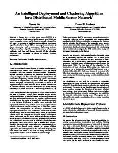
Methods Motivation To improve the accuracy of dose estimates and related analytics, it is important to identify the correct DLP for a particular body part before applying the anatomy-specific conversion factor. The k factors from DLP to whole-body effective dose are based on the following distinct regions: head, neck, chest, abdomen, pelvis, and lower extremity. Additional conversion factors are provided for combinations of contiguous regions, such as head and neck or chest, abdomen, and pelvis. Figure 2 illustrates some common combinations of body parts that can be imaged during a CT examination. However, a number of less common combinations can exist, as dictated by the needs of the patient, and these exams can be more difficult to characterize. In order to develop the sorting algorithm, we randomly selected 1,000 CT exams from the PACS and reviewed their dose sheets to determine the most common combinations of body regions imaged. We used these combinations to develop the sorting algorithm. Algorithm Design Before the sorting algorithm is applied, studies to be sorted have been processed with RADIANCE and added to the RADIANCE database. The first step in the algorithm is then to select a study from the database and identify any concurrent CT exams and series. Using the study accession number, we determine the patient’s medical record number and query our PACS database to identify all CT exams performed on that patient. From this list, those exams with a time stamp within 10 min of the index study were identified. The 10-min window was implemented to identify concurrent examinations of different body parts that are sometimes sent to the PACS at slightly different times. Ten minutes was empirically chosen based on a random sampling of studies in the PACS. The algorithm then proceeded to match the available CT series with the individual exam accession numbers. The simplest scenario is that in which only one or the equivalent of one CT examination is performed. A CT examination of one of the following body regions would constitute a single
181
exam: head, neck, cervical spine, chest, thoracic spine, abdomen, lumbar spine, pelvis, and upper or lower extremity. Similarly, multiple CT series imaging the same body region can be treated as a single exam. Examples of this include multiphase abdominal studies, such as those performed to detect hepatic masses or CT urograms in which the urinary tract is imaged before and after contrast administration. Combinations of body regions that have the same conversion factor, such as an abdominopelvic CT, can also be treated as a single study. Coronal and sagittal reformats or reconstructions of the raw data using different kernels are ignored as these do not contribute additional dose to the patient. Additional sorting is required in cases where body parts with different k factors are combined. In these cases, we apply a search tree in order to identify these combinations. When the index study is a chest CT, we search for a concurrent abdomen or abdominopelvic CT. If these are found, we look for the CT series associated with the chest to specifically identify chest-related scans. As these are not consistently named, regular expressions are used to search for the terms within the series labels that could indicate a chest CT exam (Table 1). The remaining series are assigned to the abdomen and/or pelvis. Similarly, if the index study is an abdomen or pelvis CT, we search for a concurrent chest CT. In the case of an abdominal or abdominopelvic CT, multiple series labels also exist. The series that do not match the abdominopelvic study are assigned to the concurrent chest CT. A similar search tree is applied when a neuroradiologic index study is identified. Associated concurrent examinations may include head, neck, maxillofacial, orbital, temporal bone, or cervical spine CTs. The head and neck are assigned unique k factors, but a combined head/neck k factor is also provided. In most cases, the series label for a head CT included the word “head,” sometimes as part of the label “routinehead.” A variety of series labels are used for maxillofacial CTs, including “face” and “sinus.” Dedicated imaging of the thoracic and lumbar spine is not commonly performed and instead appears in the PACS as reconstructions from existing raw data of the chest, abdomen, and pelvis. Conversely, dedicated imaging of the cervical spine is frequently performed, particularly in trauma patients. For all spine CTs, the series labels include the word spine plus an initial indicating the portion of the spine imaged (e.g., c for cervical, t for thoracic, and l for lumbar). For angiographic studies that span multiple body regions (e.g., head/neck, chest/abdomen/pelvis), the DLP for each series may not be separated according to body part but rather is reported for each enhancement phase. Additionally, regardless of the body part being imaged, the series labels may be generic: “unenhanced,” “arterial,” and “venous.” In
182
J Digit Imaging (2012) 25:179–188
Fig. 2 Sample dose sheets showing different combinations of body parts imaged as part of the same CT examination. From top to bottom: head, cervical spine, and face; chest, abdomen, and pelvis; thoracic and lumbar spines; neck and chest; abdomen and pelvis angiogram with extremity runoffs. To correctly estimate the dose for each of these exams, the individual body parts must be separated for the appropriate conversion factors to be applied
cases where the series are not named according to the anatomy being imaged, an estimate of the DLP for each body region is made. For the head and neck examinations, half the total study DLP is attributed to the head and the other half is assigned to the neck. For the chest, abdomen, and pelvis CT angiograms, one third of the total DLP is assigned to the chest and the remaining two thirds to the abdomen and pelvis. The latter proportions are empirically chosen by reviewing a sample of studies from the PACS and identifying the ratio of DLPs for the individual body parts. However, the ratio can vary with patient body
habitus, and these proportions may have to be revised once this information is readily available. In addition, these approximations become problematic when extremity runoffs are also performed but not explicitly identified by either series label or separate accession number. Algorithm Testing To test the robustness of the algorithm, we attempt to sort 1,000 randomly selected CT exams acquired at our institution and processed by RADIANCE in 2010. We
J Digit Imaging (2012) 25:179–188
183
Table 1 CT exams and sample corresponding series labels Body part or study type
Series labels
Head/face/neck
routinehead neck
spiralface face sinus
axialhead Chest
Abdomen/pelvis
Urogram Angiogram
Upper/lower extremity
headw/o chest chest+c
inspiration
chestnc
expiration PE
thorax abd abd+c
trachea abdpel
abdomen
pelvis+c
abdomen+c
abd/pel delayedpel
pelvis+c
boneypelvis
nephrophase pre excretion angio arterial venous
delays nc delayXmina routinew/o dissection
delays ltknee rtankle
unenhanced delaylegs rtknee
rightankle rleg lfootankle
lshoulder rshoulder
extremiti rthipfemur
lwrist rwrist legs lthipfemur
The CT specified in the first column can be identified on a dose sheet using one or more of the labels in the second column, thus complicating the series separation problem The X represents a numerical value indicating the number of minutes after injection the series was obtained and can be customized by the technologist
a
calculate the number of exams requiring sorting, i.e., not consisting of or equivalent to an exam of a single body region. We compare the number of exams flagged for manual sorting to the total number of sorted exams. We review a subset of these examinations to determine under what conditions the algorithm fails. In addition, we evaluate known instances in which doses to multiple body regions are attributed to a single body part, such as combined chest, abdomen, and pelvis CTs as well as pulmonary embolism (PE) protocol chest CTs with delayed imaging through the pelvis. We randomly select 1,000 CT chest exams from the RADIANCE database and apply the sorting algorithm to determine how many are actually combined exams spanning multiple body parts. We then compare the dose estimates for these studies before and after sorting to assess the effect of reporting the incorrect and often falsely elevated dose estimates.
Results Table 1 lists some of the series labels identified for each type of CT exam performed at our institution and illustrates the complexity of the series separation problem. The algorithm is able to correctly sort studies that fit templates for simple, commonly performed studies, such as a single-phase (i.e., one series) chest or abdominopelvic CT. The dose-related parameters and DLPs for examinations such as these are easily parsed from their dose sheets and converted to estimated whole-body effective dose. In addition, the algorithm easily identifies more elaborate studies that rarely deviate from a certain protocol, such as CT urograms or CT angiograms of the chest, abdomen, and pelvis with extremity runoffs. Series separation for CT urograms is typically straightforward as these are rarely combined with other studies. However, for the angiograms with lower extremity runoffs, correct assignment of DLP to the irradiated body parts is complicated when an entire series that scans from the abdomen to the toes is labeled as “angio” or “delayed.” In addition, complicated studies that deviate from expected templates or report unusual combinations of anatomic regions (e.g., head, neck, and chest) are sometimes flagged for manual review as the algorithm is unable to effectively process these combinations. Of the 1,000 randomly selected CT exams processed by the algorithm, only 12 were flagged for manual review. This was primarily as a result of imaging unexpected combinations of body parts, such as a head or cervical spine concurrently with an abdomen and pelvis or an abdomen or pelvis followed by a lower extremity. These combinations were not incorporated into the search tree and thus were not correctly sorted by the algorithm. However, they represent just over 1% of the sampled exams, suggesting that on a daily basis, only a few CTs would have to be manually reviewed and sorted with the current implementation. Figures 3, 4, and 5 illustrate the need for the application of automated series sorting to common exam combinations. In these graphs, we plot the result of sorting a subset of exams within the PACS and the RADIANCE database identified as chest CTs. One thousand chest CTs performed in 2010 were randomly selected for series sorting. Of these, 785 were exclusively chest CTs and 1 was effectively an exam of a single body part (a combination of an upper airway and a chest CT). However, the remaining 214 exams were combinations of a chest and abdomen/pelvis or a chest and pelvis. These constituted 20% of the sorted examinations. Figure 3 plots the estimated whole-body effective dose for the 1,000 chest CTs before series sorting. The average dose for one of these exams was 11.1±6.72 mSv. After sorting, the average dose dropped to 8.54±3.63 mSv (Fig. 4), reflecting the correct attribution of only the chest DLP to the chest exams. The additional DLP initially
184
J Digit Imaging (2012) 25:179–188
Fig. 3 Whole-body effective dose estimates for chest CTs before sorting, which include scans of the abdomen and/or pelvis
falsely attributed to the chest but subsequently sorted to the abdomen and/or pelvis is reflected by the exams in Fig. 5 and averages 14.7±6.67 mSv. We observe similar results when sorting series for a subset of chest CTs performed to diagnose PE. These socalled PE protocol chest CTs sometimes include delayed imaging through the pelvis to look for clots in the large pelvic veins. However, these series are not necessarily separated from the chest CT and can falsely elevate the dose estimates for the chest exams. We selected a subset of 1,000 PE protocol CTs from the PACS performed in 2010 and sorted them using our algorithm. The doses before sorting are shown in Fig. 6. Of these studies, 871 exams were exclusively scans of the chest. The remaining 129 studies were combinations of either PE protocol chest exams or Fig. 4 After sorting, the doses drop significantly, reflecting removal of the fictitious abdomen/pelvis series and demonstrating doses attributable only to imaging of the chest
abdomen and/or pelvis studies. For this group of exams, the combination study was less common than in the case of a routine enhanced or unenhanced chest CT; however, correct series separation still influenced the dose estimates, as shown in Figs. 7 and 8. Incorrectly sorting the studies led to a falsely elevated dose estimate of as much as 50 mSv, more than five times the average dose for a single-phase chest CT (60 mSv
body region to the total scan length and multiply this factor by the total DLP of the study. In future versions of the algorithm, we plan to incorporate scan length information which is currently not included in the database to perform a more accurate separation. This is particularly important for the CT angiograms which include extremity runoffs as the estimated whole-body effective dose for these studies can be artificially inflated by multiplying the DLP contributed by the extremities by the k factor of 0.015 for the chest, abdomen, and pelvis. Our algorithm fails when it cannot match a set of exams to an expected pattern, such as a CT of the head or neck followed by a CT of the abdomen and pelvis or a CT of the
186
J Digit Imaging (2012) 25:179–188
Fig. 7 After sorting, the dose estimates for the PE studies become much more consistent, reflecting the removal of the additional scans of the abdomen and/or pelvis that had previously been included
airway followed by a CT of the upper extremity. This motivates the need to develop a larger search tree with an even greater number of combinations than is currently included, although this may affect algorithm performance. Future work in this area will be devoted to reconciling these unusual combinations more effectively than in the current implementation. One of the biggest challenges in correctly sorting exam series by scanned body part is the lack of consistency in naming series within protocols. Series names differ across institutions as well as vendors and can change with firmware or protocol updates. At our institution alone, there is wide variation in series labels for a particular body Fig. 8 Additional abdomen/pelvis CTs originally attributed to the PE protocol exams falsely elevated the dose estimates for the chest CTs by as much as 50 mSv, demonstrating the need for accurate series sorting
part. In looking at chest CTs, the series on the dose sheet indicating the chest parameters could be labeled any one of the following: “chest,” “thorax,” “inspiration,” “expiration,” “PE,” “trachea”; the series labels vary with the type of study performed. An abdomen or abdomen/pelvis combination CT could be labeled “abdomen,” “abdpel,” “abd/pel,” “liver,” “renal,” “arterial” depending on the indication for the study. In CT angiograms, regardless of the anatomy being imaged, the arterial phase series is often called “arterial” or “angio,” while the delayed phase could be called “delayed,” “venous,” “delayedlegs,” or some variant of these. In order to more effectively and efficiently estimate CT dose, we need to standardize the series labels
J Digit Imaging (2012) 25:179–188
according to the anatomy being imaged. In addition, we need to minimize the addition of customized series label names and instead encourage the use of standard labels. The RadLex Playbook is making strides in this area in an effort to create a dictionary of standardized labels to be used not only for series names but also for study names [19]. Another challenge lies in how concurrent CT exams are sorted for the purposes of generating dose sheets. Some vendors’ dose sheets will group certain body parts, like the head and cervical spine or the chest, abdomen, and pelvis, and report a total DLP. The grouping system differs for angiographic and non-angiographic CTs of the same body regions. This is not problematic for the purposes of deriving an estimated whole-body effective dose as there are combined conversion factors for these body regions. However, PACS assigns the total DLP for multiple body regions to a single region’s accession number, fictitiously elevating the dose estimate. This further motivates the algorithm described above; however, as previously discussed, its ability to correctly sort concurrent CT series can be limited by the lack of consistent series labels. In addition to the challenge posed by inconsistent series naming, there is considerable variation in how vendors calculate and report dose parameters. For example, when tube current modulation is used to adjust the tube current (milliampere) to the density of the patient, the milliampere reported on the dose sheet represents the average value for the entire study and does not reflect the inherent fluctuation in current that was used to form the images. Furthermore, the size of the cylindrical acrylic phantom (either 16 or 32 cm) used to calculate the CTDI and DLP for an examination can affect the ultimate estimated dose. This information must be accounted for when comparing dose estimates for the same study as the dose estimate will be higher when using the 16-cm phantom instead of the 32-cm phantom [20]. As protocols vary between institutions, determination of what constitutes a body region can change. For example, at some institutions, a chest CT requires inclusion of all 12 thoracic ribs (automatically including the upper abdomen), while other institutions scan through the hemidiaphragms but do not include the upper abdomen within the scan length for a chest CT. In future work, the anatomy imaged could be determined by intelligently analyzing the topogram or scout image acquired before cross-sectional imaging is initiated. Alternatively, the ability to automatically identify the body region from the anatomy depicted on the axial image slice would be extremely valuable in more accurately quantifying CT dose estimates. This would account for the effect of dose modulation, which customizes the tube current to the perceived density of the patient at each location along the Z-axis in order to decrease dose
187
when possible. Ultimately, a dose estimate based on DLP is really estimating dose to a phantom rather than dose to the patient. The ability to estimate dose in a regional fashion would also facilitate the eventual calculation of organ doses, which can be more effectively correlated to cancer risk than whole-body dose extrapolated from dose to a standard-sized phantom. In the current environment, it is not possible to automatically sort all CT series for a set of concurrent examinations because of the lack of consistency in series labeling and protocols across imaging centers. For common examinations, this is a solvable problem, as demonstrated by our algorithm. Furthermore, it is critical to correctly assign DLP to a scanned body region to minimize the number of examinations with fictitiously higher-thanexpected dose estimates. This decreases the time that would be spent to investigate each of these exams for patient and/ or technical factors contributing to higher doses. In addition, it facilitates protocol optimization by providing radiologists, physicists, and vendors with dose estimates that more accurately contributed to the estimated wholebody dose. The goal of careful dose tracking and monitoring is to adhere more closely to the ALARA principle—as low as reasonably achievable—and ultimately improve patient care by decreasing exposure to unnecessary levels of medical radiation.
References 1. Maitino AJ, Levin DC, Parker L, Rao VM, Sunshine JH: Nationwide trends in rates of utilization of noninvasive diagnostic imaging among the medicare population between 1993 and 1999. Radiology 227:113–117, 2003 2. Levin DC, Rao VM, Parker L, Frangos AJ, Sunshine JH: Recent trends in utilization rates of abdominal imaging: the relative roles of radiologists and nonradiologist physicians. JACR 5:744–747, 2008 3. Sinclair WK, Adelstein SJ, Carter MW, Harley JH, Moeller DW: Ionizing radiation exposure of the population of the United States. Tech. Rep. 93, National Council of Radiation Protection, 1987 4. Kase KR et al: Ionizing radiation exposure of the population of the United States. Tech. Rep. 160, National Council of Radiation Protection, 2009 5. Brenner DJ, Hall EJ: Computed tomography: an increasing source of radiation exposure. NEJM 357(22):2277–2284, 2007 6. Brody A, Frush D, Huda W, Brent R: Radiation risk to children from computed tomography. Pediatrics 120(3):677, 2007 7. de Gonzalez AB, Mahesh M, Kim K, Bhargavan M, Lewis R, Mettler F, Land C: Projected cancer risks from computed tomographic scans performed in the United States in 2007. Arch Intern Med 169(22):2071–2077, 2009 8. Martin D, Semelka R: Health effects of ionising radiation from diagnostic CT. Lancet 367(9524):1712–1714, 2006 9. Amis JES, Butler PF, Applegate KE, Birnbaum SB, Brateman LF, Hevezi JM, Mettler FA, Morin RL, Pentecost MJ, Smith GG, Strauss KJ, Zeman RK: American College of Radiology white paper on radiation dose in medicine. JACR 4:272–284, 2007
188 10. DICOM Standards Committee: DICOM standard supplement 127: CT radiation dose reporting, 2007 11. DICOM Standards Committee: DICOM standard part 16: Content mapping resource, 2008 12. Radiation Exposure Monitoring: http://wiki.ihe.net/index.php? title=Radiation_Exposure_Management, accessed March 15, 2010 13. Goske M, Applegate K, Boylan J, Butler P, Callahan M, Coley B, Farley S, Frush D, Hernanz-Schulman M, Jaramillo D, et al: The Image Gently campaign: increasing CT radiation dose awareness through a national education and awareness program. Pediatr Radiol 38(3):265–269, 2008 14. National Radiology Data Registry: http://www.acr.org/Secondary MainMenuCategories/quality_safety/NRDR.aspx, accessed March 15, 2010 15. White Paper: Initiative to Reduce Unnecessary Radiation Exposure from Medical Imaging. http://www.fda.gov/RadiationEmittingProducts/RadiationSafety/RadiationDoseReduction/ ucm199994.htm#_Toc253092884, accessed March 15, 2010
J Digit Imaging (2012) 25:179–188 16. Neumann RD, Bluemke DA: Tracking radiation exposure from diagnostic imaging devices at the NIH. JACR 7(2):87–89, 2010 17. Cook T, Zimmerman SL, Maidment AD, Kim W, Boonn WW: Automated extraction of radiation dose information for CT examinations. J Am Coll Rad 7(11):871–877, 2010. doi:10.1016/ j.jacr.2010.06.026 18. Christner JA, Kofler JM, McCollough CH: Estimating effective dose for CT using dose-length product compared with using organ doses: consequences of adopting International Commission on Radiological Protection Publication 103 or dual-energy scanning. Am J Roentgenol 194(4):881–889, 2010. doi:10.2214/ AJR.09.3462 19. RadLex Playbook: http://www.rsna.org/Informatics/radlex_playbook. cfm, accessed January 11, 2011 20. McCollough CH, Leng S, Yu L, Cody DD, Boone JM, McNittGray MF: CT dose index and patient dose: they are not the same thing. Radiology 259(2):311–316, 2011. doi:10.1148/ radiol.11101800