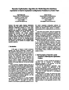
AN ALPHA-STABLE BASED BAYESIAN ALGORITHM FOR SPECKLE NOISE REMOVAL IN THE WAVELET DOMAIN Alin Achim ∗
Anastasios Bezerianos
Biosignal Processing Group Dept. of Biomedical Engineering Dept. of Medical Physics Johns Hopkins University University of Patras 720 Rutland Av. 265 00 Rio, GREECE Baltimore, MD 21205, USA
[email protected] [email protected]
Panagiotis Tsakalides † Dept. of Electrical and Computer Engineering University of Patras 261 10 Rio, GREECE
[email protected]
ABSTRACT This paper introduces a novel speckle noise removal method for medical ultrasound images. First, the logarithmic transform of the original image is analyzed in the wavelet domain. We show that the subband decompositions of ultrasound images have signi£cant non - Gaussian statistics that are best described by families of heavy-tailed distributions such as the alpha-stable. Consequently, we design a Bayesian estimator that exploits these statistics. Using the alpha-stable model we develop a blind noise-removal processor that performs a non-linear operation on the data. Finally, we compare our proposed technique to current stateof-the-art speckle reduction methods and we quantify the achieved performance improvement. 1. INTRODUCTION Ultrasonography is a widely used medical imaging modality because it is low-cost, relatively safe, portable, and versatile. However, one of its main disadvantage is the poor quality of images, which are affected by speckle noise. The presence of speckle is undesirable since it degrades image quality and it affects the tasks of human interpretation and diagnosis. As a result, speckle £ltering is a critical preprocessing step for feature extraction, analysis, and recognition from medical imagery measurements. Several methods for speckle mitigation have been already proposed. Among them, the classical Wiener £lter is not adequate since it is designed mainly for additive noise suppression. To address the multiplicative nature of speckle noise, Jain developed a homomorphic approach, which by taking the logarithm of the image, converts the multiplicative into additive noise, and consequently applies the Wiener ∗ Supported by the State Scholarship Foundation of Greece (IKY) under grant 542 / 1999. † Supported by the Greek General Secretariat for Research and Technology under Program EΠET II, Code 97EΛ - 152.
Fig. 1. Block diagram of the proposed multiscale homomorphic Bayesian-based algorithm for speckle suppression.
£lter [1]. Recently, there has been considerably interest in using the wavelet transform as a powerful tool for recovering signals from noisy data [2, 3, 4]. For the purpose of speckle reduction, Zong et al. [4] use the above described homomorphic approach to separate the noise from the original image. Subsequently, they adopt regularized soft thresholding (wavelet shrinkage) to remove noise energy within the £ner scales and nonlinear processing of feature energy for contrast enhancement. Simoncelli et al. [3] developed nonlinear estimators, based on formal Bayesian theory, which outperform classical linear processors and simple thresholding estimators in removing noise from visual images. They used a generalized Laplacian model for the subband statistics of the signal and developed a noise-removal algorithm, which performs a “coring” operation to the data. In a recent work, Tsakalides et al. showed that alpha-stable distributions, a family of heavy-tailed densities, are suf£ciently ¤exible and rich to appropriately model wavelet coef£cients of images in coding applications [5]. The processor described in this work consists of two major modules: (i) a subband representation function that utilizes the wavelet transform, and (ii) a Bayesian denoising algorithm based on an alpha-stable prior for the signal. The details of the proposed method are outlined in the following.
To appear in Proc. NSIP-01, June 03-06, 2001, Baltimore, Maryland USA
c IEEE 2001 °
1
50
10
0
Probability
10
α=2
α=1.95
0 −1
10
α=1.5 −2
10
α=1.05 −2.5
−2
−1.5
−1
−0.5
0 Data, x
0.5
1
1.5
2
−50 −50
2.5
0
50
Fig. 2. Comparison between SαS and “generalized Laplacian”
Fig. 3. Bayesian processor input-output curves for alpha-stable
density functions depicted in solid and dashed lines, respectively. SαS has characteristic exponent α = 1.244 and dispersion γ = 0.095 while the “generalized Laplacian” has parameters p = 0.541, and s = 0.035. The dotted line corresponds to the empirical pdf of the data.
signal (1 < α ≤ 2) and Gaussian noise prior distributions. The case α = 2 corresponds to a Gaussian distribution of the signal.
2. METHODS
Φd (ω) = Φs (ω) · Φξ (ω),
Three are the main processing stages of our approach (see Figure 1). First, the logarithm of the image is decomposed into several scales through a multiresolution analysis employing the 2-D wavelet transform [6]. The second and third steps differentiate our technique from existing ones. After decomposing the original image, the signal and noise components in various scales are modeled as symmetric alphastable (SαS) and Gaussian processes, respectively. The choice of the SαS prior is motivated by the modeling results obtained using a series of 44 noise-free abdominal ultrasound images (DICOM format, 4-MHz transducer, 256 gray-level resolution). In order to estimate the parameters of the presumptive SαS model, we used the maximum likelihood method described by Nolan in [7], which gives reliable estimates and provides the most tight con£dence intervals. Naturally, the real question was whether the stable £t describes the data more accurately than other probability density functions (pdf) proposed in the literature. In particular, we have found the class of SαS distributions to be superior to generalized Laplacian densities [3] because it provides a better £t to both the mode and the tails of the empirical density of the actual data. Figure 2 shows an example of a vertical wavelet subband coef£cients of an ultrasound image modeled using these two density functions. In experiments with noisy data the observed signal is a mixture of SαS signal and Gaussian noise. Thus, the parameters of the distributions can be estimated in this case by means of a least-square £tting in the characteristic function domain. Speci£cally, since the pdf of the measured coef£cients (d) is the convolution between the pdfs of the signal (s) and noise components (ξ), the associated characteristic
We estimate the parameters αs , γs , and σ by £tting the Fourier transform of the empirical pdf of the measured coef£cients with function Φ d (ω). In practice, we £rst estimate the level of noise σ as in [2], and we perform the optimization only with respect to the SαS parameters αs and γs . In the third step, a Bayesian processor based on a SαS signal prior is built at each scale. The Bayes risk estimator under a quadratic cost function minimizes the mean-square error (MSE) and is given by the conditional mean of s, given d: R Z Pξ (d − s)Ps (s)s · ds sˆ(d) = s Ps|d (s |d) · ds = R (2) Pξ (d − s)Ps (s) · ds
function of the measurements is given by the product of the characteristic functions of the signal and noise: (1)
In this work, the signal component of the wavelet coef£cients is modeled as an alpha-stable random variable that does not have £nite second-order statistics and thus the MSE is not de£ned. For this reason we consider instead the estimator that minimizes the mean absolute error and that can be shown to be the conditional median of s, given d. But, since the conditional density Ps|d (s |d) is symmetric around zero, the conditional median coincides with the conditional mean. Hence, the Bayesian estimator for the absolute error cost function is again given by equation (2). Figure 3 illustrates the processor dependency on the parameter α of the signal prior pdf. Speci£cally, for a given ratio γs /σ, the amount of shrinkage decreases as α decreases. The intuitive explanation for this behavior is that the smaller the value of α, the heavier the tails of the signal pdf and the greater the probability that the measured value is due to the signal. 2
Table 1. Quantitative image enhancement measures obtained by the 3 denoising methods. The S/MSE is given in dB. Without Filtering Homomorphic Wiener Soft Thresholding Bayesian Denoising
4.19 11.33 11.01 12.18
9.67 13.66 14.60 16.11
16.95 18.00 18.17 20.58 (a)
Table 2. Values of the correlation measure, β obtained by the 3 denoising methods. Without Filtering Homomorphic Wiener Soft Thresholding Bayesian Denoising
0.2455 0.1926 0.3701 0.4716
0.4326 0.2428 0.6034 0.6352
0.7461 0.6341 0.8106 0.8304
3. SIMULATION RESULTS We compared the results of our approach with the homomorphic Wiener £ltering, and wavelet shrinkage denoising using soft thresholding. We applied all these techniques to a real kidney ultrasound image for three different levels of log-normal multiplicative noise. The soft thresholding scheme was developed using Daubechies’ Symmlet 8 mother wavelet as suggested in [4]. Moreover, in order to minimize the effect of pseudo-Gibbs phenomena, we have embedded both wavelet-based methods into the cycle spinning algorithm [8]. The maximum number of wavelet decompositions we used was 5. In order to quantify the achieved performance improvement, two measures were computed based on the original and the denoised data. For quantitative evaluation, we used the signalto-MSE (S/MSE) ratio, de£ned as: S/M SE = 10 log10 (
K X i=1
Si2 /
K X
(b)
(c)
(d)
(e)
Fig. 4. Results of various speckle suppressing methods. (a) Original image. (b) Image degraded with simulated speckle noise (S/M SE = 9.67dB). (c) Homomorphic Wiener £ltering. (d) Soft thresholding. (e) Bayesian denoising. approximation of the Laplacian operator, and
(Sˆi − Si )2 )
(3) Γ(S1 , S2 ) =
i=1
K X
S 1i · S 2i .
(5)
i=1
This measure corresponds to the classical SNR in the case of additive noise. Remember that in ultrasound imaging, we are interested in suppressing speckle noise while at the same time preserving the edges of the original image that often constitute features of interest for diagnosis. Thus, we also consider a qualitative measure for edge preservation [9]:
The correlation measure, β should be close to unity for an optimal effect of edge preservation. The results are summarized in Tables 1 and 2 respectively. It can be seen that our proposed Bayesian approach exhibits the best performance according to both metrics. Figure 4 shows a representative result from the processing of the noisy kidney image. All the methods that we tested achieved a good speckle suppression performance. However, the homomorphic Wiener £lter looses many of the signal details and the resulting image is blurred. On the other hand, the image processed by soft thresholding is oversmoothed. Clearly, our proposed Bayesian processor
d − ∆S) d Γ(∆S − ∆S, ∆S β=q d − ∆S, d ∆S d − ∆S) d Γ(∆S − ∆S, ∆S − ∆S) · Γ(∆S (4) d are the high-pass £ltered versions of where ∆S and ∆S S and Sˆ respectively, obtained with a 3 × 3-pixel standard 3
effectively reduces speckle, it preserves step edges, and it enhances £ne signal details, better than the other methods.
[4] X. Zong, A. F. Laine, and E. A. Geiser, “Speckle reduction and contrast enhancement of echocardiograms via multiscale nonlinear processing,” IEEE Trans. Med. Imag., vol. 17, pp. 532–540, Aug. 1998.
4. CONCLUSION The main advantage of our method is that the obtained inputoutput shrinkage functions are optimal in the Bayesian sense. As a result, statistically optimal signal feature extraction and speckle suppression is achieved in each scale.
[5] P. Tsakalides, P. Reveliotis, and C. L. Nikias, “Scalar quantization of heavy-tailed signals,” IEE Proc. - Vision, Imag. Sign. Proc., vol. 147, pp. 475–484, Oct. 2000.
5. ACKNOWLEDGEMENTS
[6] S. G. Mallat, “A theory for multiresolution signal decomposition: the wavelet representation,” IEEE Trans. Pattern Anal. Machine Intell., vol. 11, pp. 674–692, July 1989.
The authors would like to thank Dr. C. Frank Starmer and the IT Lab at the Medical University of South Carolina for providing the ultrasound images used in this paper.
[7] J. P. Nolan, “Maximum likelihood estimation and diagnostics for stable distributions,” tech. rep., Dept. of Math. and Stat., American University, June 1999.
6. REFERENCES [1] A. K. Jain, Fundamental of Digital Image Processing. NJ: Prentice-Hall, 1989.
[8] R. R. Coifman and D. L. Donoho, “Translationinvariant de-noising,” in Wavelets and Statistics (A. Antoniadis, ed.), Springer-Verlag Lecture Notes, 1995.
[2] D. L. Donoho and I. M. Johnstone, “Ideal spatial adaptation by wavelet shrinkage,” Biometrika, vol. 81, pp. 425–455, 1994.
[9] X. Hao, S. Gao, and X. Gao, “A novel multiscale nonlinear thresholding method for ultrasonic speckle suppressing,” IEEE Trans. Med. Imag., vol. 18, pp. 787– 794, Sep. 1999.
[3] E. P. Simoncelli and E. H. Adelson, “Noise removal via Bayesian wavelet coring,” Third Int’l Conf. on Image Processing, vol. 1, pp. 379–382, September 1996.
4