ISSN 1392 – 1215
ELECTRONICS AND ELECTRICAL ENGINEERING
T 115
ELEKTRONIKA IR ELEKTROTECHNIKA
2010. No. 2(98)
MEDICINE TECHNOLOGY MEDICINOS TECHNOLOGIJA
An Approach for Computed Tomography Images Enhancement V. M. Georgieva
Faculty of Telecommunication, Technical University - Sofia, 8, “Kliment Ohridsky” str., 1000 Sofia, Bulgaria, e-mail:
[email protected] Introduction
Basic stages in CT image processing
CT presents images of cross-sectional slices of the body. CT images have a lower resolution as X-ray images, typically 512x512 pixels in digital format. The quality of CT images varies depending on penetrating X-rays in a different anatomically structures. The noise problem arises from the fundamentally statistical nature of photon production. The quantum noise is dominant and comes from the quantization of energy into photons. This noise is not independent of the signal. It’s Poisson distributed and independent of the measurement noise [1]. We cannot assume that, in a given pixel for 2 consecutive but independent observation intervals of length T, the same number of photons will be counted. The measurement noise is additive Gaussian noise and usually negligible relative to the quantum noise. It comes from the motion of patient [1]. Image enhancement is one of the categories of image processing, attempt to make diagnostic more obvious. In this work is presented an approach for selecting regions of interest, increasing the image contrast for selected ROI and noise suppression and detail preservation abilities of the selected ROI, based on morphological processing and wavelet transformations. By properly choosing of opening, closing filtration and top & bottom hat filtration and suitable form of structuring element, local structures can be eliminated or local geometry of the investigated object can be modified [2]. The reduction of noise components is made on the base of 2D wavelet packet transformations. To improve the diagnostic quality of the selected object are optimized some parameters of the wavelet transforms such as: determination of the wavelet packet function, determination of best shrinkage decomposition, threshold of the wavelet coefficients and value of the penalized parameter of the threshold. In the paper are analyzed some quantitative estimation parameters: Coefficient of noise reduction (CNR), Signal to noise ratio in the noised image ( SNRY ), Signal to noise ratio in the filtered image
Image enhancement techniques are applied to real digital grayscale CT images of the head and abdominal tissues that exhibited diverse pathology. In this paragraph are presented the three basic stages of the algorithm, used to improve image quality. The first stage in CT image processing is to define a ROI from the image. It can be selected in interactive procedure from the operator. The result of ROI image is written in a file format that can be used in next processing. The second stage is increasing the contrast. For that step is necessary first to increase the gray level contrast between the pixels, using gamma correction. This procedure can be applied to Y component of the selected image that is processing in YUV system. The next step in the processing included morphological operators: opening, closing and top & bottom hat filtering, which are used to enhance contrast in the image. The morphological operators are compared together and one of them is estimated as a most effective method. The top & bottom hat method is a well-suited. It increased the contrast of the object by means of increasing the details in the dark regions and near by contours. The top & bottom hat filtering extracts the original image from the morphologically closed version of the image. For this operation is used a disk-shaped structuring element. The third stage of the algorithm is noise reduction. It is based on the wavelet packet methods. The wavelet packet analysis is a generalization of wavelet decomposition that offers a richer image analysis. Based on the organization of the wavelet packet library, it can be determinate the decomposition issued from a given orthogonal wavelets. A signal of length N = 2 L can be expand in α different ways, where α is a number of binary sub trees of a complete binary tree of a depth L . The result is α ≥ 2 N 2 [4]. As this number may be very large, it is interesting to find an optimal decomposition with respect to a conventional criterion. The classical entropy-based criterion is a common concept. It’s looking for minimum of the criterion from three different entropy criteria: the
( SNR F ), Effectiveness of filtration ( E FF ), Peak signal to noise ratio (PSNR) [3].
71
energy of the transformed in wavelet domain image, entropy by Shannon and the logarithm of the entropy by Shannon [5]. By looking for best shrinkage decomposition to noise reduction two important conditions must be realized together [6]. The conditions (1) and (2) are following:
E K ( S ) = min,
for K = 1,2,3...n ,
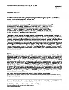
40 35
where
s ij
PSNR [dB]
Gaussian noise reduction, based on WPT
20 15
Poison noise reduction, based on WPT
5 0 1
3
5
7
9
11
13
15
17
19
Images
(2) – the wavelet coefficients of S
25
10
(1)
where E K – the entropy in the level K for the best tree decomposition of the image S
s ij ≥ T ,
Contrast increasing and morph. filtering
30
Fig. 1a. The graphical presentation of PSNR for investigated CT images
in an 3
orthonormal basis, T – the threshold of the coefficients.
2,5
By determination of the threshold it is used the strategy of Birge-Massart [7]. This strategy is flexibility and used spatial adapted threshold that allows to determinate the threshold in three directions: horizontal, vertical and diagonally. In addition the threshold can be hard or soft. The noise reduction is applied on Gaussian and Poison distributed noise components.
Contrast increasing and morph. filtering
Eff [dB]
2
Gaussian noise reduction, based on WPT
1,5
Poison noise reduction, based on WPT
1 0,5 0 1
3
5
7
9
11
13
15
17
19
Images
Experimental results
Fig. 1b. The graphical presentation of EFF for investigated CT images
The formulated stages of processing are realized by computer simulation in MATLAB environment by using IMAGE PROCESSING TOOLBOX and WAVELET TOOLBOX. In analysis are used 20 grayscale CT images of the head and abdominal tissues that exhibited diverse pathology. The original images are in different file format: jpeg, tiff and bmp, but all of them are converted into bmp. The obtained average results from simulation are presented in Table 1. The best results are obtained by noise reduction of Poisson noise on the base of WP transformation. The CNR is minimum (0.3) and shows that the noise is three times reduced. The values of PSNR and Effectiveness of filtration ( EFF ) are more sufficient. The graphical presentations of the obtained results are shown in Fig. 1.
1 0,9 Contrast increasing and morph. filtering
0,8
CNR
0,7 0,6
Gaussian noise reduction, based on WPT
0,5 0,4
Poison noise reduction, based on WPT
0,3 0,2 0,1 0 1
3
5
7
9
11
13
15
17
19
Images
Fig. 1c. The graphical presentation of CNR for investigated CT images
In Fig. 2 the original CT image of size 832x659 pixels is illustrated.
Table 1. Simulation results
Fig. 2. The original CT image
72
Fig. 3 presents selected ROI from the original CT image of size 196x152 pixels. In fig.4 is shown the selected ROI with contrast increasing. In Fig. 5 the result from the following wavelet filtration of Gaussian noise is presented. Fig. 6 illustrates the following wavelet filtration of Poison noise.
Fig. 6. The selected ROI after Poison noise reduction
Conclusion In the paper a new and effective approach for CT image enhancement is proposed. The complex processing has an effect of contrast enhancement, noise reduction and contours determination for selected ROI of different parts of diagnostic CT images. The implemented studying and obtained results by using of real images attempt to make diagnostic more precise.
Fig. 3. The selected ROI of CT image
References 1. Smith 2.
3. Fig. 4. The selected ROI with increased contrast
4. 5. 6.
7.
M., A. Docef. Transforms in telemedicine applications. – Kluwer Academic Publishers, 1999. Athhanasiadis T., Wallace M., Kapouzis K., Kollias S. Utilization of evidence theory in the detection of salient regions in successive CT images // Oncology reports. – 2006. – Vol.15. – P. 1071–1076. Gonzalez R., Woods R. Digital Image Processing. – Addison Wesley Publishing. – 1992. Donoho D., Johnston I. Adapting to unknown smoothness via wavelet shrinkage // Am. Stat. Assoc. – 1995. – No. 90. – P. 1200–1224. Coifmann R., Wickerhauser M. Entropy based Algorithms for best basis selection // IEEE Transaction on information theory. – 1992. – Vol. 38. – P. 713–718. Georgieva V., Kountchev R. An influence of the wavelet packet decomposition on noise reduction in ultrasound images // Proceedings of International Scientific Conference on Information, Communication and Energy systems and Technology. – Sofia, Bulgaria. – 2006. – P. 185–188. MATLAB User’s Guide. Accessed at: www.mathwork.com. Received 2009 08 26
Fig. 5. The selected ROI after Gaussian noise reduction V. M. Georgieva. An Approach for Computed Tomography Images Enhancement // Electronics and Electrical Engineering. – Kaunas: Technologija, 2010. – No. 2(98). – P. 71–74. A new approach for computed tomography (CT) images enhancement is presented. It consists of the following stages for image processing: selecting of the region of interest (ROI), contrast increasing by gamma correction, morphological processing of the selected ROI and noise reduction on the base of 2D wavelet packet transformations. Treatment on the base of investigation on the most suitable morphological operators and on optimization of some parameters of the wavelet transformations was the most suitable algorithm. The
73
aim is to improve the quality of the diagnostic computed tomography images. Some results of the experiments are presented, which were made by computer simulation in MATLAB environment. Ill. 8, bibl. 7 (in English; summaries in English, Russian and Lithuanian). В. М. Георгиева. Метод улучшения качества образов компьютерной томографии // Электроника и электротехника. – Каунас: Технология, 2010. – № 2(98). – С. 71–74. Представлен новый подход для улучшения образов компьютерной томографии. Он состоит из следуюших этапов: обработки образа, выделения области обработки (ROI), увеличения контраста посредством „гамма“ корекции, морфологической обработки выделеного ROI и уменьшения шума, базирующееся на двухмерных вэйвлетных пакетных преобразованиях. Эффективным алгоритмом оказалась обработка, основывающаяся на исследовании самых подходящих морфологических операторов и на оптимизации некоторых параметров вэйвлетных преобразований. Целью является увеличение качества диагностического компьютерного томографского образа. Некоторые из представленных результатов были получены при помощи компьютерного моделирования в среде MATLAB. Ил. 8, библ. 7 (на английском языке; рефераты на английском, русском и литовском яз.). V. M. Georgieva. Kompiuterinės tomografijos vaizdų kokybės pagerinimas // Elektronika ir elektrotechnika. – Kaunas: Technologija, 2010. – Nr. 2(98). – P. 71–74. Pateiktas naujas kompiuterinės tomografijos vaizdų kokybės pagerinimo metodas. Jis susideda iš dominančio regiono pasirinkimo, kontrasto padidinimo gama korekcijos būdu, morfologinio pasirinkto regiono apdorojimo ir triukšmo sumažinimo naudojant dvimates paketines vilnelių transformacijas. Parenkami geriausi morfologiniai operatoriai, optimizuojami kai kurie bangelių transformacijos parametrai. Tikslas – pagerinti diagnostinių kompiuterinės tomografijos vaizdų kokybę. Pateikti kai kurie eksperimentiniai rezultatai, gauti naudojant kompiuterinį modeliavimą MATLAB aplinkoje. Il. 8, bibl. 7 (anglų kalba; santraukos anglų, rusų ir lietuvių k.).
74