An automated method for determining the cytoadhesion of ...
Recommend Documents
The complex-step method for calculating sensitivi- ... eral methods that made use of complex variables, .... difference estimates eventually yields zero and then.
were measured by using a Hall effect device and an isolation amplifier respectively, which present small amplitude voltage signals to the DAQ board. A PC bus ...
An automated enzymatic method was developed for the measurement of D-arabinitol in human ... candidiasis have higher serum D-arabinitol (DA) levels (2, 7,.
Forman [5] have observed in tests with steel plates containing ... the elastic-plastic boundary around the crack tip is a circle, the radius of which was independent of work-hardening for small- ... (3). For the elastic plate under conditions of gene
Dec 12, 1975 - 1934), on the function of haemoglobin in the animal kingdom and the adaptations ... of the oxygen carrying properties of the blood. ... collodion membrane, and consequently free oxygen is made available. .... Caution: effects of electr
Aug 2, 2014 - e-mail: [email protected], [email protected], .... First, Belasi and Tukel [39] concluded that capability of the resources ownership have .... with a number of colleagues including at least 5 doctoral students ...
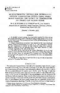
o m. (Kg/cm2). Fig.9 Edge-sliding mode stress-intensity factor(Kn) curves versus applied stress (Ta, for plexiglas plates containing an oblique internal slit forming.
injection technique cannot represent the actual reservoir fluids (Li et al., 2005). ... enough production data points to be certain and therefore long samples are ..... Haugen, à ., Fernø, M., Mason, G., and Morrow, N. R., Capillary Pressure And ...
Vis. 62(3) 249-65. [10] Alfimtsev A N, Loktev D A, Loktev A A 2012 Vestnik MGSU, 11 242â52. [11] Jiwani M A, Dandare S N 2013 Int. J. Sc. Res. Publ. 3(6) 1-6.
May 28, 2017 - Figure 1: Stages of force distribution in bolt connections [6]. These failures .... bolt holes, fine mesh was utilized in order to transfer the .... Fixed-free. Fixed-free ...... [7] G. L. Kulak, J. W. Fisher, and J. H. Struik, A Guide
b The George W. Woodruff School of Mechanical Engineering, Georgia Institute of Technology Atlanta, GA 30332- ... AAI's Shadow is a small Unmanned Aerial Vehicle (UAV) for ...... Proceedings of the 2002 SPIE Modeling, San Diego, CA, pp.
Nov 26, 2016 - quantification of labeled sugar complexes in biological samples. .... as detected with phenol-sulfuric acid, but no salts (chemical reagents) were ...
Abstract Existing methods for estimating mean grain size of sediment in an ..... intermediate axis, but that both automated and manual techniques measure the.
May 16, 2014 - Benjamin Haibe-Kains, Princess ...... [Epub ahead of print]. Zhang ... Received: 06 January 2014; paper pending published: 31 January 2014; ...
ournal of Analytical Toxicology, Vol. 29, July/August 2005. An Automated Alcohol Dehydrogenase Method for. Ethanol Quantification in Urine and Whole Blood.
automated vision system for fatigue crack growth measurement. ... Up to recently, the image acquisition apparatus with satisfactory quality for detecting thin short.
Jun 6, 2007 - also beneficial in preventing the occurrence of cerebral apoplexy in high- risk patients. The lacunar infarct regions are classified into the follow-.
Automated sensitivity analysis approaches in system dynamics focus ... automation of sensitivity analysis on functions was proposed by Hearne (2010), but the.
J NucÃ-Med 24: 529-531, 1983. The comparison of two scintigraphic images of the same organ explored under varying conditions (different tracers, various.
10 Apr 2006 - Journal of Automated Methods and Management in Chemistry ... Zhaoping He,1 Laura Bolling,1 Dalal Tonb,1 Tracey Nadal,1 and ... from Sigma (St. Louis, Mo). .... initial purchasing investment, the automated assay will re-.
Dec 16, 2011 - response of a condenser microphone. Dorel Homentcovschia) and Ronald N. Miles. Department of Mechanical Engineering, State University of ...
good indicators of regional maximum and minimum compres- sion directions. It is well known, however, that the P and T axes from a sin- gle fault plane solution ...
Azi = 46 m, z = 18 m and z,~ = 84 m. The more correct z0 and d values are those calculated through Equations (5) and (6). These values correspond to the.
An automated method for determining the cytoadhesion of ...
Casper HempelEmail author; Ida M Boisen; Akinwale Efunshile; Jørgen AL Kurtzhals ... In the automated assay, binding was quantified spectrophotometrically in ...
Hempel et al. Malaria Journal (2015) 14:112 DOI 10.1186/s12936-015-0632-4
METHODOLOGY
Open Access
An automated method for determining the cytoadhesion of Plasmodium falciparum-infected erythrocytes to immobilized cells Casper Hempel1*, Ida M Boisen1,2, Akinwale Efunshile3,4, Jørgen AL Kurtzhals1,2 and Trine Staalsø2
Abstract Background: Plasmodium falciparum exports antigens to the surface of infected erythrocytes causing cytoadhesion to the host vasculature. This is central in malaria pathogenesis but in vitro studies of cytoadhesion rely mainly on manual counting methods. The current study aimed at developing an automated high-throughput method for this purpose utilizing the pseudoperoxidase activity of intra-erythrocytic haemoglobin. Methods: Chinese hamster ovary (CHO) cells were grown to confluence in chamber slides and microtiter plates. Cytoadhesion of co-cultured P. falciparum, selected for binding to CHO cells, was quantified by microscopy of Giemsa-stained chamber slides. In the automated assay, binding was quantified spectrophotometrically in microtiter plates after cell lysis using tetramethylbenzidine as peroxidase-catalysed substrate. The relevance of the method for binding studies was assessed using: i) binding of P. falciparum-infected erythrocytes to CHO cells over-expressing chondroitin sulfate A and ii) CHO cells transfected with CD36. Binding of infected erythrocytes including field isolates to primary endothelial cells was also performed. Data was analysed using linear regression and Bland-Altman plots. Results: The manual and automated quantification showed strong, positive correlation (r2 = 0.959, p