Research Article
ISSN: 2321-2969
Int. J. Pharm. Biosci. Technol.
Received: 14 July 2013, Accepted: 31 July 2013
To cite this Article: Click here International Journal of Pharma Bioscience and Technology. 2013; 1(3): 118-129
Journal home page: www.ijpbst.com
AN EFFICIENT AND RAPID IN VITRO PROPAGATION SYSTEM OF THYMUS HYEMALIS LANGE, A WILD MEDICINAL AND AROMATIC PLANT OF MEDITERRANEAN REGION Aicha Nordine*, Dalila Bousta , Abdesalem El Khanchoufi , Abdelmalek El Meskaoui Unit of Plant Biotechnology, National Institute of Medicinal and Aromatic plants; Taounate. University of Sidi Mohamed Ben Abdellah, Fez, Morocco. Corresponding Author* E-mail address-
[email protected]
ABSTRACT: The objective of this study was to develop an in vitro regeneration protocol of Thymus hyemalis Lange. Initially, shoots were obtained from in vitro seedlings grown on Murashige & Skoog (MS) basal medium containing 3% (w/v) sucrose and 0.4% (w/v) gellan gum. Four basal media were tested for establishing their capacity for in vitro cultivation of T. hyemalis. Then several factors, namely explant type, plant growth regulators, genotype and various concentrations and type of sugar were tested to assess the shoots proliferation capacity. The optimal duration for shoot proliferation was also determined. Shoots were excised from proliferation medium and transferred in to rooting medium containing various auxins. Compared to White medium, B5 or ½ MS media, the results indicated that MS basal salt medium was found to be optimal for in vitro establishment. After 4 weeks, the maximum proliferation) was observed (corresponding to 6.58± 0.22 number of shoots) for nodal explants cultured on MS medium containing 3% (w/v) of sucrose and supplemented with 1.8 µM of kinetin (KIN). The results also showed that in vitro culture was genotype dependant. Extension of the culture period up to 5 weeks improved the number of shoots up to 9.33 ± 1.01. The best rooting of shoots was obtained on auxin-free MS medium or supplemented with 7.4 µM indole-3-butyric acid (IBA). Well-rooted plants were successfully established in dimpled plates filled by peat and vermiculite (2/3:1/3 v/v) mixture, with a survival rate of 90%. The regenerated plants were morphologically uniform and exhibited similar growth characteristics and vegetative morphology. Key words: In vitro tissue culture, Medicinal and aromatic plants, Plant growth regulators, Plant regeneration, Thymus hyemalis INTRODUCTION The use of medicinal and aromatic plants (MAPs) is increasing worldwide. Herbs belonging to the Lamiaceae family are rich in phytochemicals [1]. Thymus spp provides a stable economic return to local communities especially through the sale of wild-harvested material [2]. This genus is known in several countries as a spice and food preservative as well as a protective and curative remedy for many ailments [3]. It is reported that thyme possesses numerous biological activities including antifungal, antioxidant, insecticidal and antibacterial activities [4-11]. Thymus species are Nordine et al
widely distributed and found in several Mediterranean regions [12]. It is widely collected since its essential oil with a high proportion of phenols, which is greatly appreciated by exporters [13]. The chemical variability of the essential oil from wild T. hyemalis of the Southeastern Iberian Peninsula has been reported [13, 14]. These researchers stated that thymol, carvacrol, borneol, and linalool were the chemotypes most abundant in this area. Thymus hyemalis Lange, winter thyme, is an endemic shrub of Morocco, Algeria and Iberian Peninsula Pg. 118
Int. J. Pharm. Biosci. Technol. [12, 15]. Indeed, in Morocco, T. hyemalis was mentioned as endangered and rare species [16].
stored at 4°C experiments.
To the best of our knowledge, the major part of Thymus included T. hyemalis is harvested from the wild populations, except the Thymus vulgaris which is operated under cultivation. However, it there is not possible to collect the chemically homogeneous and standardized raw material of Thymus species in the natural habitat. Because the chemical polymorphism is characteristic for the plants belonging to this genus [12, 17]. It was also reported that the chemical composition of thyme species is influenced by soil, climatic and ecological factors, leading to a chemical variability [18]. This imposes many disadvantages such as the heterogeneity of plant material, the difficulty of predicting supplies for industry and lack of control. In addition, the harvest of medicinal plants on a mass scale from their natural habitats, is leading to a depletion of plant resources [19]. Therefore, there is an urgent need to look for alternate means of production, which could ensure large-scale and high quality plant materials to fulfill the growing demand [20]. Thus, the domestic cultivation and plant breeding offers the opportunity to adapt wild MAPs to the specific demands of their users, improving the prerequisites to high quality, profitable and sustainable production.
Stage I: Germination and in vitro establishment
The rapidness of tissue culture techniques can be advantageous for the continuous provision of a plantlet stock for domestic cultivation [21, 22] and MAPs breeding programmes. Some thyme species have been previously micropropagated by this technique such as Thymus vulgaris [23-25], Thymus piperella [26], Thymbra spicata L. var. spicata L. [27] and Thymus lotocephalus [28]. To the best of our knowledge, there are no reports on the micropropagation of T. hyemalis. Therefore, we investigated the most suitable in vitro propagation protocol for conservation and production of large number of genetically uniform plantlets of this species.
until
the
beginning
of
the
Seeds were sorted out for uniform size and similar external characteristics, discarding those with obvious alterations or malformations. Then, they were germinated either by ex vitro by sowing of about 200 seeds in plates with wells of 3 cm in diameter filled with peat and vermiculite (2/3:1/3 v/v) mixture or in vitro after sterilization according to the following protocol; seeds were incubated in 70% (v/v) alcohol for 3 min, followed by 10 min in a solution of 1% (v/v) sodium hypochlorite. They were then rinsed thrice with sterile distilled water, and finally dried on sterile filter paper. After decontamination, seeds were placed on Petri dish (9 cm of diameter) containing 25 ml of MS medium [29] (3% w/v of sucrose and 0.4% w/v of gellan gum) devoid of plant growth regulators (PGRs). A seed was considered to have germinated at the emergence of the radical (radical > 1 mm) [30]. The germination was recorded for a period of 6 weeks. The germination parameter evaluated was germination rate and it was expressed as the percentage of seeds germinated. For in vitro germination, four replicates (30 randomly selected seeds) were used. After the seed’s germination, to assess the effect of basal media on in vitro establishment of T. hyemalis, four media were tested. Nodal segments and shoot tips (1-1.5 cm long) that did not show contamination were aseptically excised from in vitro seedlings and cultured in glass flasks (175 ml) containing 30 ml of MS, ½ MS, Gamborg B5 (B5) [31] or White [32] media without hormones. After 4 weeks, the regeneration capacity was evaluated based on the regeneration percentage, shoot number and shoot length (cm). Subcultures were performed on the selected basal medium (MS medium without hormones) every 4 weeks until a sufficient stock for subsequent experiments was available. Stage II: Growth and in vitro multiplication.
MATERIALS AND METHODS Seeds source Seeds of wild Thymus hyemalis were offered graciously by the MAPs Beni Boufrah association located in Al hoceima, Centre Nord of Morocco. The plant in the flowering stage was identified by Dr. Ennabili A. and a voucher specimen (INP. 786) was deposited at the herbarium of the National Institute of Medicinal and Aromatic Plants (NIMAP); Taounate. University of Sidi Mohamed Ben Abdellah Fez Morocco. Seeds were separated from the inflorescence (Fig. 1a), cleaned and dry Nordine et al
Experiment 1: Effect of explant type and PGRs on rate multiplication. The experiment consisted of two main factors namely explant type and PGRs. Two types of explants; nodal segments (1 cm long) with a pair of axillary buds and apical segments (1–1.5 cm long shoot tips) were excised from uniform microshoots (one genotype). The explants were cultured on MS medium containing two cytokinins at different concentrations [6-benzylaminopurine, BAP (2.2, 4.4 and 8.8 µM) and kinetin, KIN (1.8, 4.6, 6.9 and 9.3 µM)] and one auxin α-naphthalene acetic acid, NAA at two concentrations [0.5 or 1 Pg. 119
Int. J. Pharm. Biosci. Technol. µM]. MS basal medium without PGRs was included as control. Data of the evaluation parameters such as regeneration percentage, number of shoots per explant and shoot length were recorded after 4 weeks. Experiment 2: Effect of genotype The objective of the second experiment was to investigate the effect of genotype on in vitro multiplication of T. hyemalis. The nodal explants (1 cm long) of three genotypes of this species designated as genotypes G1, G2 and G3 were tested. They were implanted into MS proliferation medium (MS + 1.8 µM of KIN) during 4 weeks. The evaluated parameters were regeneration rate percentage rate (%), number and length of shoots. Experiment 3: Effect of type and concentration of carbon sources The objective of the third experiment was to study the effect of type and concentration of sugar on multiplication rate of T. hyemalis. Based on the results of the previous experiments, MS medium with 1.8 µM of KIN and nodal segments were used in this experiment. The proliferation medium was supplemented with five concentrations (0, 15, 30, 45 and 60 g/l) of sucrose, glucose, sorbitol, fructose and mannitol. The MS medium lacking sugar was used as control. Regeneration percentage rate (%), number and length of shoots were evaluated after 4 weeks of culture. Experiment 4: Determination interval for shoot proliferation
of
optimal
Stem nodal segments (1 cm long) were cultured on MS proliferation medium for 1, 2, 3, 4, 5, 6 and 7 weeks, without change of medium. For each growing period, 14 explants were evaluated for their shoot number and shoot length. The time of browning medium was also determined. Stage III& IV: In vitro rooting and ex vitro transfer to sol For rooting, plantlets growing in MS proliferation medium were cultured on full-strength MS medium supplemented with indole-3-acetic acid, IAA (2.8, 3.6, 7.3 or 10.9 µM), indole-3-butyric acid, IBA (1, 2.5, 5 or 7.4 µM) and NAA (0.5, 1, 1.5 or 2 µM). The auxin-free MS medium was included as control. After 4 weeks, data of various parameters were recorded which included the percentage of root formation, number and the length of roots. In vitro-rooted shoots were removed from the in vitro containers and were washed in water to remove the agar from the roots. The plantlets were then transplanted into plates with wells of 3 cm in diameter filled by peat and vermiculite (2/3:1/3 v/v) mixture. In the first stage of acclimatization, plantlets were covered Nordine et al
with a plastic for providing the condition of high humidity. The humidity was maintained between 60 and 70% by successive (several time per day) and manual irrigation during the first 3 days, and were irrigated once daily thereafter for 10 days. When new leaves developed in the micropropagated plants inside the plastic tent, the plastic cover was removed periodically and progressively whenever leaves appeared water soaked. Then, the plants were transferred to large pots filled with peat and vermiculite (2/3:1/3 v/v) mixture and were regularly irrigated by water. After 3 months of growth in pots, the plantlets were transferred to soil in open field. Environmental conditions For all culture media, pH was adjusted to 5.8 before being autoclaved at 121°C and 100 KPa for 15 min. All the cultures were incubated in a growth chamber at temperature of 23 ± 2°C e, with illumination provided by cool white florescent lamps at 60 µmol m-2 s-1 with a 16-h light photoperiod. Statistical analysis The design of all the experiments was a complete randomized block. Each experiment was consisted by 6 replicates with 5 explants per replicate. Statistical analysis of data was carried out by means of the software “SPSS for Windows”. The homogeneity was carried out by leven’s test and the mean values were calculated and were compared by Duncan’s multiple range tests at P≤0.05. RESULTS AND DISCUSSION Stage I: Germination and in vitro establishment Germination The protocol used for disinfection was found to be effective, and 99% of in vitro seedlings did not manifest symptoms of bacterial and fungal contamination. Seeds were germinated either ex vitro or in vitro for a period of 6 weeks. The result showed that in vitro germination rate achieved was 24.91% and the seedlings showed a normal appearance (Fig. 1b), however germination ex vitro was poor and produced germination rate of only 6%. In vitro establishment In this experiment, the explants (nodal segments and shoot tips) were transferred to MS, ½ MS, B5 and White media for seedlings development. In this stage, the results showed that basal media influenced strongly the morphogenetic capacity of explants (Table 1). In term of regeneration, no statistical difference was observed between MS, ½ Pg. 120
Int. J. Pharm. Biosci. Technol. MS and B5 media, while the White medium gave a significantly lower result which was limited to 41.66%. On MS medium shoot number achieved was 5.79 ± 0.27. However, this parameter decreased significantly to 4.26 ± 0.40, 3.32 ± 0.47 and 1.49 ± 0.18 on ½ MS, B5 and White media respectively. The results obtained on B5 (3.32 ± 0.47) and ½ MS (4.26 ± 0.40) were not statistically different. The average length of shoots was also evaluated; the maximum length of shoots (5.67 ± 0.75 cm) was obtained on MS medium followed by
½ MS (4.9 ± 0.75 cm). However the B5 and White media gave the short stems compared to MS and ½ MS media (Table 1). Following this result, the MS basal medium proved to be superior to ½ MS, B5 and White for in vitro establishment of T. hyemalis. This result is in agreement with those reported by several researchers and further it also revealed the superiority of MS medium compared to other media [28, 33]. This is can be a result of several mineral salts especially macronutrients in full-strength MS medium [34, 35].
Table 1 Effect of basal media on regeneration (%), shoot number and shoot length (cm) of T. hyemalis micropropagated shoots. Basal media
Regeneration (%)
Shoot number
Shoot length (cm)
MS
91.66a
5.79 ± 0.27a
5.67 ± 0.75a
MS/2
83.33a
4.26 ± 0.40b
4.9 ± 0.75a
B5
75a
3.32 ± 0.47b
2.93 ± 0.57b
W
41.66b
1.49 ± 0.18c
0.8 ± 0.12c
Data indicate mean ± SE. Values followed by the same letter within the same column are not significantly different at P ˂ 0.05. Data recorded after 4 weeks of culture. Stage II: Growth and in vitro multiplication. Effect of explant type and multiplication of T. hyemalis.
PGRs
on
During the proliferation stage, the results showed that the best regeneration obtained was 100% on hormone-free MS medium or when supplemented with 4.6 or 6.9 µM of KIN. This parameter decreases significantly with high concentrations (4.4 and 8.8 µM) of BAP alone or with 2.2 µM of BAP and 1 µM of NAA combination; this is true for nodal explants. However, apical explants showed the highest regeneration capacity in all the tested media, except MS medium containing a high concentration of KIN (9.3 µM) which gave a significantly lower regeneration rate (58.33%) (Table 2). Using BAP in the medium, the average number of shoots in nodal and apical explants was 5.12 ± 0.63 and 4.25 ± 0.62 respectively on MS medium with BAP (2.2 µM). This parameter was decreased thereafter by increasing BAP at highest concentration. The use of KIN (1.8 µM) alone in nodal explants gave the highest shoot number reaching 6.58 ± 0.22, whereas this concentration gave 3.37 ± 0.37 shoots per explant only in apical explants. The use of NAA and BAP combinations in the MS medium did not improve the multiplication rate in both the explants. However, KIN and NAA combinations significantly decreased the shoot number in nodal explants, but a slight increase of shoot number was observed in apical explants Nordine et al
(Table 2). The average length of shoots was also affected by the explant type and hormones. On hormone-free MS medium, average length of shoots achieved was 4.38 ± 0.26 and 4.87 ± 0.85 cm in nodal and apical explants respectively. In both explants, BAP has generally decreased shoot length. While KIN relatively increased the shoot length (5.25 ± 2.25) in apical explants on MS medium with 1.8 µM of KIN. Following these results, the ability of T. hyemalis explants to form new shoots varied with the explant type and hormones. In our study, the nodal explant was found to be the better explant. This result is in agreement with those obtained by Zuzarte et al. (2010) [36] and Arikat et al (2004) [33], who showed that the nodal explants gave better results than the apical explants in micropropagated plants. The different responses of both types of explant were probably due to the endogenous hormone balance in the plant tissue [37]. In this study, the KIN proved to be the effective cytokinin of T. hyemalis proliferation. This result is in agreement with those obtained by Ozudogru et al. (2011) [25] who showed that KIN is the best cytokinin for regeneration of T. vulgaris. While our results indicated that the BAP (whatever at all the tested concentration) did not improve the rate of multiplication which is. Contrary, to the observations of other researchers, who have reported BAP as the most effective cytokinin for micropropagation of T. lotocephalus [28] and T. pepirella. [26]. Pg. 121
Int. J. Pharm. Biosci. Technol.
Table 2 Effect of plant growth regulators and explants type on regeneration (%), shoot number and shoot length (cm) of T. hyemalis Nodal explants
Apical explants
PGRs (µM) Regeneration Shoot number Shoot length Regeneration (%) 100a
Control
(cm) 5.62 ± 0.12ab 4.38 ± 0.26a
(%)
Shoot
Shoot length
number
(cm)
83.33ab
2.08± 0.84abc 4.87 ± 0.85ab
100a
4.25± 0.62abc 3.83 ± 0.98abc
75ab
1.83 ± 0.54bc 1.54 ± 0.34c
BAP 2.2
83.33ab
4.4
37c
8.8
33c
1 ± 0.50f
0.83 ± 0.33e
66.66ab
1.5 ± 0.28c 2.06 ± 0.34bc
1.8
83.33ab
6.58 ± 0.22a
3 ± 0.14bc
100a
3.37± 0.62abc 5.25 ± 2.25a
4.6
100a
100a
3.58± 0.87abc 3.58 ± 0.54abc
6.9
100a
91.66ab
4.33 ± 0.36ab 3.83 ± 0.44abc
9.3
83.33ab
2.58 ± 0.71def 2 ±0 .57cde
58.33b
3.25± 0.94abc 4.12 ± 0.76abc
2.2 + 0.5
75ab
2.87 ± 1.12def 1.62 ± 0.37de
100a
4.62 ± 0.62ab 2.62 ± 0.12abc
2.2 + 1
50bc
2.33 ± 0.36ef 1.41 ± 0.36de
75ab
4.25 ± 1.75abc 3.23 ± 1.28abc
1.8 + 0.5
83.33ab
3.42 ± 0.87cd 2.41 ± 0.36bcd
91.66ab
4.16 ± 0.58abc 3.13 ± 1.07abc
1.8 + 1
75ab
3.25 ± 0.25cde 1.75 ± 0.25de
91.66ab
4.75 ± 0.28a 3.91 ± 0.36abc
5.12± 0.63abc
3 ± 0.28bc
1.12 ± 0.62ef 0.86 ± 0.37e
KIN
5.25± 0.38abc 3.41 ± 0.22ab 4 ± 1bcd
2.50 ± 0.5bcd
BAP+ NAA
KIN + NAA
Data indicate mean ± SE. Values followed by the same letter within the same column are not significantly different at P ˂ 0.05. Data recorded after 4 weeks of culture. Effect of genotype In this experiment, the genotypes had shown different development responses on in vitro culture (Table 3). G1 gave the highest percentage of regeneration (95.83%), but no statistical difference was observed among the three genotypes in term of regeneration. However, the number of shoots (6.16 ± 0.73) was significantly higher in G1 compared with G2 and G3. Length of shoots was also affected by the effect of genotype. Indeed, shoots length Nordine et al
of G1 (5.37 ± 0.84 cm) and G3 (3.83 ± 0.75 cm) were statistically identical, but they were different to shoot length obtained in G2 (1.27 ± 0.5 cm) (Table 3). The effect of genotype on morphogenesis responses of in vitro plant has been previously demonstrated by several studies [38-41]. It is plausible to assume that the genotypes have different levels of endogenous auxins and/or cytokinins that influence their in vitro behavior [42].
Pg. 122
Int. J. Pharm. Biosci. Technol. Table 3 Effect of genotype on regeneration (%), shoot number and shoot length of T. hyemalis Genotypes Regeneration (%) Shoot number Shoot length (cm) G1
95.83a
6.16 ± 0.73a
5.37 ± 0.84a
G2
68.75a
0.93 ± 0.21b
1.27 ± 0.5b
G3
71a
2.37 ± 0.57b
3.83 ± 0.75a
Data indicate mean ± SE. Values followed by the same letter within the same column are not significantly different at P ˂ 0.05. Data recorded after 4 weeks of culture. Effect of type and concentration of carbon sources The responses of in vitro culture to different carbon sources added to the medium were also tested in this work. Regeneration (%), multiplication rate and shoot length (cm) were clearly affected by the concentration and type of sugar (Table 4). In the medium without sugar, shoots regeneration was limited to 33.33%, while their growth is almost inhibited. The addition of carbohydrate sources to the medium is indispensable, since in vitro cultures are unable to perform photosynthesis to sustain organ growth, induction and differentiation [43, 44]. It was reported that carbohydrates provide the metabolic energy and carbon skeletons of all organic compounds required for cell growth and development [45]. The highest regeneration percentage was 100% obtained on media enriched with 30 g/l of sucrose or glucose and 15 g/l of fructose, followed by 91.67% obtained on 15 g/l of sorbitol. The effect of type of carbon source and its concentration on adventitious shoots regeneration was previously found by Jain et al. (1997) [46]. The concentration of 30 g/l of sucrose provided a higher number of shoots (6.33 ± 0.88) with an average length reached 4.92 ± 0.33 cm. In medium supplemented with 15 g/l of sucrose measured parameters were statistically identical with those obtained on MS medium containing 30 g/l of the same sugar, but we observed that the leaves of the shoots were stained yellow after 20 days of culture. This may be due to a lack of photosynthesis because the green tissues are not sufficiently autotrophic in in vitro culture. Using sucrose concentrations more than 30 g/l, all measured parameters were significantly decreased. Elevated levels of sucrose may result in: (1) higher osmotic pressure potential in media inhibiting water and mineral uptake by explants, and (2) higher levels of the respiratory CO2 which is toxic due to the poor gas exchange in culture vessels [47]. It was reported that the sucrose concentration usually used in the in vitro culture varied between 10 to 50 g/l [34]. The effect of sucrose concentration on plant growth was
Nordine et al
reported for in several plants [26, 48]. Shoot number obtained with glucose (30 g/l) and fructose (15 g/l) was 4.92 ± 0.38 and 4.42 ± 0.82 respectively. Mannitol and sorbitol whatever their concentrations used were unnecessary for regeneration and proliferation of T. hyemalis (Table 4). Mannitol has often been added to culture media to mimic osmotic stress, as it is assumed to be only occasionally metabolized by in vitro cultured plants [49]. According to our result, sucrose is almost universally used as the most suitable energy source for plant micropropagation [50]. Contrary, in some works sorbitol has proven to be the most effective carbon source for in vitro growth of several species [44, 51]. Determination of optimal interval for shoot proliferation The number of multiple shoots formed from each explant increased with longer culture time. The explants produced an average of 5.96 ± 0.21 shoots per explant on the proliferation medium within 4 weeks and the number of multiple shoots increased almost 1.5-fold (9.33 ± 1.01 shoots per explant) after a further cultivation for one more week (Table 5, Fig. 1c). After the fifth week, the number of shoots was increased slightly reaching a number of 10.83 ± 0.1 shoots per explant at the end of 7th week. In the case of prolonged cultures, the nutrients in the medium are gradually exhausted, and at the same time, the relative humidity in the vessels decreases leading to the drying of the culture medium. Subculturing decreases the effect of competition of the developing shoots for nutrients [52]. In our study the browning of the medium was also observed, it started from the 6th week, and the quality of the most shoots was lower. Indeed, yellowing of the leaves was observed from this period (sixth week). It was reported that browning of media occurred as a result of oxidation of polyphenols exuded from explants [53]. Similar effects of subculture interval has been previously reported in several reports [43, 54].
Pg. 123
Int. J. Pharm. Biosci. Technol. Table 4 Effect of type and concentration of carbon sources on regeneration (%), number and length of shoots of T. hyemalis micropropagated shoots. Carbon sources Concentration g/l Regeneration (%) Shoot number Shoot length (cm) Contrôl
0
33.33cde
0.58 ± 0.22c
0.37 ± 0.25cd
15
83.33ab
4.92 ± 0.55ab
4.62 ± 0.18a
30
100a
6.33 ± 0.88a
4.92 ± 0.33a
45
33.33cde
1.58 ± 1.02c
1.31 ± 1.10c
60
16.67de
1.75 ± 1.63c
0.08 ± 0.08cd
15
83.33ab
4.08 ± 1.47b
3.17 ± 0.71b
30
100a
4.92 ± 0.38ab
3.25 ± 0.14b
45
50bcd
1.58 ± 1.02c
1.08 ± 0.58cd
60
33.33cde
0.75 ± 0.25c
0.54 ± 0.21cd
15
100a
4.42 ± .82ab
3.50 ± 0.38b
30
50.00bcd
1.08 ± 0.65c
0.75 ± 0.52cd
45
75.00abc
1.42 ± .30c
1.33 ± 0.30c
60
0e
0c
0d
15
0e
0c
0d
30
16.67de
0.25 ± 0.25c
0.08 ± 0.08cd
45
16.67de
0.25 ± 0.25c
0.08 ± 0.08cd
60
0e
0c
0d
15
91.67ab
1.67 ± 0.33c
1 ± 0.14cd
30
75.00abc
1.33 ± 0.44c
0.67 ± 0.22cd
45 60
83.33ab 8.33de
1.83 ± 0.3c 0.5 ± 0.5c
0.92 ± 0.08cd 0.33 ± 0.33cd
Sucrose
Glucose
Fructose
Mannitol
Sorbitol
Data indicate mean ± SE. Values followed by the same letter within the same column are not significantly different at P ˂ 0.05. Data recorded after 4 weeks of culture. Table 5 Effect of different culture durations on shoot number of T. hyemalis Durations 1 2 3 4 5 6 7
Shoot number 2.25 ± 0.35c 3.00 ± 0.41c 3.17 ± 0.32c 5.96 ± 0.21b 9.33 ± 1.01a 10.67 ± 0.50a 10.83 ± 0.47a
Data indicate mean ± SE. Values followed by the same letter within the same column are not significantly different at P ˂ 0.05.
Nordine et al
Stage III&IV: in vitro rooting and ex vitro transfer to sol In this experiment, the result showed that all tested auxins can induce rooting in T. hyemalis with a percentage ranging from 81.25% to 100% (Table 6). Auxin-free MS medium also induced rooting with a maximum rooting of 100%. This result was similar with those obtained by Lê (1989) [23] and Ozudogru et al. (2011) [25] with T. vulgaris who obtained 100% rooting on hormonefree MS medium. The maximum number of roots obtained was 21.38 ± 1.95 on 2 µM of NAA, with a very short average length (0.23 ± 0.07 cm) (Table 6). These roots were formed on surface of medium and they were very thin. Roots produced by the Pg. 124
Int. J. Pharm. Biosci. Technol. IAA were similar to those produced by NAA except that they were accompanied by callus at the base of explant. However, IBA at 7.4 μM was capable of producing principal roots with secondary roots with a maximum number of 6.50 ± 0.38 roots per explant with an average length of 0.67 ± 0.08 cm. The control medium also gave the thicker and well penetrated roots into medium with a number of 6.06 ± 0.37 roots per explant (Fig. 1d). This result was similar with those obtained by Furmanowa and Olszowska (1992) [24] who found that T. vulgaris rooted easily in medium with IBA.
In the same species (T. vulgaris), Ozudogru et al. (2011) [25] tested the effect of several auxins [IAA, IBA, NAA and 2,4-dichlorophenoxyacetic (2,4-D)] on its in vitro rooting and they observed that 2,4-D gave the best result of rooting. In T. lotocephalus, the best rooting was achieved with IAA [28]. The differences in rooting response between thyme species cited above could be related to multiple factors, such as the genotype, the endogenous cytokinin/auxin ratio, the influence of shoot multiplication medium and the sensitivity of tissues to absorb or use the exogenous auxin, among others [55, 56].
Table 6 Effect of IAA, IBA and NAA on in vitro rooting (%), root number and root length (cm) of T. hyemalis Auxins (µM)
Rooting (%)
Root number
Root length (cm)
Control
100a
6.06 ± 0.37bcd
0.99 ± 0.09a
81.25b 100a 100a 100a
6.56 ± 1.53bcd 7.69 ± 1.73bcd 9.75±1.14bc 10.50 ± 0.27b
0.45 ± 0.02cd 0.64 ± 0.07bc 0.65 ± 0.09bc 0.61 ± 0.07bc
100a 87.50ab 93.75ab 100a
4.92 ± 0.55d 4.69 ± 0.91d 5.75 ± 0.63cd 6.50 ± 0.38bcd
0.83 ± 0.08ab 0.74 ± 0.15abc 0.85 ± 0.18ab .67 ± .08bc
0.5
100a
9.63 ± 1.48bc
0.48 ± 0.08cd
1
100a
7.86 ± 2.44bcd
0.21 ± 0.06d
1.5
100a
18.75 ± 1.25a
0.22 ± 0.03d
2
93.75ab
21.38 ± 1.95a
0.23 ± 0.07d
IAA 2.8 3.6 7.3 10.9 IBA 1 2.5 5 7.4 NAA
Data indicate mean ± SE. Values followed by the same letter within the same column are not significantly different at P ˂ 0.05. Data recorded after 4 weeks of culture. Healthy rooted plantlets were successfully acclimatized to ex vitro conditions with a survival percentage of 90%. The newly produced plants presented no apparent morphological variation, and the number of new leaves and shoot length increased considerably (Fig. 1e & f).
compounds for pharmaceutical purposes. In addition, seeds from acclimatized plants were obtained in the following season and were germinated for the production of new thyme plants. The new regenerated plants of T. hyemalis have been appeared normal and no morphological variation was shown.
CONCLUSION The above protocol describes an efficient method for rapid multiplication of Thymus hyemalis by direct regeneration. This protocol can ensure a stable supply of this commercial crop in limited time and space, irrespective of seasonal variations and thus meet the global demand for its essential oil. Regenerated plants could also serve as potential sources for the extraction of active
Nordine et al
ACKNOWLEDGMENTS We thank the Beni Boufrah association for providing seeds and Dr. A. Ennabili for identification of the botanical name of Thymus species. Dr. D. Mezian for her help on statistical analysis. This research program was supported by National Institute of Medicinal and Aromatic Plants-Taounate, Morocco. Pg. 125
Int. J. Pharm. Biosci. Technol.
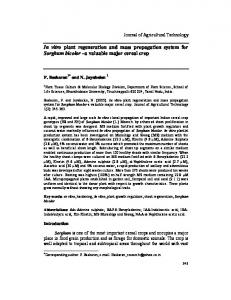
Fig. 1 Micropropagation of Thymus hyemalis a) Separate seeds from inflorescence of wild plant b) Germinated seeds on hormone-free MS medium c) Micropropagated plant on 1.8 µM of KIN after 5 weeks of culture d) Obtained roots of plants on hormonefree MS medium e) Acclimatized plants in large pots after 3 months g) Micrppropagated plants after two years of transfer into soil. Nordine et al
Pg. 126
Int. J. Pharm. Biosci. Technol. REFERENCES 1. Shan B, Cai YZ, Sun M, Corke H. Antioxidant capacity of 26 spice extracts and characterization of their phenolic constituents. Journal of agricultural and food chemistry. 2005; 53(20):7749–7759. 2. Hamilton A. Medicinal plants, conservation and livelihoods. Biodiversity and conservation. 2004; 13(8):1477−1517. 3. Bellakhadar J, Claiss R, Fleurentin J, Younos C. Repertory of standard herbal drugs in the Moroccan pharmacopoeia. Journal of ethnopharmacology. 1991; 35(2):123–143. 4. Daferera DJ, Ziogas BN, Polissiou MG. GC/MS analysis of essential oils from some Greek aromatic plants and their fungitoxicity on Penicillium digitatum. Journal of agricultural and food chemistry. 2000; 48(6):2576–2581. 5. Saad A, Fadli M, Bouaziz M, Benharref A, Mezrioui N-E, Hassani L. Anticandidal activity of the essential oils of Thymus maroccanus and Thymus broussonetii and their synergism with amphotericin B and fluconazole. Phytomedicine. 2010; 17(13):1057–1060. 6. Nieto G, Bañón S, Garrido MD. Effect of supplementing ewes’ diet with thyme (Thymus zygis ssp. gracilis) leaves on the lipid oxidation of cooked lamb meat. Food Chemistry. 2011; 125(4):1147–1152. 7. Ramchoun M, Harnafi H, Alem C, Büchele B, Simmet T, Rouis M, Atmani F, Amrani S. Hypolipidemic and antioxidant effect of polyphenol-rich extracts from Moroccan thyme varieties. e-SPEN Journal. 2012; 7(3):e119−e124. 8. Zarzuelo A, Crespo E. The medicinal and non-medicinal uses of thyme. In: Stahl-Biskup E, Sáez F, editors. Thyme: The genus Thymus. Medicinal and aromatic plants—Industrial profiles. London: Taylor & Francis; 2002. p. 263–292. 9. Kalemba D, Kunicka A.Antibacterial and antifungal properties of essential oils. Current medicinal chemistry. 2003; 10(10):813–829. 10. Fadli M, Saad A, Sayadi S, Chevalier J, Mezrioui N, Pagès J-M, Hassani L. Antibacterial activity of Thymus maroccanus and Thymus broussonetii essential oils against nosocomial infection – bacteria and their synergistic potential with antibiotics. Phytomedicine. 2012; 19(5):464–471. Nordine et al
11. Gutierrez-Larrainzar M, Rua J, Caro I, de Castro C, de Arriaga D, Garcia-Armesto MR, del Valle P. Evaluation of antimicrobial and antioxidant activities of natural phenolic compounds against foodborne pathogens and spoilage bacteria. Food Control. 2012; 26(2):555–563. 12. Stahl-Biskup E, Sáez F. Thyme. The genus Thymus. medicinal and aromatic plants— Industrial profiles. London: Taylor & Francis; 2002. 13. Sáez F. Essential oil variability of Thymus hyemalis growing wild in Southeastern Spain. Biochemical Systematics and Ecology. 1995; 23(4):431−438. 14. Sotomayor JA. Estudio sobre plantas aromàticas de los géneros Salvia y, espontáneas en el Sureste Ibérico, para su establecimiento como cultivo. Doctoral thesis, departamento de biologıa vegetal (Botànica), University of Murcia, Murcia, Spain; 1998. 15. Fennane M, Ibn Tattou M, Ouyahya A, El Oualidi J. Flore Pratique du Maroc. 2nd ed. Rabat: Institut Scientifique; 2007. 16. Fennane M, Ibn Tattou M. Catalogue des plantes rares, menacées ou endémiques du Maroc. Palermo Bocconea; 1998. 17. Santos PA, Barroso JG, Figueiredo AC, Pedro LG, Salgueiro LR, Fontinha SS, Deans SG, Scheffer JC. Chemical polymorphism of populations of Thymus caespititius grown on the Islands Corvo, Flores, São Miguel and Terceira (Azores) and on Madeira, assessed by analysis of their essential oils. Plant Science. 2005; 169(6):1112−1117. 18. Sáez F. Volatile oil variability in serpylloides ssp. gadorensis growing wild in Southeastern Spain. Biochemical Systematics and Ecology. 2001; 29(2):189−198. 19. Sen J, Sharma AK. Micropropagation of withania somnifera from germinating seeds and shoot tips. Plant Cell, Tissue and Organ Culture. 1991; 26(2):71−73. 20. Lange D. The German foreign trade in medicinal and aromatic plants during the 1990s (Newsletter of the IUCN species survival commission) Medicinal Plant Conservation. 2004; 9/10:38–46. 21. Reddy PS, Rodrigues R, Rajasekharan R. Shoot organogenesis and mass propagation of Coleus forskohlii from leaf derived callus.
Pg. 127
Int. J. Pharm. Biosci. Technol. Plant Cell, Tissue and Organ Culture. 2001; 66(3):183–188.
32. White PR. The cultivation of animal and plant cells. New York: Ronald Press; 1963.
22. Tiwari V, Tiwari KN, Singh BD. Shoot bud regeneration from different explants of Bacopa monniera (L.) Wettst. by trimethoprim and bavistin. Plant cell reports. 2006; 25(7):629–35.
33. Arikat NA, Jawad FM, Karam NS, Shibli RA. Micropropagation and accumulation of essential oils in wild sage (Salvia fruticosa Mill.). Scientia Horticulturae. 2004; 100(14):193−202.
23. Lê CL. Microbouturage in vitro du thym (Thymus vulgaris). In: Stahl-Biskup E, Saéz F, editors. Thyme: the genus Thymus, medicinal and aromatic plants—industrial profiles. London: Taylor & Francis , 1989. p. 177–196.
34. Pierik RLM. In vitro culture of higher plants. Netherlands: Kluwer academic publishers; 1987.
24. Furmanowa M, Olszowska O. Micropropagation of thyme. In: Bajaj YPS, editors. Bioteclmology in agriculture and forestry. Berlin Heidelberg: Springer-Verlag; 1992. p. 230−243. 25. Ozudogru EA, Kaya E, Kirdok E, IsseverOzturk S. In vitro propagation from young and mature explants of thyme (Thymus vulgaris and T. longicaulis) resulting in genetically stable shoots. In Vitro Cellular and Developmental Biology – Plant. 2011; 47(2):309–320. 26. Sáez F, Sknchez P, Piqueras A. Micropropagation of Thymus piperella. Plant Cell, Tissue and Organ Culture. 1994; 39(3):269−272. 27. Daneshvar-Royandezagh S, Khawar KM, Ozcan S. In vitro micropropagation of garden thyme (thymbra spicata L. var. spicata L.) collected from Southeastern Turkey using cotyledon node. Biotechnology & Biotechnological Equipment. 2009; 23(3):1319−1321. 28. Coelho N, Gonçalves S, González-Benito ME, Romano A. Establishment of an in vitro propagation protocol for Thymus lotocephalus, a rare aromatic species of the Algarve (Portugal). Plant Growth Regulation. 2012; 66(1):69−74. 29. Murashige T, Skoog F. A revised medium for rapid growth and bioassays with tobacco tissue cultures. Physiologia Plantarum. 1962; 15(3):473–497. 30. Bewley JD, Black M. Seeds: Physiology of development and germination. 2nd ed. London: Plenum Press; 1994. 31. Gamborg OL, Miller RA, Ojima K. Nutrient requirements of suspension cultures of soybean root cells. Experimental cell research. 1968; 50(1):151–158.
Nordine et al
35. Shimomura K, Kitazawa T. Tanshinone production in adventitious roots and regenerates of Salvia miltiorrhiza. Journal of Natural Products. 1991; 54(6):1583–1587. 36. Zuzarte MR, Dinis AM, Cavaleiro C, Salgueiro LR, Canhoto JM. Trichomes, essential oils and in vitro propagation of Lavandula pedunculata (Lamiaceae). Industrial Crops and Products. 2010; 32(3):580–587. 37. Grattapaglia D, Machado MA. Micropropagação. In: Torres AC, Caldas LS, Buso JA, editors. Cultura de tecidos e transformação genética de plantas. Brasília: Embrapa; 1998. p. 183−260. 38. Ostrolucká MG, Gajdošová A, Libiaková G, Ondrušková E. Protocol for micropropagation of Vaccinium vitis-idaea L. In: Jain SM, Haggman H, editors. Protocols for micropropagation of woody trees and fruits. Netherlands: Springer; 2007. p. 457– 464. 39. Gahan PB, George EF. Adventitious regeneration. In: George EF, Hall MA, Klerk GJ, editors. Plant propagation by tissue culture. Netherlands: Springer; 2008. p. 335– 401. 40. Bhau BS, Wakhlu AK. Effect of genotype, explant type and growth regulators on organogenesis in Morus alba. Plant Cell, Tissue and Organ Culture. 2001; 66(1):25–29. 41. Pati PK, Rath SP, Sharma M, Sood A, Ahuja PS. In vitro propagation of rose −A review. Biotechnology Advances. 2006; 24(1):94−114. 42. George EF, Debergh PC. Micropropagation: uses and methods. In: George EF, Hall MA, Klerk GJ, editors. Plant propagation by tissue culture. Netherlands: Springer; 2008. p. 29– 64. 43. George EF. Plant propagation by tissue culture: the technology. 2nd edition. England: Exegetics; 1993.
Pg. 128
Int. J. Pharm. Biosci. Technol. 44. Kozai T. Micropropagation under photoautotrophic conditions. In: Debergh PC, Zimmerman RH, editors. Micropropagation: Technology and application. Netherlands: Kluwer academic publishers; 1991. p. 447−469. 45. Caldas LS, Haridasan P, Ferreira ME. Meios nutritivos. In: Torres AC, Caldas LS, editors. Técnicas e aplicações da cultura de tecidos de plantas. Brasília: EMBRAPA/CBAB; 1988. p. 87−132. 46. Jain RK, Davey MR, Cocking EC, Wu R. Carbohydrate and osmotic requirements for high-frequency plant regeneration from protoplast-derived colonies of indica and japonica rice varieties. Journal of Experimental Botany. 1997; 48(3):751−758. 47. Ebrahim MKH. Comparison, determination and optimizing the conditions required for rhizome and shoot formation, and flowering of in vitro cultured calla explants. Scientia Horticulturae. 2004; 101(3):305−313. 48. Kanji M, Yuji S. Effects of sugar concentration and strength of basal medium on conversion of somatic embryos in Asparagus officinalis L. Scientia Horticulturae. 2000; 84(1-2):15−26. 49. Lambardi M, Rugini E. Micropropagation of olive (Olea europaea L.). In: Jain SM, Ishii K, editors. Micropropagation of woody trees and fruits. Dordrecht: Kluwer; 2003. p. 621– 646.
temperature storage of shoot cultures of apricot. Scientia Horticulturae. 2010; 126(4):434–440. 51. Marino G, Bertazza GP, Magnanini E, Doro AA. Comparative effects of sorbitol and sucrose as main carbon energy sources in micropropagation of apricot. Plant Cell, Tissue and Organ Culture. 1993; 34(3):235– 244. 52. Rout GR, Samantaray S, Das P. In vitro manipulation and propagation of medicinal plants. Biotechnology Advances. 2000; 18(2):91–120. 53. George EF, Sherrington PD. Plant propagation by tissue culture. England: Exegetics Ltd., versley; 1984. 54. Lee WL, Chan LK. Plant regeneration from stem nodal segments of Orthosiphon stamineus Benth., a medicinal plant with diuretic activity. In Vitro Cellular and Developmental Biology – Plant. 2004; 40(1):115−118. 55. de Klerk G-J, van der Krieken W, de Jong JC. The formation of adventitious roots: new concepts, new possibilities. In Vitro Cellular & Developmental Biology - Plant. 1999; 35(3):189–199. 56. de Klerk G-J. Rooting of microcuttings: theory and practice. In Vitro Cellular & Developmental Biology - Plant. 2002; 38(5):415–422.
50. Marino G, Paola N, Antonio C, Andrea M. Effect of carbohydrates on in vitro low-
Nordine et al
Pg. 129
Int. J. Pharm. Biosci. Technol.
How to cite this article APA style Nordine, A., Bousta, D., El Khanchoufi, A., & El Meskaoui, A. (2013). An efficient and rapid in vitro propagation system of Thymus hyemalis Lange, a wild medicinal and aromatic plant of Mediterranean region. International Journal of Pharma Bioscience and Technology, 1(3), 118–129. Elsevier Harvard style Nordine, A., Bousta, D., El Khanchoufi, A., El Meskaoui, A., 2013. An efficient and rapid in vitro propagation system of Thymus hyemalis Lange, a wild medicinal and aromatic plant of Mediterranean region. Int. J. Pharm. Biosci. Technol. 1, 118–129. Vancouver Style Nordine A, Bousta D, El Khanchoufi A, El Meskaoui A. An efficient and rapid in vitro propagation system of Thymus hyemalis Lange, a wild medicinal and aromatic plant of Mediterranean region. Int J Pharm Biosci Technol. 2013; 1(3):118–29. To receive bibliographic information in RIS format (For Reference Manager, ProCite, EndNote): Send request to:
[email protected]
To Return to the first Page: Click Here