cell Cycle Feature
Cell Cycle 10:3, 364-365; February 1, 2011; © 2011 Landes Bioscience
An example of functional interaction between NFAT5/TonEBP and nuclear factor κB by hypertonic stress Aquaporin-2 transcription Udo Hasler Department of Cellular Physiology and Metabolism; University of Geneva; Geneva, Switzerland
Aquaporins (AQPs) are channels that facilitate the passage of water across lipid bilayers.1 Numerous AQPs have been identified in almost all organisms. In mammals, the AQP family consists of 13 members. AQP2 was first identified in principal cells of the kidney collecting duct (CD), and later in many other tissues as well, such as the central nervous system, gastrointestinal tract and pancreas.2 In the kidney, AQP2 plays a major role in the urinary concentrating mechanism. In response to hormonal stimulation, principally vasopressin (VP), CD water permeability dramatically increases due to insertion of AQP2 in the apical membrane of principal cells. Water is then returned to the circulatory system via AQP3 and AQP4 inserted in the basolateral membrane. Deficient AQP2 expression leads to diabetes insipidus. Although this clinical entity is not usually lethal in humans, the need to drink large quantities of water is a major obstacle for quality of life. Transport of water across the CD epithelia requires osmotic gradients between the tubule lumen and the interstitium. In the renal medulla the interstitium is hypertonic relative to plasma. Cells of the CD medulla are therefore subjected to hypertonic stress, the magnitude of which varies according to water intake and which increases along the length of the CD. Hypertonic stress has important consequences on AQP2 expression. In the absence of VP it promotes AQP2 insertion in the apical membrane immediately following challenge.3 After longer periods of
stimulation it also induces AQP2 insertion in the basolateral membrane.4 In addition to its effects on AQP2 trafficking, hypertonic stress modulates AQP2 transcription independently of VP.5 As suggested by observations made in cultured CD principal cells, this event is likely mediated by a time-dependent interaction between two major transcription factors, Tonicity-Responsive Enhancer BindingProtein (TonEBP or NFAT5) and Nuclear Factor-κB (NFκB). This interaction is described below. Hypertonic stress induces time-dependent variations of AQP2 transcription in vitro. Transcription first decreases during the first hours of challenge (3 h) then increases after longer periods of time (24 h).5 Decreased AQP2 transcription is at least party mediated by NFκB. Several NFκB subunits exist, and they assemble as dimers. Different combinations of DNA-bound dimers differentially affect the transcriptional activity of numerous genes under their control.6 As depicted in Figure 1, two κB sites are contained in the first 2.1 kbp of the AQP2 promoter, both of which bind the NFκB subunits p65, p50 and p52. Deletion of either site increases AQP2 transcriptional activity under baseline conditions, indicating that they act as transcriptional repressors.7 NFκB activation by cytokines or lipopolysaccharides (LPS) decreases AQP2 transcription. Similar to classical NFκB stimulation, hypertonic stress increases NFκB activity and NFκB activation is at least party responsible for AQP2
transcriptional repression on the onset of hypertonic stress.7 This effect, however, is transient7,8 and appears to be mediated by p65 release and binding of p50 and p52 to both κB sites. Numerous genes are under the control of TonEBP. TonEBP is only partly active under isotonic conditions and both its activity and nuclear expression are increased in response to an increase of environmental tonicity.9 TonEBP has recently been shown to interact with p65 in response to hypertonic stress, but not TNFα or cytokines.8 Increased nuclear translocation of TonEBP immediately following hypertonic stress (3 h) increases its interaction with p65. ‘Quenched’ nuclear p65 in turn might allow binding of free p50- and p52-containing complexes to the AQP2 promoter that in turn repress AQP2 transcription even further. After longer periods of hypertonic stimulation (24 h), NFκB activity decreases and TonEBP activity increases.8,10 As depicted in Figure 1, the AQP2 promoter contains at least one TonEBP binding site (TonE). Binding of active TonEBP to this site is responsible for increased AQP2 transcription after long periods of hypertonic stimulation.10 Such activity would supersede the repressive effects of NFκB manifested at the onset of hypertonic stress. Interestingly, in vitro overexpression of TonEBP decreases AQP2 expression four fold under baseline conditions independently of VP (unpublished observations by the author). Under these artificial (isotonic) conditions, TonEBP may forcibly
©201 1L andesBi os c i enc e. Donotdi s t r i but e.
Correspondence to: Udo Hasler; Email:
[email protected] Submitted: 12/08/10; Accepted: 12/09/10 DOI: 10.4161/cc.10.3.14520 Feature on: Roth I, et al. Mol Biol Cell 2010; 21:3459–74. 364
Cell Cycle
Volume 10 Issue 3
cell Cycle Feature
cell Cycle Feature
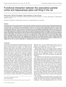
Figure 1. A model of time-dependent effects of hypertonic stress on AQP2 transcription. Under basal (isotonic) conditions, p65-containing complexes bind to κB elements of the AQP2 promoter while TonEBP bound to TonE elements is minimal. The overall effect is repressed AQP2 transcription. Hypertonic stress induces three major events, depicted as encircled numbers. (1) Immediately following hypertonic stress, TonEBP nuclear translocation increases. This increases the interaction between TonEBP and p65-containing complexes, leading to p65 release from κB elements of the AQP2 promoter. (2) p65 ‘quenching’ by TonEBP allows p50- and p52-containing complexes to bind to κB elements of the AQP2 promoter. This represses AQP2 transcription even further. (3) Binding of TonEBP to TonE elements of the AQP2 promoter increases over time of stimulation. Increased AQP2 transcription by TonEBP eventually supersedes the repressive effect of p50/p52. The overall effect is an increase of AQP2 transcription. The localization of κB and TonE elements of the mouse AQP2 promoter relative to the transcription start site are shown.
©201 1L andesBi os c i enc e. Donotdi s t r i but e.
interact with p65 but not TonE elements. On the other hand, AQP2 transcription is highest in cells challenged 24 h with hypertonic medium that overexpress TonEBP (unpublished observations by the author). In this case, an artificial increase of TonEBP abundance would enhance AQP2 transcription via increased binding of TonEBP to TonE elements. Together, these observations show that AQP2 expression, and thereby water permeability of AQP2-expressing cells, is adjusted by hypertonicity in a time-dependent manner via an interaction between NFκB and TonEBP. Both NFκB and TonEBP play anti-apoptotic roles. It is therefore reasonable to speculate that timely downregulation of AQP2
www.landesbioscience.com
expression may be crucial for survival of cells expressing this AQP isoform following hypertonic stress. Possibly, decreased AQP2 expression may help protect cells against recurrent hypertonic challenge. On the other hand, once challenged cells adapt to its hypertonic environment, increased AQP2 expression would help maximize water retention. Hypertonic stress was also found to increase AQP1 expression in a TonEBP-dependent manner.11 Contrary to AQP2, AQP1 expression did not decrease on the onset of challenge (unpublished observations by the author) indicating that time-dependent transcription events regulating AQP expression described here may be specific for AQP2.
Cell Cycle
References 1. 2. 3. 4. 5. 6. 7. 8. 9.
Preston GM, et al. Science 1992; 256:385-7. Mobasheri A, et al. J Mol Histol 2005; 36:1-14. Hasler U, et al. J Biol Chem 2008; 283:26643-61. van Balkom BWM, et al. J Biol Chem 2003; 278:1101-7. Hasler U, et al. J Am Soc Nephrol 2005; 16:1571-82. Hayden MS, et al. Cell 2008; 132:344-62. Hasler U, et al. J Biol Chem 2008; 283:28095-105. Roth I, et al. Mol Biol Cell 2010; 21:3459-74. Miyakawa H, et al. Proc Natl Acad Sci USA 1999; 96:2538-42. 10. Hasler U, et al. J Am Soc Nephrol 2006; 17:1521-31. 11. Lanaspa MA, et al. J Biol Chem 2010; 285:31694-703.
365