Renibacterium salmoninarum were also sectioned for immunohistochemistry. Paraffin wax embedded tissue samples from P. salmonis-infected, RLO-infected, ...
SHORT COMMUNICATION
An immunohistochemical diagnostic test for rickettsial disease V. ALDAY-SANZ, H. RODGER, T. TURNBULL, A. ADAMS & R. H . R I C H A R D S Institute of Aquaculture, University of Stirling, Stirling, Scotland
Piscirickettsia salmonis is a major pathogen of farmed salmonids in Chile and infection with this organism can result in significant mortalities (Fryer, Lannan, Giovannoni & Wood 1992). Fanned salmonids exhibiting clinical pathology, histopathology and rickettsia-like organisms (RLOs) resembling that seen in Chile have also been observed in Canada (Brocklebank, Speare, Armstrong & Evelyn 1992) and Ireland (Rodger & Drinan 1993). The recommended methods for presumptive detection of P. salmonis include cell culture or detection in acridine orange (Lannan & Fryer 1991). After detection by these methods, confirmatory identification of the organism can be achieved using an indirect fluorescent antibody test (IFAT) (Lannan, Ewing & Fryer 1991). An alternative rapid confirmation using immunohistochemistry has been demonstrated and is described in this communication. Formalin fixed tissues obtained from farmed Atlantic salmon, Salmo salar L., in Ireland affected by RLOs were embedded in paraffin wax and specimens cut at 5jt/m for immunohistochemistry. Wax-embedded tissue samples of Atlantic salmon, which were experimentally infeeted with P. salmonis in Chile, were obtained from the Veterinary Sciences Faculty, University of Chile, Santiago, Chile. Antisera to Piscirickettsia salmonis, isolated from coho salmon, Oncorhynchus kisutch (Walbaum), in Chile (Fryer, Lannan, Garces, Larenas & Smith 1990) was obtained from the Department of Microbiology, Oregon State University, Oregon, USA. Wax-embedded tissues from Atlantic salmon infected with Aeromonas salmonicida and Vibrio anguillarum, and rainbow trout, Oncorhynchus mykiss (Walbaum), infected with Renibacterium salmoninarum were also sectioned for immunohistochemistry. Paraffin wax embedded tissue samples from P. salmonis-infected, RLO-infected, bacterialpathogen-infected and non-diseased control salmonids were all screened using a methodology adapted from Adams, Richards & Marin de Mateo (1992). Sections were dewaxed in xylene (5min), dehydrated in 100% alcohol (5min) and 70% alcohol (3 min), then rinsed in distilled water. Tissues were then ringed with a PAP pen (BDH Ltd, Poole, Dorset, UK) and endogenous peroxidase activity blocked by incubating slides in 5% hydrogen peroxide in methanol overnight. Following a 15-min wash in tris buffered saline (TBS), the slides were incubated with normal donkey serum, diluted 1:10 in TBS, for 10min. This was poured off and slides blotted dry. Rabbit antirickettsia-sera, diluted 1:800 in TBS, was added to slides and incubated at room temperature for 1 h in a moist chamber. Duplicates of the slides were incubated without the addition of antisera and acted as a further control. AU slides were then washed three times with TBS. Samples were incubated with donkey antirabbit-horseradish peroxidase conjugate (1.50 in TBS) for 30min in a humid chamber. Slides were again washed three times with TBS. Samples were then incubated with 200|tiI of Correspondence: H. D. Rodger, Institute of Aquaculture, University of Stirling, Stirling FK9 4LA, Scotland.
189
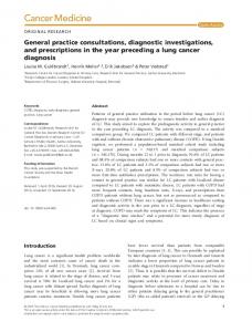
Figure 1. (a) Spleen from Atlantic salmon infected with rickettsia-Uke organisms (RLO), demonstrating darkpigmented positive staining cells infeeted with RLOs ( | ) eounterstained with haematoxylin (x400). (b) Liver from Atlantic salmon infected with (RLOs), demonstrating dark-pigmented positive-staining eells infeeted with RLOs { I ) counterstained with haematoxylin (x400).
Immtmohistochemistry
oj rtckettsial dtsease
IVl
1% hydrogen peroxide in 1-5mg 3,3-diaminobenzidine tetrahydrochloride and 10ml TBS for lOmin. The reaction was stopped by immersing the slides in tap water and the sections were counter stained for 3min with haematoxylin. Slides were left in tap water for 10min, dehydrated in 70% alcohol (3 min), 100% alcohol (15 min) and rinsed twice in xylene (5 min each). Coverslips were adhered using pertex and slides observed under a light microscope. Sections from Chile and Ireland presented specific reactions with distinct dark brown colouration of affected cells and organisms within cells in the kidney of the experimentally infected fish, and in the spleen, liver, heart, kidney and pancreas of naturally infected fish from Ireland (Fig. 1). This reaction was not observed in the A. salmonicida-, V. anguillarum-, and R. salmoninarum-infeded fish, in control fish, nor in those incubated without antisera. The results indicate that rickettsia can be detected immunohistochemically in formalinfixed tissue of Atlantic salmon. The possibility that there is a cross reaction between P. salmonis antisera and other rickettsial species cannot be ruled out, and is the subject of further investigation. Comparing the immunoreactivity with histopathologieal lesions, a specific reaction was demonstrated in degenerating cells of the liver, spleen, kidney, heart and pancreas. The use of immunohistochemistry for confirmation of rickettsia has some advantages over existing methods including the ease of use of formalin-fixed or wax-embedded samples, concomitant evaluation of histopathologieal lesions and no special equipment requirements. Acknowledgments We thank Professor John Fryer for antisera and background information. Dr Pedro Smith is thanked for providing the tissue specimens of experimentally infected fish. References Adams A., Richards R. H. & Marin de Mateo M. (1992) Development of monoclonal antibodies to PK"X\ the causative agent of proliferative kidney disease. Journal of Fish Diseases 15, 515-521. Brocklebank J. R., Speare D. J., Armstrong R. D. & Evelyn T. (1992) Septicaemia suspected to be caused by a rickettsia-like agent in farmed Atlantic salmon. Canadian Veterinary Journal 33, 407—408. Fryer J. L., Lannan C. N., Garces L. H., Larenas J. J. & Smith P. A. (1990) Isolation of a rickettsiales-like organism from diseased coho salmon (Oncorhynchus kisutch) in Chile. Fish Pathology 25, 107—114. Fryer J. L., Lannan C. N., Giovannoni S. J. & Wood N. D. (1992) Piscirickettsia salmonis gen. nov.. sp. nov., the causative agent of an epizootic disease in salmonid fishes. International Journal of Systematic Bacteriology 42, 120-126. Lannan C. N., Ewing S. A. & Fryer J. R. (1991) A fluorescent antibody test for detection of the rickettsia causing disease in Chilean salmonids. Journal of Aquatic Animal Health 3, 229-234. Lannan C. N. & Fryer J. R. (1991) Recommended methods for inspection of fish for the salmonid rickettsia. Bulletin of the European Association of Fish Pathologists 11, 135-136. Rodger H. D. & Drinan E. M. (1993) Observation of a rickettsia-like organism in farmed Atlantic salmon, Salmo salar L., in Ireland. Journal of Fish Diseases 16, 361—369.