ORIGINAL RESEARCH & CONTRIBUTIONS
Use of Portable Ultrasound Machine for Outpatient Orthopedic Diagnosis: An Implementation Study Sean Adelman, MD; Paul Fishman, PhD
Perm J 2013 Summer;17(3):18-22 http://dx.doi.org/10.7812/TPP/12-128
Abstract Introduction: Ultrasonography and magnetic resonance imaging (MRI) are used to evaluate shoulder disorders. This implementation study investigated outpatient ultrasonography at an orthopedic practice in a managed care setting. Methods: A portable ultrasound machine was introduced at an orthopedic clinic in a group practice. An orthopedic surgeon who primarily treated shoulder disorders received 15 hours of training. The impact of physician-performed ultrasonography on subsequent MRI and other outcomes of patients with shoulder disorders from January 2011 through October 2011 was determined using automated administrative and clinical data. Comparisons were made to patients who did not undergo ultrasonography at the experimental practice and 2 orthopedic clinics in the same practice. Results: During the study, 146 ultrasound examinations were administered. Compared with patients who did not undergo ultrasonography, patients who received ultrasonography had significantly higher comorbidity. However, they were significantly less likely to have MRI (9.7% with ultrasonography vs 14.4% without; p = 0.03) although equally likely to undergo surgery (33.6% with ultrasonography vs 22.1% without, p = 0.77). Mean time to surgery was 89.3 ± 49.3 days for patients with ultrasonography vs 32.9 ± 43.3 days for patients without (p < 0.05). No ultrasonography-examined patients had an incorrect diagnosis at surgery. For patients receiving ultrasonography, an estimated 35 MRIs were avoided, saving a predicted $17,603, a 50% return in less than 1 year on a $34,897 investment for an ultrasound machine and supplies. Conclusion: Outpatient ultrasonography by an orthopedic surgeon can be useful for diagnosing shoulder disorders and might reduce MRI utilization.
Introduction The US spends more per capita on health care than other developed nations, and overuse of high-end imaging is a factor in these higher costs. The Organization for Economic Cooperation and Development, which tracks health care use and outcomes among 34 industrialized nations, found that in the US, per capita rates of magnetic resonance imaging (MRI) use rose 9.4% during 2003 to 2007.1 In 2007, more than 80 million tests were performed at a cost of more than $120 billion. Data from the Commonwealth Fund suggest that at least 15% of high-end imaging tests are considered unnecessary,2 suggesting that a large number of patients undergo expensive and time-consuming tests that do not necessarily contribute to outcomes. As a diagnostic tool for specific presentations of pathology of the upper arm
and shoulder, ultrasonography can be as sensitive and specific as more expensive, invasive, and time-consuming technologies such as MRI. Ultrasonography is superior to MRI for some presentations such as rotator cuff injuries, because it is faster and less expensive, yet it results in similar clinical outcomes.3-10 Advantages of ultrasonography include the ability to visualize both shoulders with a dynamic, low-cost examination. Ultrasonography is the modality of choice after total joint replacement because it avoids artifacts that can severely inhibit visualization of the rotator cuff tendon. Ultrasonography is also helpful for claustrophobic patients and for patients in the immediate postoperative period whose healing tissue can be difficult to differentiate on MRI.3,8 In addition to being faster and less expensive at the point of service, ultra-
sonography might reduce downstream health care costs by eliminating the need for a dedicated radiology visit and a follow-up visit with the surgeon to discuss MRI results and plan a course of treatment. Ultrasonography can also increase patient satisfaction. In a study that examined perceptions and satisfaction with MRI and ultrasonography of 118 patients with shoulder pain who underwent both procedures, patients reported less pain with ultrasonography.8 Only 2 patients thought the ultrasonography took too much time compared with 28 who had that perception of MRI. All patients were willing to repeat the ultrasonography, but only 10 were willing to repeat MRI. Most patients with shoulder pain preferred ultrasonography to MRI, with 93 preferring ultrasonography, 8 preferring MRI, and 17 stating no preference.8 With proper training, surgeons and clinical staff can introduce ultrasonography into initial consultations with patients for diagnosis of shoulder pathology. Jeyam et al11 found that surgeons are capable of diagnosing rotator cuff tears using ultrasonography in outpatient settings. Iannotti and colleagues12 found that a well-trained office staff and an experienced orthopedic surgeon can effectively use ultrasonography in conjunction with clinical examination and a review of shoulder radiographs to accurately diagnose the extent of rotator cuff tears. Moosmayer and Smith13 found that surgeons quickly become adept at performing ultrasonography, despite previous assertions that long experience was required to ensure diagnostic accuracy. This descriptive study investigated the implementation of a portable ultrasound machine in the practice of a single, boardcertified orthopedic surgeon with a sports subspecialty certification (the “experimental
Sean Adelman, MD, is an Orthopedic Surgeon with Group Health Cooperative in Seattle, WA. E-mail:
[email protected]. Paul Fishman, PhD, is a Senior Investigator in Health Services and Economics with Group Health Research Institute. E-mail:
[email protected].
18
The Permanente Journal/ Summer 2013/ Volume 17 No. 3
ORIGINAL RESEARCH & CONTRIBUTIONS Use of Portable Ultrasound Machine for Outpatient Orthopedic Diagnosis: An Implementation Study
practice”). The goal was to determine the feasibility of outpatient ultrasonography for diagnosis of rotator cuff disorders. We compared patients who received ultrasonography at the experimental practice to patients who did not receive ultrasonography at the experimental practice and at two comparable orthopedic practices that did not have portable ultrasound machines. Patients were not randomly assigned to a treatment but received ultrasonography as appropriate during normal care. We analyzed automated data to evaluate patient characteristics, subsequent use of MRI, likelihood of surgical outcome, and speed with which surgical patients completed care. We postulated that ultrasonography would reduce costs measured as subsequent MRI, whose institutional costs include the time of a radiologist, a technologist, and the orthopedic surgeon for correlation of the findings. Although direct application of ultrasonography does not replace the need for MRI, we investigated whether it could be added to the practice of an orthopedic surgeon in an effort to reduce institutional and patient costs while maintaining a high quality of care. Materials and Methods Introduction of Ultrasonography and Training Ultrasonography as a diagnostic tool for shoulder and upper arm pathologies, primarily rotator cuff pathologies, was evaluated in a single experimental orthopedic office that is part of Group Health Cooperative (Group Health), which provides health care and preventive care on a prepaid basis to more than 630,000 individuals. In Fall 2010, an orthopedic surgeon was trained over 3 months to use a portable ultrasound machine. Training was 15 hours over 1 month to establish technique and correlate anatomy using 20 healthy volunteers, guided by 4 hours of consulting from a certified musculoskeletal ultrasonographer. This period was also used to assess examination room logistics. The following 2 months were used to define pathology using patients with preexisting imaging data. Approximately 60 patients with known pathology, documented by other imaging techniques, were scanned preoperatively to correlate technique and findings. The ultrasonographer returned for a 2-hour session before the study interval started, to ensure correct technique.
The Permanente Journal/ Summer 2013/ Volume 17 No. 3
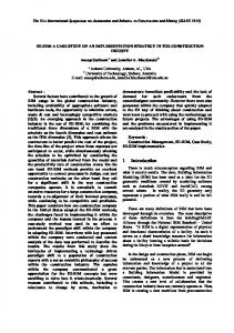
Patients and Data The enrollment interval for this study was January 1, 2011, to October 31, 2011. Included in comparisons were patients from the clinics of two other orthopedic surgeons with similar backgrounds as the surgeon and staff at the experimental practice and who worked under the same policies and workflow of the same health care system but without physicianperformed ultrasonography. Only patients with surgical consultations for diagnoses relevant to shoulder and arm pathology were included in final analyses. Patients treated in the practice that administered ultrasonography gave informed consent before each procedure. All patients were given the option of obtaining an MRI in lieu of ultrasonography when clinically appropriate. The study was reviewed by the Group Health institutional review board and was deemed a quality-improvement/ quality-assurance project and not research under the federal definition. For the experimental practice, two sets of encounters were identified: one for patients who presented with shoulder and arm pathology who received ultrasonography and one for patients presenting with similar diagnoses who did not receive ultrasonography. Patients receiving similar diagnoses were identified at the comparator practices. The pathways for this convenience sample of patients presenting with shoulder or arm pathology are depicted in Figure 1.10 Patient characteristics and diagnoses are shown in Table 1. Information on health services use among individuals included in our analysis was from Group Health automated clinical and administrative records. Through an electronic health record, Group Health information systems capture all services provided by physicians who practice in the integrated group practice. A unique procedure code was established for tracking physician-performed ultrasonography in the electronic health record. Outcome Metrics Evaluated outcomes were patient demographics, including body mass index (BMI); use of ultrasonography and MRI; primary care, orthopedic clinic, and physical therapy visits; surgery; and time to surgery. To determine subsequent services received, we examined MRIs performed and all likely
surgical events in the 180 days following the end of the study period through December 31, 2011. Surgical events were identified by examining patient records for either surgical codes or anesthesia codes that were occasionally present despite the absence of a specific surgical code. The resource utilization band measure of the Adjusted Clinical Group classification system was used to determine statistically significant baseline differences in patient mix. This method groups diagnoses received by individuals in the previous year into 1 of 6 hierarchical classes that indicate likelihood of greater future health service use, with 0 reflecting lowest and 5 reflecting the highest expected health care use based on an individual’s comorbidity profile.14-21 Diagnoses related to shoulder pathology were validated by individual chart review of all patients in the experimental practice during the study period who proceeded to surgery specifically for rotator cuff disorder, identified by surgical code. For statistical analyses, continuous variables were analyzed by t test, and discrete variables were tested by χ2 test. Results Patient Characteristics Table 1 provides a descriptive summary of patients who did and did not receive ultrasonography during their initial consultation with an orthopedic surgeon at the experimental practice or the 2 comparator practices. Compared with patients not receiving ultrasonography, patients receiving ultrasonography were as likely
Figure 1. Pathways for patients presenting with shoulder or arm pathology. MRI = magnetic resonance imaging.
19
ORIGINAL RESEARCH & CONTRIBUTIONS Use of Portable Ultrasound Machine for Outpatient Orthopedic Diagnosis: An Implementation Study
to be women but were older and had a higher comorbidity index as measured by the resource utilization band scale. Patients receiving ultrasonography were significantly more likely to have a diagnosis of shoulder arthropathy (International Classification of Diseases, Ninth Revision [ICD-9] code 719) or rotator cuff injury (code 726). Rotator cuff injury is a presentation for which ultrasonography is a superior diagnostic tool,4-7 and the surgeon at the experimental practice specializes in shoulder and arm pathology. Patient Outcomes Overall, patients who received ultrasonography were 55% less likely to have an MRI and 37% more likely to proceed to surgery without further imaging than patients who did not receive ultrasonography at either the comparator or experimental practices. Patients who were seen at the experimental practice but did not receive ultrasonography were 58% less likely to
have surgery than patients at the clinic who received ultrasonography. Compared with patients who did not undergo ultrasonography, patients who received ultrasonography were significantly less likely to have shoulder MRI regardless of whether they eventually had surgery. Patients who received ultrasonography were 34% more likely to have surgery than patients who did not receive ultrasonography. These findings suggested that ultrasonography was used for the proper indication and in a way that decreased the need for subsequent diagnostic work. To validate diagnoses of people who received ultrasonography and then proceeded to surgery, the experimental surgeon pulled cases by surgical code. This method identified 41 patients in the experimental practice who proceeded to rotator cuff surgery on the basis of ultrasonography diagnosis alone. These patients were classified into 4 categories: 1) correct diagnosis; 2) cor-
rect diagnosis but missed glenohumeral pathology (which ultrasonography is poor at recognizing); 3) incorrect diagnosis; or 4) correct diagnosis but inaccurate assessment (eg, partial rotator cuff tear that was actually a full tear, or a small tear that was larger than predicted with ultrasonography). Of the 41 patients who proceeded to rotator cuff surgery based on ultrasonography diagnosis alone, 31 patients (76%) had a correct diagnosis, including degree of pathology; 3 (7%) had the correct diagnosis with missed intra-articular pathology (all but 1 case was glenohumeral chondral pathology); and 7 (17%) correctly progressed to surgery, although with underestimated pathology. All patients in the latter category received a diagnosis of partial-thickness rotator cuff pathology and at the time of surgery had either a higher degree of partial thickness change or a small, fullthickness component. No patients during the study period were found to require
Table 1. Characteristics and outcomes of ultrasonography-examined and no-ultrasonography patients Characteristic or outcome Total participants Women, % Age in years, mean (SD) BMI, mean (SD) Comorbidity index, % resource utilization band Low Mid-range High Diagnoses at index visit, ICD-9 code, % 715 Osteoarthritis 718 Dislocation of joint of shoulder region 719 Shoulder pain 726 Injury to rotator cuff 727 Calcium deposits in tendon and bursa 812 Fracture of greater tuberosity of humerus 831 Closed dislocation of shoulder 840 Sprains and strains of shoulder and upper arm 959 Other and unspecified injury to shoulder and upper arm Mean (SD) Orthopedic Department clinical visits in study interval Mean (SD) primary care visits in study interval Mean (SD) Physical Therapy Department clinical visits in study interval Orthopedic physician-performed shoulder ultrasonography Shoulder MRI, % Shoulder surgery, % Mean (SD) days to MRI after initial Orthopedic Department evaluation Mean (SD) days to surgery after initial Orthopedic Department evaluation
Ultrasonography 143 51.4 62.4 (12.1) 27.1 (6.5)
No ultrasonography 830 45.1 55.3 (16.6) 27.6 (6.5)
13.1 55.5 34.5
13.5 67.8 18.7
8.8b 2.0 8.1 46.6 27.0 0 2.1 1.4 3.4 3.2 ( 2.3) 4.6 (3.3) 5.2 (4.1) 146b 9.7 33.6 17.5 (39.4) 89.3 (49.3)
9.5 6.5 35.5 23.1 5.8 1.4 1.1 5.3 11.2 2.7 (2.1) 3.8 (3.2) 4.6 (3.6) 0 14.4 22.1 80.0 (45.7) 32.9 (43.3)
p valuea NS NS NS
< 0.05
< 0.05
NS NS NS NA 0.028 NS < 0.05 < 0.05
p < 0.05 criterion for statistical significance. Discrepancies in numbers are because full utilization records were not always available for all patients. BMI = body mass index; ICD-9 = International Classification of Diseases, Ninth Revision; MRI = magnetic resonance imaging; NA = not available; NS = not significant; SD = standard deviation. a b
20
The Permanente Journal/ Summer 2013/ Volume 17 No. 3
ORIGINAL RESEARCH & CONTRIBUTIONS Use of Portable Ultrasound Machine for Outpatient Orthopedic Diagnosis: An Implementation Study
surgery following the initial assessment for which no surgery was indicated. Of the 33 patients who underwent surgery on the basis of MRI diagnosis, 22 patients (67%) had the correct diagnosis, 3 patients (9%) had the correct diagnosis but missed glenohumeral pathology, 1 patient (3%) had an incorrect diagnosis, and 7 patients (21%) had the correct diagnosis with underestimated tear size/pathology. Table 1 reports the mean time from the initial consultation to a subsequent surgical procedure for patients who eventually had surgery. These data suggested that patients who had ultrasonography during their initial consultation did not progress to surgery faster than patients in the comparator practices. Of note, except for a single patient who underwent surgery at 16 days, the subgroup of patients who received ultrasonography who were classified as correctly progressing to surgery with underestimated pathology underwent surgery at an average of 112 days, more than twice the average of the other groups. The counterintuitive result of longer times to surgical procedures for patients who had ultrasonography led to an examination of whether this trend was constant over the course of the 10-month evaluation. We estimated time to surgery following the initial consultation for the comparator practices and the experimental practice. The mean days to surgery following initial consultation fell from nearly 100 in January to under 40 days in October for the experimental practice, whereas the wait times in the comparator practices fell from approximately 75 to 40 days. These results are probably based on 2 factors: surgeons typically start the year with a backlog of cases, and the surgeon at the experimental practice had recently joined the group practice and thus started the evaluation period with a larger patient set than at the other practices. Thus, the overall mean times to surgery among the patients who experienced ultrasonography probably do not reflect a steady state. Discussion Our implementation study demonstrated the clinical and economic value of use of a portable ultrasound machine in an orthopedic practice. Analysis of automated data and surgeon review of patient records confirmed that the use of ultrasonography
The Permanente Journal/ Summer 2013/ Volume 17 No. 3
provided valid, sensitive, and specific diagnosis of shoulder pathology when used for patients who presented with symptoms appropriate to rotator cuff disorders. The use of ultrasonography in lieu of MRI for specific shoulder and arm pathology is expected to reduce costs subsequent to the initial consultation. The ultrasound machine, operating supplies, and maintenance contracts cost $34,897. Using a national, weighted, Centers for Medicare and Medicaid Services resource-based relative value scale, fully loaded professional and facility-based reimbursement for an MRI was between $1350 and $1500.22 By comparing the experiences of patients in the experimental and comparator practices, we estimated that 35 MRIs were avoided by using ultrasonography. Therefore, the 35 avoided MRIs saved between $12,353 and $17,603, for a 35% to 50% return on investment in less than 1 year. Although this does not include training costs, it also does not include the costs of follow-up visits to interpret the MRI and plan a course of treatment, or out-of-pocket costs to patients for additional visits. Although Group Health is reimbursed on a capitated basis, it is appropriate to consider the cost of ultrasonography relative to MRI for clinicians and systems that reimburse on a fee-for-service basis. The national average reimbursement for an ultrasonography of the upper torso, including the full professional and facility charge, is $114. The net savings, therefore, if each MRI that was avoided were replaced by a reimbursed ultrasonography, would be reduced to a range of $8362 to $13,613. This still reflects a strong return on investment to the purchase of the portable ultrasound machine. None of the 41 patients in the experimental practice who received surgery on the basis of ultrasonography diagnosis alone required a follow-up visit to discuss the imaging results. This equates to fewer patient copays and less time off from work. It also equates to 41 more clinic appointments available for the surgeon to evaluate other patients. Even a follow-up phone visit for MRI review requires time and effort from both the patient and surgeon that are not required if diagnosis is by ultrasonography. Our report is limited by differences in patient sets. The trend toward patients
with increasing age and comorbidities in the ultrasonography group can be explained by patient selection and referral patterns. The practice that implemented ultrasonography tended to see patients with more severe shoulder disorders or previously failed treatments; this explains why the ultrasonography-examined patients were generally older, with greater comorbidity, … a follow-up and had more orthopedic, visit … even a primary care, and physical phone visit … therapy visits than patients [is] not required without ultrasonography. if diagnosis is by For diagnosis of shoulder ultrasonography. disorders, ultrasonography is most useful for evaluation of rotator cuff disorders. Patients with rotator cuff disorders tend to be older and have more medical issues. Patients with glenoid labral pathology and other shoulder disorders excluding arthritis tend to be younger and healthier as a population. In addition, as the ultrasonography evaluation period progressed in this study, referral patterns increased, specifically for ultrasonography. Many of these referrals were for primary ultrasonography indications, including failed rotator cuff surgery or arthroplasty, and were frequently patients with pacemakers. Patients with pacemakers cannot have MRI and tend to be older with more clinical comorbidities than the pooled MRI population. Patients receiving ultrasonography also had a greater length of time between the initial orthopedic consultation and eventual surgery; however, this is possibly because of a greater patient queue in the experimental practice, or administrative issues such as vacations and backlogs that were not related to the use of ultrasonography. In any case, many of the time- and cost-saving advantages of ultrasonography over MRI would apply to all practices. Our cost-reduction calculations do not include other musculoskeletal conditions for which ultrasonography can be used. Within 15 months of the start of the study, more than 300 ultrasound examinations were performed for rotator cuff conditions in this single practice. Ultrasonography could be done in less than 15 minutes and did not change visit durations or patient interval times; the experimental surgeon saw more patients overall during the study period than either of the comparator clinic sur-
21
ORIGINAL RESEARCH & CONTRIBUTIONS Use of Portable Ultrasound Machine for Outpatient Orthopedic Diagnosis: An Implementation Study
geons. Extrapolating these figures across a small group practice, the ultrasound machine can easily pay for itself within a year in cost savings to the institution that are directly related to decreased MRI use. Despite the advantages of ultrasonography as a diagnostic tool in orthopedic practice, several barriers must be overcome before it can be widely implemented. First, orthopedic surgeons are not routinely trained in ultrasonography administration, although this study found this barrier to be surmountable. Second, most orthopedic offices do not have a readily available ultrasound machine, in particular a mobile device that is easily moved between examination rooms. Mobile ultrasound devices currently cost $30,000 to $40,000. Finally, surgeons must overcome the worry that using ultrasonography will take too much time, effectively limiting the ability to efficiently see patients in the clinic. Conclusion In this study, we showed that an orthopedic surgeon can be trained to perform musculoskeletal ultrasonography at substantial institutional and patient cost savings without compromising patient care. Our results were observed in a single practice at a single specialty center, with patients presenting with conditions ideally suited for ultrasonography as a diagnostic tool. Ultrasonography was administered by a physician with specific expertise in diagnosing shoulder pathology, but no substantial training in ultrasonography. In the interest of specificity, we did not include the use of ultrasonography for other musculoskeletal conditions or for needle localization procedures. Given the other proven uses of musculoskeletal ultrasonography in an orthopedic practice, a case can be made for even more dramatic cost savings. The implication of our findings is that additional training in musculoskeletal ultrasonography should be considered for orthopedic residents and fellows. Widespread review of radiographs, MRIs, and computed tomography images are currently part of training. Adding didactics in ultrasonography is feasible and desirable based on the results presented here and elsewhere.2-6 Ultrasonography is not the first-line imaging technique for all shoulder and arm pathologies; however, when used
22
correctly, it provides fast, inexpensive, and accurate diagnosis of rotator cuff injuries. On the basis of our experience, we are currently developing a didactic series to help train practicing surgeons in the use of ultrasonography. v
10.
11.
Disclosure Statement The author(s) have no conflicts of interest to disclose. Acknowledgments This work was made possible in part by a grant from the Group Health Foundation. We thank Linda Wehnes, MA, for project management; Kay Theis, MA, MS, for programming; Benjamin Betteridge, MD, for expert consultation; Chris Tachibana, PhD, for editorial assistance; and Jamie Antoine, MD and Stephen Walker, MD, for critical reading of the manuscript. Kathleen Louden, ELS, of Louden Health Communications provided editorial assistance. References
1. OECD health data 2012—frequently requested data [Web page on the Internet]. Paris, France: The Organisation for Economic Co-operation and Development; 2012 [cited 2013 Apr 29]. Available from: www.oecd.org/els/healthpoliciesanddata/oecdhealthdata2012-frequentlyrequesteddata.htm. 2. Squires DA. Explaining high health care spending in the United States: an international comparison of supply, utilization, prices, and quality. Issue Brief (Commonw Fund) 2012 May;10:1-14. 3. Teefey SA, Rubin DA, Middleton WD, Hildebolt CF, Leibold RA, Yamaguchi K. Detection and quantification of rotator cuff tears. Comparison of ultrasonographic, magnetic resonance imaging, and arthroscopic findings in seventy-one consecutive cases. J Bone Joint Surg Am 2004 Apr;86-A(4):708-16. 4. Frei R, Chládek P, Trc T, Kopecný Z, Kautzner J. Arthroscopic evaluation of ultrasonography and magnetic resonance imaging for diagnosis of rotator cuff tear. Ortop Traumatol Rehabil 2008 Mar-Apr;10(2):111-4. 5. Chang CY, Wang SF, Chiou HJ, Ma HL, Sun YC, Wu HD. Comparison of shoulder ultrasound and MR imaging in diagnosing full-thickness rotator cuff tears. Clin Imaging 2002 JanFeb;26(1):50-4. DOI: http://dx.doi.org/10.1016/ S0899-7071(01)00323-0 6. Dubs B. [Ultrasound study of the shoulder]. [Article in German]. Praxis (Bern 1994) 2001 Apr 19;90(16):667-71. 7. Bachmann GF, Melzer C, Heinrichs CM, Möhring B, Rominger MB. Diagnosis of rotator cuff lesions: comparison of US and MRI on 38 joint specimens. Eur Radiol 1997;7(2):192-7. DOI: http://dx.doi.org/10.1007/s003300050133 8. Middleton WD, Payne WT, Teefey SA, Hildebolt CF, Rubin DA, Yamaguchi K. Sonography and MRI of the shoulder: comparison of patient satisfaction. AJR Am J Roentgenol 2004 Nov;183(5):1449-52. 9. Parker L, Nazarian LN, Carrino JA, et al. Musculoskeletal imaging: medicare use, costs,
12.
13.
14.
15. 16.
17.
18.
19.
20.
21.
22.
and potential for cost substitution. J Am Coll Radiol 2008 Mar;5(3):182-8. DOI: http://dx.doi. org/10.1016/j.jacr.2007.07.016 Seagger R, Bunker T, Hamer, P. Surgeon-operated ultrasonography in a one-stop shoulder clinic. Ann R Coll Surg Engl 2011 Oct;93(7):52831. DOI: http://dx.doi.org/10.1308/14787081 1X13137608454939 Jeyam M, Funk L, Harris J. Are shoulder surgeons any good at diagnosing rotator cuff tears using ultrasound?: A comparative analysis of surgeon vs radiologist. Int J Shoulder Surg 2008 Jan;2(1):4-6. DOI: http://dx.doi. org/10.4103/0973-6042.39580 Iannotti JP, Ciccone J, Buss DD, et al. Accuracy of office-based ultrasonography of the shoulder for the diagnosis of rotator cuff tears. J Bone Joint Surg Am 2005 Jun;87(6):1305-11. DOI: http://dx.doi.org/10.2106/JBJS.D.02100 Moosmayer S, Smith HJ. Diagnostic ultrasound of the shoulder—a method for experts only? Results from an orthopedic surgeon with relative inexperience compared to operative findings. Acta Orthop 2005 Aug;76(4):503-8. DOI: http://dx.doi.org/10.1080/17453670510041484 Starfield B, Weiner J, Mumford L, Steinwachs D. Ambulatory care groups: a categorization of diagnoses for research and management. Health Serv Res 1991 Apr;26(1):53-74. Verhulst L, Reid RJ, Forrest CB. Hold it—my patients are sicker! BC Medical Journal 2001 Jul-Aug;43(6):328-33. Weiner JP, Starfield BH, Steinwachs DM, Mumford LM. Development and application of a population-oriented measure of ambulatory care case-mix. Med Care 1991 May;29(5):45272. DOI: http://dx.doi.org/10.1097/00005650199105000-00006 Reid R, Evans R, Barer M, et al. Conspicuous consumption: characterizing high users of physician services in one Canadian province. J Health Serv Res Policy 2003 Oct;8(4):215-24. DOI: http://dx.doi. org/10.1258/135581903322403281 Reid RJ, MacWilliam L, Verhulst L, Roos N, Atkinson M. Performance of the ACG case-mix system in two Canadian provinces. Med Care 2001 Jan;39(1):86-99. DOI: http://dx.doi. org/10.1097/00005650-200101000-00010 Reid RJ, Roos NP, MacWilliam L, Frohlich N, Black C. Assessing population health care need using a claims-based ACG morbidity measure: a validation analysis in the Province of Manitoba. Health Serv Res 2002 Oct;37(5):1345-64. DOI: http://dx.doi.org/10.1111/1475-6773.01029 Rivara FP, Anderson ML, Fishman P, et al. Healthcare utilization and costs for women with a history of intimate partner violence. Am J Prev Med 2007 Feb;32(2):89-96. DOI: http://dx.doi. org/10.1016/j.amepre.2006.10.001 Bonomi AE, Anderson ML, Rivara FP, Thompson RS. Health care utilization and costs associated with physical and nonphysical-only intimate partner violence. Health Serv Res 2009 Jun;44(3):1052-67. DOI: http://dx.doi. org/10.1111/j.1475-6773.2009.00955.x Medicare Physician Fee Schedule [database on the Internet]. Baltimore, MD: Centers for Medicare & Medicaid Services; updated 2013 Mar 18 [cited 2013 Apr 29]. Available from: www.cms. gov/apps/physician-fee-schedule/overview.aspx [click on “Click here to begin your Physician Fee Schedule look-up”].
The Permanente Journal/ Summer 2013/ Volume 17 No. 3