(From the Imperial Cancer Research Fund and Department of Pathology, .... The air space below the guard cells has not collapsed and the chloroplasts are ...
275
An Improved and Rapid Embedding Method By J. CHAYEN and P. B. GAHAN (From the Imperial Cancer Research Fund and Department of Pathology, Royal College of Surgeons, Lincoln's Inn Fields, London, W.C. 2) With one plate (fig. 1) SUMMARY Plant tissues may be embedded in half the time taken for paraffin embedding if acetone is used as the dehydrating and infiltrating agent, and the tissue is embedded in ester wax. This procedure retains appreciably more lipid material inside the cells than does the more usual technique, even when the tissue has been fixed in osmium tetroxide or in La Cour's 2BE. INTRODUCTION
W THEN fixed plant tissues are to be sectioned, they are usually dehydrated V V by passing through graded concentrations of alcohol and are then immersed in chloroform to allow infiltration of the cells by paraffin wax. When performed most meticulously, this process may take as long as 9 days; the least time we have been able to use is about 3 days. Moreover, this procedure, especially the treatment in mixtures of chloroform and alcohol, is likely to remove lipid material from the tissues and so cause distortion. Hence to decrease the time spent in embedding and in an attempt to reduce the amount of phospholipid extracted during this process, the use of acetone as a dehydrating agent and as an infiltrating solution has been investigated. Ester wax, being more readily soluble in acetone than is paraffin wax, has yielded satisfactory results and so has been used in this study. METHOD
Root tips and pieces of leaves of Viciafaba were investigated. After fixation in an aqueous fixative such as osmium tetroxide solution, and suitable washing, the tissue was treated as follows: 50% acetone in water 1 h 70% acetone in water 2 h 90% acetone in water 2 h absolute acetone 1 h It was transferred to fresh absolute acetone. Small pieces of ester wax were added, and it was left overnight in a container which was corked to prevent evaporation of the acetone. The tissue, in this solution in the corked container, was placed in an embedding oven (about 500 C will suffice) and more solid wax was added, the container being shaken so that the molten wax dissolved in the acetone. It was left for \ h. [Quarterly Journal of Microscopical Science, Vol. 100, part 2, pp. 275-277, June 1959.]
276
Chayen and Gahan—An Improved and
The cork was then removed to allow the acetone to evaporate. This was completed in about i\ h. The tissue was transferred to a bath of fresh molten ester wax for 10 min and then embedded. Sections were cut at 7/x, but there seems to be no greater limitation on the thickness that can be cut after this procedure than after chloroform has been used. The wax may be removed from the sections by immersion in acetone and, when required, they may then be mounted directly in euparal. RESULTS
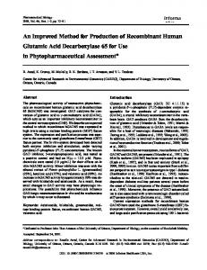
Sections of roots fixed in acetic-alcohol (1:3 by volume) and embedded by the acetone / ester wax method were identical with those which had been prepared by the usual chloroform / paraffin wax procedures. The embedding of roots fixed in La Cour's 2BE solution (Darlington and La Cour, 1947) was better by the former than by the latter technique. Such sections were stained with either the acid haematein (Baker, 1946) or the orange G / aniline blue (La Cour and Chayen, 1958) methods for lipids; those embedded by the acetone / ester wax procedure contained appreciably more stainable matter, especially on the chromosomes (fig. 1, c). Indeed, these sections showed as great retention of lipids, and the same localization of these substances, as did sections fixed in Lewitsky's (1931) fluid (equal volumes of 1% chromic acid and 10% formalin) and embedded by the chloroform / paraffin wax process (La Cour and Chayen, 1958). More striking were the results obtained with roots fixed in a 2% aqueous solution of osmium tetroxide (not buffered) for 1 h. The preservation of root-cells fixed in this manner and embedded by the chloroform / paraffin wax method was extremely poor (also see Chayen and Jackson, 1957) and no granules of any kind were found (fig. 1, A). When embedded by the acetone / ester wax procedure, however, an array of osmiophil granules of various shapes and sizes was found, particularly in the elongating cells behind the meristem (fig. 1, B). Cells of the different regions of the root were distinguishable by their cytoplasmic organization and the vacuoles were FIG. I (plate), A, part of an unstained longitudinal section through a root of the broad bean, Viciafaba, which was fixed in a 2% solution of osmium tetroxide and embedded in paraffin wax by the conventional procedure. The cortex and central cylinder are shown; in the cells of neither are cytoplasmic organelles visible. B, part of an unstained longitudinal section through a root of V.faba fixed in 2% solution of osmium tetroxide and embedded in ester wax after dehydration in acetone. Cells of the cortex and central cylinder, from just behind the true meristematic region, are shown; in the latter many osmiophil granules are visible. C, part of a longitudinal section through a root of V.faba which had been fixed in La Cour's 2BE fixative and embedded in ester wax after dehydration in acetone. The section was stained by a modified acid haematein test (La Cour and Chayen, 1958). The embedding is as good as by the conventional procedure but the retention of staining matter, particularly in nucleoli and mitotic chromosomes, is better by this method. D, transverse section through a leaf of V. faba, which had been fixed in Lewitsky's fluid, dehydrated in acetone, and embedded in ester wax. Photographed by phase contrast microscopy. The air space below the guard cells has not collapsed and the chloroplasts are large and distinct.
*W-' "'ITSSC
FIG. I
J. CHAYEN and P. B. GAHAN
Rapid Embedding Method
277
well delimited. The results obtained by this method of fixation, dehydration, and embedding were comparable with those described by Guillermond (1941). The retention of the osmiophil granules or droplets by the acetone / ester wax method but not by the alcohol / chloroform / paraffin wax technique might indicate that the former procedure does retain more lipid material. The results obtained by both embedding methods with pieces of leaves of Viciafaba, fixed under reduced pressure in 2% osmium tetroxide for 1 h or in Lewitsky's fluid for 6 h, were rather similar (see fig. 1, D). The chloroplasts after treatment with alcohol and chloroform seemed to be more shrunken and pressed against the cell-walls than they were by the acetone method. Moreover, the birefringence of the cell-walls of the outer layers of parenchymatous cells was preserved only by fixation in Lewitsky's fluid and embedding by the acetone / ester wax procedure. DISCUSSION
The results obtained by staining for lipid after fixation in 2BE and the occurrence of considerable osmiophil organization after osmium fixation and embedding by the acetone / ester wax method, suggest that this embedding procedure removes less lipid than does the usual chloroform / paraffin wax technique. Furthermore, although these tissues were embedded more rapidly by this process than by that in which chloroform is utilized (i\ against 3 days), the morphological preservation was rather better. The granules preserved by fixation in osmium tetroxide and embedding by the acetone / ester wax procedure deserve further study. All that need be noted at present is that they would seem at least to represent osmiophil matter which is preserved by this embedding technique but not by that involving the use of alcohol and chloroform. It is possible that this embedding method may also be of use in the histochemical study of enzymes because of the use of acetone and of the lower temperature of embedding which is theoretically possible by this procedure, especially if purer ester waxes are used (see Chesterman and Leach, 1956). We are grateful to Professor J. T. Randall, F.R.S., in whose Laboratory this method was first designed, and to Dr. H. B. Fell, F.R.S., for her kind advice. We are indebted to Mr. A. L. E. Barron for the photomicrography. REFERENCES BAKER, J. R., 1946. Quart. J. micr. Sci., 87, 441. CHAYEN, J., and JACKSON, S. F., 1957. S.E.B. Symposium, 10, 134. CHESTERMAN, W., and LEACH, E. H., 1956. Quart. J. micr. Sci., 97, 593. DARLINGTON, C. D., and LA COUR, L. F., 1947. The handling of chromosomes, znd edit. London (Allen & Unwin). GUILLERMOND, A., 1941. The cytoplasm of the plant cell, translated by L.R.Atkinson. Waltham, Mass. (Chronica Botanica). LA COUR, L. F., and CHAYEN, J., 1958. Exp. Cell Res., 14, 462.
LEWITSKY, G. A., 1931. Bull. appl. Botany Genet. Plant Breeding, 27, 175.