May 19, 1971 - W. WOODBURY'. A. K. SPENCER. M. A. STAHMANN. Depwtment of Biochemtit,ry. Pollege of Agricultural and Life Sciences. Urliversity.
SHORT
301
COMMUNICATIONS
3. M. ROVERY, C. FABRE, AND P. DESNUELLE, Biochh. 1. G. BISERTE, J. IV. HOLLEMAN, J. HOLLEMAN-DEHOVE, Rev. 2, 59 (1960). 5. M. R. HEINRICH AND E. BUGNA, Anal. Biochem.
Biophys. Acta 12, 547 (1953). ANI P. SAUTIERE, ChrOmntOgr. 28,
1 (1969). HENRY LEONARD
Department of the Army Biological Sciences Laboratories Fort Detrick, Frederick, Maryland IZeceived May 19, 1971
An
M.
JACOBY
SFERO
21701
Improved. Procedure Using Ferricyanide for Detecting Catalase lsozymes
Catalase is a widely occurring enzyme which converts hydrogen peroxide to oxygen and water very rapidly (8). Recently interest has developed in genetic variants or isozymes of this enzyme in plants (9) and animals (5). Isozymes of catalase are usually detected by starch or acrylamide gel electrophoresis. After electrophoretic separation in starch gel, catalase bands usually are detected by incubating the gel briefly in a solution of hydrogen peroxide followed by a solution of acidified potassium iodide. Oxidation of the iodide by hydrogen peroxide produces iodine, which forms a dark blue complex with the starch. Where catalase activity has destroyed the peroxide, a clear zone is observed in the blue gel. With a&amide gel systems, 0.3% soluble starch is incorporated into the gel mixture before polymerization and t,he same staining procedure as for starch gel can be used (2,9). This starch-iodide method has several limitations: First, the starchiodine reaction is a complex equilibrium. The catalase bands are not permanent; they darken continuously until indistinguishable from background. The pattern requires five to ten minutes to become visible and is then discernible only about ten minutes. This unstable nature of the bands requires that the gels be inspected or photographed within ,minutes of staining and, since the pattern is constantly darkening, a comparison between gels is difficult. Such rigid timing requirements are especially inconvenient when many gels are processed simultaneously. Second, some migration of starch occurs during electrophoresis. This problem is especially severe in isoelectric focusing with acrylamide gels (4,12) ;
302
SHORT
COMMUNICATIONS
we have observed irregularities in starch density in the region of the pH gradient occupied by catalase isozymes. Third, at least two enzymes interfere with the starch-iodide staining method. Peroxidase catalyzes the oxidation of iodide to iodine by hydrogen peroxide (3). Iodine produced very dark areas of the starch-iodine complex, which obscured some light catalase b,ands when a mixture of catalase and peroxidase was applied to the starch gels. Amylase in the sample may hydrolyze the starch and produce clear zones or smearing. The new method based on the reaction of hydrogen peroxide with ferricyanide(II1) is devoid of these limitations. Hydrogen peroxide reduces potassium ferricyanide (III) to potassium ferrocyanide (II) (10) ; the peroxide is oxidized to molecular oxygen. Ferric chloride reacts with ferrocyanide (II) to form stable, insoluble Prussian Blue pigment (6). These reactions are the bases for the following procedure, which has been found to be very sensitive for the det,ection of catalase activity in acrylamide gels. After acrylamide gel electrophoresis (1)) the gels are washed in three changes of distilled water for a total of about 45 ,min to remove the buffer from the gel’s outside surface where staining occurs. Washing is done in stoppered large (60 ml) test tubes on an inclined turntable rotating at 4 rpm. The gels are then transferred to 12 X 125 mm test tubes containing 0.003% hydrogen peroxide (Merck 30% hydrogen peroxide diluted lo4 with distilled water) ; the tubes are closed with rubber stoppers and placed on the rotating turnt.able for 10 min. The gels are next briefly rinsed with distilled water and placed in test tubes containing a 10% solution of ferric chloride and potassium ferricyanide(II1) (freshly prepared as described below) ; these tubes are again stoppered and placed on the turntable for 10 min. The staining solution is then poured off; the gels are briefly rinsed and stored in distilled water. They should remain in the dark except when being studied. The catalase bands are yellow on a dark green held and are stable for at least several hours. We attempted to buffer the peroxide solution to rigidly control the conditions for the enzymic hydrogen peroxide destruction. Phosphate, bis-tris, and cacodylate buffers interfered with staining so that no bands appeared, and acetate and maleate buffers shortened band lifetime. When the hydrogen peroxide solution was buffered at either pH 5 or pH 7 with 0.1 M sodium maleate, bands identical to those with unbuffered hydrogen peroxide in distilled water were observed. Thus buffering with maleate or acetate is allowed, but the unbuffered hydrogen peroxide solution is sufficient and seems desirable. Catalase has a very broad pH optimum (3). The effects of different hydrogen peroxide concentrations were also
SHORT
COMMGNICATIONS
303
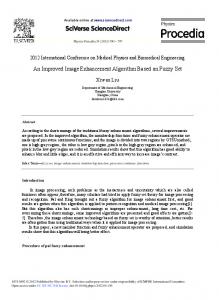
studied. Decreasing peroxide concentration to 0.001% resulted in ins&Iicient background intensity ; higher concentration of peroxide, 0.03% applied for 1 min, allowed better definition and a very dark blue black background, but reduced the sensitivity. An attempt was made to increase the sensitivity of the method by incubating the gel, after hydrogen peroxide treatment, for an additional 10 min in distilled water before application of ferric chloride/potassium ferricyanide (III) solut.ion. This alteration produced slightly increased sensitivity, but much inferior definition and reduced background staining (Fig. 1). The 1% ferric chloride/l% potassium ferricyanide(II1) solution should be mixed immediately before use from equal volumes of 2% stock solutions of each. The ferric chloride solution may need to be
FIG.
mercial contain
1. Catalase
staining with ferricyanide(II1). Gel 1 contains 1 unit of comcatalase (Sigma crystalline-suspension, 30,000 units/mg). Gels 2 and 3 0.3 unit of catalase. Gel 4 was prepared with 30 pl crude bacterial extract,. 2. and 4 were stained as described in above procrdure. Gel 3 was subjected min incubation in distilled water aft.er hydrogen prrosidct treatment, heforc c:llloricie/potsssium ferricyunide(II1) treatment. The large-pore gels were rcprior to photography. The bright spot, in the center of the cxtalase band is due to thr fact, that the: Prussian k)lw pigmr>nt is formed only in 11~ of the gel so that, when photographed by kansmit tcxd ligllt ( 1l1rrt3 by the center of the gel than by the edges.
304
SHORT
COMMUNICATIONS
filtered after preparation to remove suspended iron oxides. Both stock solutions should be stored in brown bottles in the dark, where they are stable for two to three months. The staining solution is a powerful oxidant and excessive contact with skin should be avoided. Dark blue stains on metal or glass can be removed by wiping with acid. The band intensity with the ferricyanide(II1) catalase stain is not strictly proportional to cat,alase activity of the band; this is a drawback it shares with the starch-iodide catalaee stain and other negative stains. This is because a band means that there was enough catalase to decompose the hydrogen peroxide at this particular position; it does not indicate how much additional enzyme was present in excess of the amount needed, although large excesses of activity produce wider but not lighter bands. Preparations of greater than 100 catalase units per milliliter activity should be diluted before application fpr this reason. A catalase unit will decompose one micromole of hydrogen peroxide per minute using 0.059M hydrogen peroxide and 0.05 M phosphate buffer, pH 7.0, at 25°C. Since 0.1 catalase unit (3 X 1W gm) per band can be detected, one can conveniently observe in the same gel, isozymes of different Rf's whose activities differ by a factor of 100. Ferricyanide(II1) is a strong oxidizing agent known to react with phenols, indole derivatives, tertiary amines, furans, hydroxylamines, and glycols (11). Although the above reactions are predominantly observed at high pH, such materials in the gel could possibly cause dark areas when the ferricyanide(II1) stain is employed. However, such small molecular weight substances do not appear to present a problem, as they migrate with the dye band or are removed by the washing. Reactive groups on proteins or on protein-linked carbohydrates in very high concentration might cause Iocalized darkening. We have successfuhy applied the ferricyanide stain to acetone powders and fresh crude extracts of fungus, bacteria, and various plant tissues. We have compared this new procedure involving ferricyanide with the starch-iodine method for demonstrating catalase isozymes. The two methods showed the same patterns of isozymes. Ferricyanide has the advantage of providing a stable pattern with very high contrast between enzyme bands and background. The method is free from interference of peroxidase. Since starch need not, be incorporated into the gel, difficulties due to starch migration and amylase activity are avoided. ACKNOWLEDGMENTS
This work was supported by grants AI Infectious GM00236
Diseases, the Herman BCH from National
Frasch Institute
101 from the National Institutes Foundation, and NIH Training Grant of General Medical Sciences.
of No.
SHORT
305
COMMUNICATIONS
REFERENCES 1.
DAVIS,
2. HONOLD.
3.
IIONG,
B. J., Axn. N. I-. Acad. Sci. 121, 321 (1964). G. R., AND STAHMANN, M. A., Cereal Chem. 45, 99 (1968). C., “Biochemist’s Handbook.” p. 383. Van Nostrand, New York,
1961.
Hoppe-seyler’s
917
4. M.~cKo,
V.,
AND
STEGMANN,
H..
Z.
Physiol.
Chem.
350,
(1969,). 5. MARKERT,
C.
L.,
AND
WHITT,
G.
S.,
Ezpelde?Llin
24,
977
(1968).
J., Z. Anorg. Chem. 9, 126 (1895). L., Ann. N. Y. Acad. Sci. 121, 274 (1968). SAI~NDERS, B. C., HOLMES-SEIDLE, A. C., AND STARK, B. P., “Peroxidase.” worth, London, 1964.
6. MESSTER. 7. ORNSTRIN. 8.
9. S~ANUALIOS. J. C., Ann. N. Y. Acad. Sci. 151, 274 (1968). 10. SRIKANTTAN. B. S., AND RAO, *-I. RANGO. J. Zndinn Chem. Sot. 11. THYOGARAJAN, B. S.. ChevL. Rev. 58, 439 (1958). 12. WRKLET, C., A%. Tools 15, 17 (1968). W.
110,
Butter-
301 (1933)
WOODBURY’
A. K. SPENCER M. A. STAHMANN Depwtment of Biochemtit,ry Pollege of Agricultural and Life Urliversity of Wisconsin Madi.soq Wisconsin 55706 Received December 21, 1970
‘Present address: Department nipeg 19, Manitoba. Canada.
Use of Urea
Sciences
of Plant
Sciences, University
in the Chromatography
on Reversed-Phase
Column
of Manitoba,
of Transfer
Win-
RNA
RPC-2l
Reversed-phase column chromatography can be used for both preparative and analytical separations of tRNA’s (for a description of the various reversed-phase systems, refer to the series of papers by Novelli and his colleagues (1) ) . RPC-2 is a good system for general use since it gives good resolution across the entire elution profile (1). However, the quality of the elution profile can be varied by changing parameters of the chromatographic system. For instance, divalent metal ions have aSupported by PHS Research Grant 1 ROl GM-17421-01 PC from the National Institute of General Medical Sciences and by a Brown-Hazen Grant from the Research Corporation.