An Integrated Discrete-Event/System Dynamics Simulation. Model of Breast
Cancer Screening for Older US Women. (Under the direction of Julie S. Ivy.
ABSTRACT TEJADA, JEREMY JOHN. An Integrated Discrete-Event/System Dynamics Simulation Model of Breast Cancer Screening for Older US Women. (Under the direction of Julie S. Ivy and James R. Wilson).
The objective of this research is to develop, validate, and exploit a simulation modeling framework for evaluating the effectiveness of breast cancer screening policies in the near future (that is, over the period 2012–2020) for US women who are at least 65 years old. This work includes an examination of key components in the breast cancer screening process for older women, and an approach to defining and modeling those components using simulation. In the near future, it is expected that half of newly diagnosed breast cancer cases will be in women 65 and older. This development, along with the aging US population, is evidence that older women will become the prevalent patient cohort in the breast cancer population of the United States. This research utilizes a two-phase simulation modeling approach. The first simulation is a natural history model of breast cancer incidence and progression in randomly sampled individuals from the designated population of older US women. The second simulation is an integrated screening-and-treatment model that uses knowledge about the genesis of breast cancer within the same population gained from the natural history model to estimate the benefits of different policies for screening the designated population and treating the relevant individuals. Both simulation models are composed of interacting submodels that represent key aspects of the incidence, progression, screening, treatment, survival, and cost of breast cancer in the population of older US women as well as the overall structure of the system for detecting and treating this disease. We discuss the rationale for combining the discrete-event and system-dynamics modeling techniques for the analysis of this problem, with an underlying goal of identifying the benefit of using this integrated approach. Our methodology is “individualized” in the sense that we simulate the lives of individual women who are representative of the designated population, and each woman’s risk of being diagnosed with breast cancer is based on her individual risk factors. Other problem areas are explored in this research, including the development of techniques for input modeling, general systems modeling, and output analysis that are
specifically adapted to address the special needs of simulation-based health care decision making.
© Copyright 2012 by Jeremy John Tejada All Rights Reserved
An Integrated Discrete-Event/System Dynamics Simulation Model of Breast Cancer Screening for Older US Women
by Jeremy John Tejada
A dissertation submitted to the Graduate Faculty of North Carolina State University in partial fulfillment of the requirements for the degree of Doctor of Philosophy
Industrial Engineering
Raleigh, North Carolina 2012
APPROVED BY:
_______________________________ Dr. Julie Ivy Committee Co-Chair
______________________________ Dr. James R. Wilson Committee Co-Chair
________________________________ Dr. Russell E. King
________________________________ Dr. Peter Bloomfield
DEDICATION To my family, friends, professors and staff who helped me achieve my goal of completing my Ph.D.
ii
BIOGRAPHY Jeremy Tejada is a Ph.D. student in the Edward P. Fitts Department of Industrial and Systems Engineering at North Carolina State University. He was born on February 24, 1987, in Tucson, AZ, and raised in Garland, TX. He is the son of John Tejada and Andrea Tejada, and brother of Kristen Tejada. In May 2008, he graduated from Texas Tech University with a Bachelor of Science degree in industrial engineering with a minor in mathematics. His undergraduate academic achievements include memberships in Alpha Pi Mu and Tau Beta Pi National Honor Societies. In August 2008, he came to North Carolina State University to pursue graduate studies. He received his Master of Industrial Engineering degree in December 2009. His research interests include simulation modeling, health care applications, breast cancer, statistics, and operations research. His hobbies include playing rugby, watching sports, cooking, and eating. While working on his Ph.D., he served as a research assistant, and as a teaching assistant for graduate and undergraduate simulation and production classes.
iii
ACKNOWLEDGEMENTS First and foremost, I would like thank my advisor, Dr. James R. Wilson, for his insightful perspective, his ability to work with my personality, his willingness to go out of his way to help me prepare for my qualifying exam, and his guidance throughout my entire time at NC State. Special thanks go to Dr. Julie Ivy for serving as my committee co-chair, developing my interest in breast cancer research, and the advice she has given me throughout my graduate studies. I thank Dr. Bonnie Yankaskas and Dr. Kathleen Diehl for providing their guidance on breast cancer related matters. I thank Dr. Russell King for recruiting me to come to NC State, serving on my committee, and helping me maintain a long-term perspective. I thank Dr. Peter Bloomfield for serving on my committee, and for his guidance in statistics. I thank Dr. Thom Hodgson for his input on teaching, and his general guidance throughout my graduate studies. Finally, I especially thank my parents and my sister for their support during both my undergraduate and graduate studies. Their advice and patience was a great help while I was working on this dissertation. I would also like to thank my friends who kept me smiling throughout my time in graduate school.
iv
TABLE OF CONTENTS LIST OF TABLES ................................................................................................................. vi LIST OF FIGURES ............................................................................................................... ix CHAPTER 1: INTRODUCTION.......................................................................................... 1 1.1 Problem Statement and Research Objectives .............................................................. 1 1.2 Overview of the Dissertation ....................................................................................... 3 CHAPTER 2: LITERATURE REVIEW AND RESEARCH MOTIVATION ................ 9 2.1 Simulation in Health Care ........................................................................................... 9 2.2 Breast Cancer: The Disease, Screening, Treatment, and Modeling Approaches ...... 14 2.3 DES Modeling ........................................................................................................... 27 2.4 SD Modeling.............................................................................................................. 28 2.5 Integration of DES and SD ........................................................................................ 34 2.6 Research Motivation .................................................................................................. 43 CHAPTER 3: THE NATURAL HISTORY SIMULATION MODEL............................ 48 3.1 Cancer Incidence Submodel ...................................................................................... 50 3.2 Disease Progression Submodel .................................................................................. 61 3.3 Survival and Mortality Submodel .............................................................................. 82 3.4 Population Growth Submodel.................................................................................... 84 3.5 Summary of Model Parameters, Assumptions, and Logic ........................................ 86 3.6 Important Results, Discussion, and Validation Considerations................................. 99 3.7 Relationship with Integrated Screening Simulation Model ..................................... 121 CHAPTER 4: INTEGRATED DES/SD SCREENING SIMULATION MODEL........ 122 4.1 DES Submodel......................................................................................................... 125 4.2 SD Submodel ........................................................................................................... 150 4.3 Linkage between DES and SD Submodels .............................................................. 180 4.4 User Interface........................................................................................................... 184 4.5 Analysis of Results, Discussion, and Validation Techniques.................................. 196 CHAPTER 5: CONCLUSIONS AND FUTURE WORK ............................................... 259 5.1 Conclusions.............................................................................................................. 259 5.2 Limitations ............................................................................................................... 261 5.3 Future Work ............................................................................................................. 263 BIBLIOGRAPHY ............................................................................................................... 265 APPENDICIES ................................................................................................................... 275 Appendix A: SAS Code for Barlow Risk Model........................................................... 276 Appendix B: SAS Results from Barlow Risk Model .................................................... 277 Appendix C: Raw Data for Graphical Results from the Natural History Model........... 283 Appendix D: Inferred Fitted Distributions from Natural History Model ...................... 291 Appendix E: Distributions Fitted to Costing Data ......................................................... 305 Appendix F: Method for Sampling a Pearson Type VI Distribution............................. 319
v
LIST OF TABLES Table 2-1. Summary Data on Papers Appraised [33] ............................................................. 10 Table 2-2. Responses: Current Trends of Simulation in Health Care [30] ............................. 11 Table 2-3. Detailed Stages of Breast Cancer [105]................................................................. 17 Table 2-4. Local, Regional, and Distant Stages of Breast Cancer [105] ................................ 17 Table 2-5. Breast Cancer Screening, Diagnostic, and Monitoring Tests ................................ 20 Table 2-6. Mammography Screening Guidelines [77] ........................................................... 21 Table 2-7. Technical Differences between DES and SD [63] ................................................ 35 Table 2-8. Conceptual Differences between DES and SD [63] .............................................. 36 Table 2-9. Comparison of DES and SD Model Literature [96] .............................................. 37 Table 2-10. Comparison Between DES and SD Model Experiments and Results [96] ......... 38 Table 3-1. Risk Factor Attributes of Individuals in the BCSC Risk Model Data set ............. 53 Table 3-2. Percentiles for Original Distribution of Tumor Growth Constant b .................... 67 Table 3-3. Relationship between Tumor Size at Mammographic Detection and Tumor Growth Constant b ................................................................................................................. 70 Table 3-4. Maximum Likelihood Estimates of Parameters, with Asymptotic Confidence Intervals Conditioned on α = β [75] ..................................................................................... 78 Table 3-5. Distribution of Breast Cancer Cases by Size and Lymph Node Status [17] ......... 81 Table 3-6. Probabilities of Lymph Node Involvement Used in the Natural History Model .. 81 Table 3-7. Method for Determining Detailed Stage as a Function of Tumor Size and Lymph Node Involvement ...................................................................................................... 82 Table 3-8. Abridged Life–Tables with Breast Cancer Removed as a Cause of Death [79] ... 84 Table 3-9. Global Variables Initialized at the Beginning of the Natural History Model........ 91 Table 3-10. Attributes Initialized for the Women When First Created .................................. 92 Table 3-11. US Population by Age and Sex: 2000 and 2010 [43].......................................... 93 Table 3-12. US Population by Age and Sex: 2000 Details [43] ............................................. 94 Table 3-13. Initial Age Distribution for Women 35 and Older to Women 65 and Older Conditioned on Minimum Population Age ............................................................................. 94 Table 3-14. SEER Prevalence Data by Age from 2000 Study of U.S. Population ............... 107 Table 3-15. 95% CIs on Population Characteristics Based on Natural History Model ........ 114 Table 3-16. Stage Counts and Distribution at Diagnosis: Results of Natural History Model vs SEER Data for Comparison .................................................................................. 116 Table 3-17. 95% CIs on Distribution of Invasive Cancer Outcomes from Natural History Model ....................................................................................................................... 118 Table 3-18. Phase I Tumor Growth Rate Counts and Distribution CI Data After Age Adjustment ............................................................................................................................ 118 Table 3-19. Phase I Inferred Distributions and Their Parameters ........................................ 120 Table 4-1. Percentiles for Barlow 1-Year Risk Distribution From Women in the Simulation Model.................................................................................................................. 132 Table 4-2. Table of Risk-Factors, Their Levels, and Their Categorical Variables ............... 134 Table 4-3. Method for Assigning Factor-Based Screening Intervals to Each Woman ......... 135 Table 4-4. BCSC Digital Mammography Data..................................................................... 137
vi
Table 4-5. Probability of False Positive Screening Exam by Age and Time Since Last Mammogram [14] ................................................................................................................. 139 Table 4-6. Probability of Mammographic Detection as a Function of Tumor Size [34] ...... 139 Table 4-7. Probability of False Positive Diagnostic Exam by Age and Time Since Last Mammogram [14] ......................................................................................................... 141 Table 4-8. Diagnostic Exams Distribution Percentages by Year for the Period 2000–2007............................................................................................................................. 142 Table 4-9. Work-Up Procedure Distribution ........................................................................ 142 Table 4-10. Treatment Percentages by Age and Health Status ............................................. 143 Table 4-11. Health State Utilities Used to Compute QALYs [98] ....................................... 146 Table 4-12. Exam Costing Information [98, 100]................................................................. 147 Table 4-13. Treatment Costing Information By Health State and Treatment Phase [98]..... 148 Table 4-14. Medical CPI Data for 2001–2011 [101] and Model Results for 2012–2020 .... 149 Table 4-15. Demand Scale Factor Used to Compute Total Demand .................................... 166 Table 4-16. Relationship Between Comorbidity, Age, and BMI ......................................... 176 Table 4-17. Characteristics of Individuals Directly Affecting Adherence form Gierisch [37] ......................................................................................................................... 182 Table 4-18. Results from Validation Technique ................................................................... 207 Table 4-19. Optimization 1 Costs by Category for the Period 2012–2020 .......................... 217 Table 4-20. Optimization 1 Life-Years Saved and Cost Per Life-Year Saved by Category for the Period 2012–2020 ...................................................................................... 217 Table 4-21. Optimization 1 QALYs Saved and Cost Per QALY Saved by Category for the Period 2012–2020...................................................................................................... 217 Table 4-22. Optimization 1 False Positive Costs by Category for the Period 2012–2020 ... 218 Table 4-23. Optimization 1 Cancer Deaths, Stage Distribution, and Method of Detection for the Period 2012–2020 ..................................................................................... 218 Table 4-24. Top 5 Policies Ranked in Terms of Top 5 Performance Measures for Optimization 1 Part I............................................................................................................. 219 Table 4-25. Top 5 Policies Ranked in Terms of Top 5 Performance Measures for Optimization 1 Part II ........................................................................................................... 219 Table 4-26. Optimization 2 Costs by Category for the Period 2012–2020 .......................... 220 Table 4-27. Optimization 2 Life-Years Saved and Cost Per Life-Year Saved by Category for the Period 2012–2020 ...................................................................................... 220 Table 4-28. Optimization 2 QALYs Saved and Cost Per QALY Saved by Category for the Period 2012–2020 ...................................................................................... 221 Table 4-29. Optimization 2 False Positive Costs by Category for the Period 2012–2020............................................................................................................................. 221 Table 4-30. Cancer Deaths, Stage Distribution, and Method of Detection for the Period 2012–2020 ........................................................................................................... 221 Table 4-31. Top 5 Policies Ranked in Terms of Top 5 Performance Measures for Optimization 2 Part I............................................................................................................. 222 Table 4-32. Top 5 Policies Ranked in Terms of Top 5 Performance Measures for Optimization 2 Part II ........................................................................................................... 222
vii
Table 4-33. Results from Paired t-tests on Two Best Policies.............................................. 223 Table 4-34. Average Values of Individual Characteristics by Year for Statistically Best Policy ........................................................................................................ 227 Table 4-35. SD Input Levels by Year for Statistically Best Policy ...................................... 230 Table 4-36. Intermediate SD Levels by Year for Statistically Best Policy .......................... 232 Table 4-37. Primary SD Levels by Year for Statistically Best Policy .................................. 234 Table 4-38. SD Population Results ....................................................................................... 236 Table 4-39. Incidence and Breast Cancer Death Rates for Statistically Best Policy ............ 238 Table 4-40. Incidence and Breast Cancer Death Rates for Historically Calibrated Policy .. 239 Table 6-1. Incidence Counts CI Data .................................................................................... 283 Table 6-2. Incidence Percentages CI Data ............................................................................ 284 Table 6-3. Incidence Rates Per 100,000 CI Data .................................................................. 284 Table 6-4. SEER Breast Cancer Incidence Rates Per 100,000 ............................................. 285 Table 6-5. Prevalence Counts CI Data .................................................................................. 286 Table 6-6. Prevalence Percentages CI Data .......................................................................... 286 Table 6-7. Prevalence Rates Per 100,000 CI Data ................................................................ 287 Table 6-8. SEER Prevalence Percentages by Age in U.S. Population ................................. 287 Table 6-9. Death Counts CI Data .......................................................................................... 288 Table 6-10. Death Percentages CI Data ................................................................................ 288 Table 6-11. Death Rates Per 100,000 CI Data ...................................................................... 289 Table 6-12. SEER Death Rates Per 100,000 CI Data ........................................................... 289 Table 6-13. Population Size CI Data with Census Data for Comparison ............................. 290
viii
LIST OF FIGURES Figure 1-1. Overall Structure of the Simulation Models .......................................................... 3 Figure 2-1. Diagram of a Typical Breast [103]....................................................................... 16 Figure 2-2. Casual Loop Diagram of Colon Cancer Care System [65] .................................. 30 Figure 2-3. Casual Loop and Stock-Flow Diagrams from Hospital Admissions Model [8] .. 32 Figure 2-4. DES Model (Top) and SD Model (Bottom) of Same Production Process [93] ... 40 Figure 3-1. Health-State Transitions in the Natural History Model ....................................... 50 Figure 3-2. Distribution of Lethal Tumor Size, N L ................................................................ 65 Figure 3-3. Distribution of Maximum Tumor Size, N () ..................................................... 65 Figure 3-4. Original Distribution of Tumor Growth Constant b x 1,000 ............................... 67 Figure 3-5. Distribution of Tumor Growth Constant b for a 65-Year-Old Woman x 1,000 .. 68 Figure 3-6. Distribution of Tumor Growth Constant b for a 75-Year-Old Woman x 1,000 .. 68 Figure 3-7. Distribution of Tumor Size at Clinical Detection, N (tcd ) ................................... 72 Figure 3-8. Stage Distributions for Untreated Breast Cancers [75] ........................................ 77 Figure 3-9. Population Growth for Older U.S. Women Since 2000 ....................................... 85 Figure 3-10. Actual 65 and Older US Female Population Size for the period 2001–2010 .... 86 Figure 3-11. Annual Invasive Cancer Incidence Rates Per 100,000 Women ....................... 101 Figure 3-12. Annual DICS Incidence Rates Per 100,000 Women ....................................... 101 Figure 3-13. Annual Total Cancer Incidence Rates Per 100,000 Women ............................ 102 Figure 3-14. Annual Invasive Cancer Prevalence Rates Per 100,000 Women ..................... 104 Figure 3-15. Annual Invasive Cancer Prevalence Percentages ............................................ 104 Figure 3-16. Annual DCIS Prevalence Rates Per 100,000 Women ..................................... 105 Figure 3-17. Annual DCIS Prevalence Percentages ............................................................. 105 Figure 3-18. Annual Total Prevalence Rates Per 100,000 Women ...................................... 106 Figure 3-19. Annual Total Prevalence Percentages .............................................................. 106 Figure 3-20. Annual Cancer Death Rates Per 100,000 Women with SEER Data for Comparison ........................................................................................................................... 108 Figure 3-21. Annual Non Breast Cancer Death Rates Per 100,000 Women with SEER Data for Comparison .................................................................................................. 109 Figure 3-22. Annual Total Death Rates Per 100,000 Women .............................................. 109 Figure 3-23. Annual Mean Simulated Population Size with Census Data for Comparison . 112 Figure 3-24. Simulated Stage Distribution at Diagnosis Pie Chart ...................................... 116 Figure 3-25. SEER Stage Distribution at Diagnosis Pie Chart ............................................. 116 Figure 3-26. Pie Chart of Distribution of Invasive Cancer Outcomes from Natural History Model .......................................................................................................... 117 Figure 3-27. Categorical Distribution of Tumor Growth Rate for Women over 65 ............. 119 Figure 4-1. Histogram of Barlow 1-Year Risk for Women Entering the Simulation Model.................................................................................................................. 132 Figure 4-2. Percentage of Digital Mammograms from 2001–2020 ...................................... 138 Figure 4-3. Graph of the Probability of Mammographic Detection as a Function of Tumor Size [34] ................................................................................................................ 140
ix
Figure 4-4. CDFs for Survival After Treatment by Stage at Diagnosis [66] ........................ 145 Figure 4-5. Graph of Medical CPI from Past Data [101] and Predicted Future Values ....... 150 Figure 4-6. Combined DES/SD Causal Loop Diagram ........................................................ 152 Figure 4-7. Distance to Facility as a Function of the Number of Facilities .......................... 163 Figure 4-8. Linear Model for 65 and Older Percentage of Female Population .................... 167 Figure 4-9. User Message Box Appearing Before the User Interface Launches .................. 185 Figure 4-10. User Interface Prompt for Screening Interval during the Period 2001–2011 .. 186 Figure 4-11. User Interface Prompt for Type of Screening Stopping Age during the Period 2001–2011 ........................................................................................................... 187 Figure 4-12. User Interface Prompt for Deterministic Screening Stopping Age during the Period 2001–2011 ................................................................................................ 188 Figure 4-13. User Interface Prompt for Stochastic Screening Stopping Age during the Period 2001–2011 ................................................................................................ 188 Figure 4-14. User Interface Prompt for the Type of Screening Policy during the Period 2001–2011 ................................................................................................ 189 Figure 4-15. User Interface Prompt for Screening Interval for the Period 2012–2020 ........ 190 Figure 4-16. User Interface Prompt for Screening Interval for High Risk Women for the Period 2012–2020...................................................................................................... 191 Figure 4-17. User Interface Prompt for Screening Interval for Low Risk Women for the Period 2012–2020...................................................................................................... 192 Figure 4-18. User Interface Prompt for Factor-Based Screening Intervals for the Period 2012–2020...................................................................................................... 193 Figure 4-19. User Interface Prompt for Type of Screening Stopping Age for the Period 2011-2020 ...................................................................................................... 194 Figure 4-20. User Interface Prompt for Deterministic Screening Stopping Age for the Period 2012–2020...................................................................................................... 194 Figure 4-21. User Interface Prompt for Stochastic Screening Stopping Age for the Period 2012–2020...................................................................................................... 195 Figure 4-22. User Message Box Appearing Before SD Input Prompt ................................. 195 Figure 4-23. User Interface Prompt for SD Input Levels ..................................................... 196 Figure 4-24. Q-Q Plot for Annual Screening with Stopping Age Mode of 70 ..................... 208 Figure 4-25. Q-Q Plot for Annual Screening with Stopping Age Mode of 75 ..................... 208 Figure 4-26. Q-Q Plot for Annual Screening with Stopping Age Mode of 80 ..................... 209 Figure 4-27. Q-Q Plot for Annual Screening with Stopping Age Mode of 85 ..................... 209 Figure 4-28. Distribution of Race for the 10 Sampled Populations...................................... 225 Figure 4-29. Distribution of Ethnicity for the 10 Sampled Populations ............................... 225 Figure 4-30. Distribution of Body-Mass Index for the 10 Sampled Populations ................. 225 Figure 4-31. Distribution of Breast Density for the 10 Sampled Populations ...................... 226 Figure 4-32. Averages of Individual Characteristics by Year I for Statistically Best Policy ............................................................................................................................ 228 Figure 4-33. Averages of Individual Characteristics by Year II for Statistically Best Policy ............................................................................................................................ 228 Figure 4-34. Plots of SD Input Levels for Statistically Best Policy ..................................... 231
x
Figure 4-35. Plots of Intermediate SD Levels I for Statistically Best Policy ....................... 233 Figure 4-36. Plots of Intermediate SD Levels II for Statistically Best Policy ...................... 233 Figure 4-37. Plots of Primary SD Levels for Statistically Best Policy ................................. 235 Figure 4-38. Plots of SD Outputs for Statistically Best Policy ............................................. 237 Figure 4-39. Invasive Cancer Incidence Rates for Statistically Best Policy......................... 240 Figure 4-40. Invasive Cancer Incidence Rates for Historically Calibrated Policy ............... 240 Figure 4-41. DCIS Incidence Rates for Statistically Best Policy ......................................... 241 Figure 4-42. DCIS Incidence Rates for Historically Calibrated Policy ................................ 241 Figure 4-43. Breast Cancer Death Rates for Statistically Best Policy .................................. 242 Figure 4-44. Breast Cancer Rates for Historically Calibrated Policy ................................... 242 Figure 4-45. Method of Detection and Benign Biopsy Percentages for Historically Calibrated Policy................................................................................................................... 243 Figure 4-46. Method of Detection and Benign Biopsy Percentages for Statistically Best Policy ............................................................................................................................ 243 Figure 4-47. Percentage of Women Treated for Breast Cancer ............................................ 244 Figure 4-48. Stage Distribution for Historically Calibrated Policy ...................................... 245 Figure 4-49. Stage Distribution for Statistically Best Policy................................................ 245 Figure 4-50. Costs by Procedure Type ................................................................................. 246 Figure 4-51. Costs by Method of Detection.......................................................................... 247 Figure 4-52. False Positive Costs by Procedure Type .......................................................... 247 Figure 4-53. QALYs Saved .................................................................................................. 248 Figure 4-54. Cost Per QALY Saved ..................................................................................... 248 Figure 4-55. Life-Years Saved.............................................................................................. 249 Figure 4-56. Cost Per Life-Year Saved................................................................................. 249 Figure 4-57. SD Input Levels for Worst-Case Scenario and Statistically Best Policy ......... 251 Figure 4-58. SD Input Levels for Best-Case Scenario and Statistically Best Policy............ 251 Figure 4-59. Intermediate SD Levels I for Worst-Case Scenario and Statistically Best Policy ............................................................................................................................ 252 Figure 4-60. Intermediate SD Levels I for Best-Case Scenario and Statistically Best Policy ............................................................................................................................ 252 Figure 4-61. Intermediate SD Levels II for Worst-Case Scenario and Statistically Best Policy ............................................................................................................................ 253 Figure 4-62. Intermediate SD Levels II for Best-Case Scenario and Statistically Best Policy ............................................................................................................................ 253 Figure 4-63. Primary SD Levels for Worst-Case Scenario and Statistically Best Policy..... 254 Figure 4-64. Primary SD Levels for Best-Case Scenario and Statistically Best Policy ....... 254 Figure 4-65. SD Outputs for Worst-Case Scenario and Statistically Best Policy ................ 255 Figure 4-66. SD Outputs for Best-Case Scenario and Statistically Best Policy ................... 255 Figure 6-1. Months to Minimum Size Fitted Lognormal CDF vs. Empirical CDF ............. 291 Figure 6-2. Months to Minimum Size Fitted Lognormal PDF vs. Empirical PDF .............. 292 Figure 6-3. Months to Minimum Size P-P Plot of Lognormal Fit ........................................ 292 Figure 6-4. Months to Clinical Size Fitted Pearson 6 CDF vs. Empirical CDF ................... 293 Figure 6-5. Months to Clinical Size Fitted Pearson 6 PDF vs. Empirical PDF .................... 294
xi
Figure 6-6. Months to Clinical Size P-P Plot of Pearson 6 Fit ............................................. 294 Figure 6-7. Months to Lethal Size Fitted Pearson 6 CDF vs. Empirical CDF ..................... 295 Figure 6-8. Months to Lethal Size Pearson 6 PDF vs. Empirical PDF ................................. 296 Figure 6-9. Months to Lethal Size P-P Plot of Pearson 6 Fit ................................................ 296 Figure 6-10. Minimum Mammography Detectable Size Fitted Lognormal CDF vs. Empirical CDF ...................................................................................................................... 297 Figure 6-11. Minimum Mammography Detectable Size Fitted Lognormal PDF vs. Empirical PDF ...................................................................................................................... 298 Figure 6-12. Minimum Mammography Detectable Size P-P Plot of Lognormal Fit ........... 298 Figure 6-13. Minimum Clinical Detectable Size Fitted BETA CDF vs. Empirical CDF .... 299 Figure 6-14. Minimum Clinical Detectable Size Fitted BETA PDF vs. Empirical PDF ..... 300 Figure 6-15. Minimum Clinical Detectable Size P-P Plot of BETA Fit ............................... 300 Figure 6-16. Onset Age Fitted Pearson 6 CDF vs. Empirical CDF ...................................... 301 Figure 6-17. Onset Age Fitted Pearson 6 PDF vs. Empirical PDF ....................................... 302 Figure 6-18. Onset Age P-P Plot of Pearson 6 Fit ................................................................ 302 Figure 6-19. Tumor Growth Rate Fitted Lognormal CDF vs. Empirical CDF .................... 303 Figure 6-20. Tumor Growth Rate Fitted Lognormal PDF vs. Empirical PDF ..................... 304 Figure 6-21. Tumor Growth Rate P-P Plot of Lognormal Fit............................................... 304 Figure 6-22. Cost of Film Screening Mammogram Fitted Lognormal CDF vs. Empirical CDF ...................................................................................................................... 305 Figure 6-23. Cost of Film Screening Mammogram Fitted Lognormal PDF vs. Empirical PDF ...................................................................................................................... 306 Figure 6-24. Cost of Film Screening Mammogram P-P Plot of Lognormal Fit ................... 306 Figure 6-25. Cost of Digital Screening Mammogram Fitted Lognormal CDF vs. Empirical CDF ...................................................................................................................... 307 Figure 6-26. Cost of Digital Screening Mammogram Fitted Lognormal PDF vs. Empirical PDF ...................................................................................................................... 308 Figure 6-27. Cost of Digital Screening Mammogram P-P Plot of Lognormal Fit ............... 308 Figure 6-28. Cost of Film Diagnostic Mammogram Fitted Lognormal CDF vs. Empirical CDF ...................................................................................................................... 309 Figure 6-29. Cost of Film Diagnostic Mammogram Fitted Lognormal PDF vs. Empirical PDF ...................................................................................................................... 310 Figure 6-30. Cost of Film Diagnostic Mammogram P-P Plot of Lognormal Fit .................. 310 Figure 6-31. Cost of Digital Diagnostic Mammogram Fitted Lognormal CDF vs. Empirical CDF ...................................................................................................................... 311 Figure 6-32. Cost of Digital Diagnostic Mammogram Fitted Lognormal PDF vs. Empirical PDF ...................................................................................................................... 312 Figure 6-33. Cost of Digital Diagnostic Mammogram P-P Plot of Lognormal Fit .............. 312 Figure 6-34. Cost of Diagnostic Ultrasound Fitted Lognormal CDF vs. Empirical CDF .... 313 Figure 6-35. Cost of Diagnostic Ultrasound Fitted Lognormal PDF vs. Empirical PDF ..... 314 Figure 6-36. Cost of Diagnostic Ultrasound P-P Plot of Lognormal Fit .............................. 314 Figure 6-37. Cost of CNB (Ultrasound Guided) Fitted Pearson 6 CDF vs. Empirical CDF ...................................................................................................................... 315
xii
Figure 6-38. Cost of CNB (Ultrasound Guided) Fitted Pearson 6 PDF vs. Empirical PDF ...................................................................................................................... 316 Figure 6-39. Cost of CNB (Ultrasound Guided) P-P Plot of Pearson 6 Fit .......................... 316 Figure 6-40. Cost of FNA Fitted Pearson 6 CDF vs. Empirical CDF .................................. 317 Figure 6-41. Cost of FNA Fitted Pearson 6 PDF vs. Empirical PDF ................................... 318 Figure 6-42. Cost of FNA P-P Plot of Pearson 6 Fit ............................................................ 318
xiii
CHAPTER 1: INTRODUCTION 1.1 Problem Statement and Research Objectives The objective of this research is to develop, validate, and exploit a simulation modeling framework for evaluating the effectiveness of breast cancer screening policies in the near future (that is, over the period 2012-2020) for US women who are at least 65 years old in the near future, that is, over the period 2012–2020. This work includes an examination of key components in the breast cancer screening process for older women, and an approach to defining and modeling those components using simulation. In the near future, it is expected that half of newly diagnosed breast cancer cases will be in women 65 and older. This development, along with the aging US population, is evidence that older women will become the prevalent patient cohort in the breast cancer population of the United States. This research utilizes a two-phase simulation modeling approach. The first simulation is a natural history model of breast cancer incidence and progression in random samples of women from the designated population of older US women. The second simulation is an integrated screening-and-treatment model that uses knowledge about the genesis of breast cancer within the same population gained from the natural history model to estimate the benefits of different policies for screening the designated population and treating the relevant individuals. Both simulation models are composed of interacting submodels that represent key aspects of the incidence, progression, screening, treatment, survival, and cost of breast cancer in older US women as well as the overall structure of the system for detecting and treating this disease. Both models have the "warm-up" period 2001–2011, and then they simulate the future years 2012–2020.
The natural history model is a purely discrete event simulation (DES) model that contains a population growth submodel as well as the previously mentioned submodels. The outputs of the natural history simulation model are databases of older women whose breast cancer histories are entirely known; and these histories are critical inputs to the integrated screening simulation model. Additionally, we provide point and confidence interval (CI)
1
estimates of several system-wide performance measures on an annual basis during the period 2001–2020, including: cancer incidence rates and stage distribution at diagnosis with perfect annual screening; cancer deaths without treatment; cancer prevalence; and the percentages of different invasive-cancer outcomes (breast cancer death, non–breast cancer death, or survival past 2020). We also infer the distribution of several patient-specific performance measures, such as the time for a cancer to reach lethal size when untreated.
The screening-and-treatment simulation model integrates DES and system dynamics (SD) modeling techniques into a single model where both stochastic details and populationlevel state variables are accounted for and allowed to interact. In addition to the previously mentioned submodels, the DES/SD model contains a population-level SD submodel that represents overall access to (and satisfaction with) screening as well as public awareness of the need for screening. The integrated DES/SD model provides a flexible tool for evaluating the effectiveness of a wide range of population-level screening policies that can be compared directly on individual women who are representative of the designated population. The effectiveness measures for alternative screening policies are compared using statistically designed experiments and optimization routines, and conclusions are presented about the most effective screening policies in terms of several key performance measures. We discuss the rationale for combining the discrete-event and system-dynamics modeling techniques for the analysis of this problem, with an underlying goal of identifying the benefit of using this integrated approach. Our methodology is “individualized” in the sense that we simulate the lives of individual women who are representative of the designated population, and each woman’s risk of being diagnosed with breast cancer is based on her individual risk factors. Other problem areas are explored in this research, including the development of techniques for input modeling, general systems modeling, and output analysis that are specifically adapted to address the special needs of simulation-based health care decision making. Figure 1-1 graphically depicts the overall structure of the simulation models, information about the major inputs, and information about the two simulation models and their submodels.
2
Figure 1-1. Overall Structure of the Simulation Models 1.2 Overview of the Dissertation The delivery of health care is currently failing to meet the expectations of patients, physicians, administrators and government entities [8]. The methods of operations research have been applied to the health care domain for over sixty years. Simulation is one of the most widely used modeling approaches in operations research, and many would consider it the method of choice for modeling health care systems [8]. We focus on two different approaches to simulation modeling, DES and SD. These are two of the most widely used approaches by modelers in academia and in industry [8, 9]. To this point, the two modeling approaches have coexisted, but have almost exclusively remained separate modeling techniques that are generally used independently.
The integration of these two modeling techniques will help inform guidelines for breast cancer screening, focusing on women at least 65 years of age. As a result of the baby boom generation, the US population is aging. In 1980, 11.3% of the population was at least 65 years old. In 2030, it is projected that over 20% of the population will be at least 65 years old [28, 42], a dramatic increase. Medical advances have also made it possible for people to live longer; life-tables from the Social Security Administration [87] reveal the life
3
expectancy of 65-year-old women is 19.5 years, which suggests women 65 and over still have considerable life expectancy. With the aging of the US population, larger numbers of older patients will present complex scenarios; and effective screening guidelines for these age groups will become very important [1, 28, 42, 58, 77]. Breast cancer is one of the most common cancers among North American women [42], with over 200,000 new cases of breast cancer in 2009 [105]. The benefits of mammography for middle-aged women are commonly accepted, and much work has been done in evaluating the costs and benefits of screening women in this age group for breast cancer [1, 42, 45, 59]. On the other hand, there are no well-established screening guidelines for women at least 65 years old [42, 45, 77, 86]. Furthermore, clinical trials for breast cancer screening have generally not included women at least 65 years old, and clinicians do not anticipate any clinical trials specific to breast cancer screening in the near future [1, 21, 45]. This research addresses the aforementioned gap in breast cancer screening guidelines using simulation. We provide a detailed discussion of breast cancer in Chapter 2.
DES models progress by scheduling events at discrete points in time, advancing the simulation clock to the time of the event soonest to occur, executing the relevant logic at that event epoch, and then continuing to repeat these steps until reaching the end of the simulation's time horizon. More simply, a DES model is a structured network of queues through which entities flow and services are rendered at discrete points in time. The DES approach is typically used for health care systems, where patients join some form of a waiting list for appointments, examination, and treatments [10]. This approach is also used for disease/biological process models, where it can be used to model diseases within individuals, and to determine optimal screening and treatment policies. DES is explored in further detail in Chapter 2.
In contrast to DES models, SD models do not focus on individual entities; rather, the entities are represented collectively as an amount of “fluid” that flows through different parts of the system to simulate entity movements at an aggregate level. An SD model uses state
4
variables, which are often called "stocks," to represent fluid levels in different parts of the system, with inflows and outflows from these stocks being governed by "flow rates" and other system status variables. The fundamental concept behind SD modeling is that the structure interconnecting different parts of the system determines overall system behavior. SD models typically have both continuous and discrete state variables, whose evolution over time is governed by the following: (a) differential and/or difference equations for the continuous state variables; and (b) piecewise constant functions of simulated time with jumps at event times for the discrete state variables. Typical SD models represent the evolution of these state variables over time, and the output analysis is focused on particular state variables of interest. For example, Murray [65] is an SD model of colorectal cancer, where the state variables of interest are the colorectal cancer screening rates. This model is revisited is Chapter 3. The integrated screening model has an SD submodel and several DES submodels that interact with one another. The population-level state variables evolving over time in the SD submodel are linked to stochastic details of individual women in the DES submodel using "hybrid levels" and a logistic regression model for adherence to breast cancer screening policies. Hybrid levels are SD levels (global variables) that are also used in the DES submodel to alter logic and the probabilities of certain events occurring. The logistic regression model uses a combination of attributes (local variables) that are assigned to each individual woman and primary SD levels to compute the probability of adherence to screening appointments for use in the DES screening submodel. The adherence or nonadherence of each woman alters both future discrete-events and SD levels. The development of the SD submodel required a study of the structure of the breast cancer screening system. The term “system” refers to the interdependent group of components that make up the US national infrastructure for breast cancer screening such as physicians, Medicare, patients, and doctors. SD modeling is explored in further detail in Chapter 2.
The idea of an integrated DES/SD model applied to health care is discussed briefly in the literature [11, 19], but few models have been developed; and these papers are simply a call for further research on this topic. In this research, we integrate these two modeling
5
approaches into a unified approach that has the power to give insights into specific operational details as well as the dynamics of the system's response to changes in its state variables. SD is used to represent the time-dependent response of the overall system for breast cancer screening and treatment, while DES will be used to represent screening events, treatment events, and other relevant discrete-time events for each individual woman in the designated population served by this system. For example, screening compliance of the population is a state variable of interest in the SD submodel that is linked to individual compliance in the DES submodel. Brailsford [9] states "if it were possible to develop a methodology with the advantages of both – the detailed, stochastic, individual patient level approach of DES combined with the whole-systems, strategic view of SD – this approach would have benefits far beyond health care." This research is a step towards that goal. Although the applications of this type of modeling approach could be easily extended beyond the domain of health care, breast cancer screening provides a particularly instructive example of the advantages of this approach.
Arena 13.9 produced by Rockwell Automation [48] is the software package chosen to construct both simulation models and to integrate the different modeling approaches. It has been chosen because of its versatility in that it can model both discrete and continuous variables, and it can also approximate the solution of a system of ordinary differential equations governing the behavior of smooth (continuous) system status variables over time. An arbitrary number of attributes can be assigned to entities, and these attributes can contain logical statements of arbitrary complexity using Arena's "Expression Builder." Arena is typically described as DES software, but it has the continuous modeling capabilities needed for developing SD models and has been used to create SD simulation models [65].
The health care system, and in particular breast cancer screening, will benefit from a simulation methodology that takes a holistic system view and still captures stochastic details. For health care in general and breast cancer screening in particular, there is a structure guiding the actions of the people and components that make up the system. That structure is
6
imposed by a number of sources, including government policy and insurance requirements. Although this overall structure defines the general behavior of the population, each individual has a different experience defined by a number of detailed stochastic components such as the growth rate of an individual’s tumor or the reliability of the screening test for their particular cancer. A combined DES/SD model addresses the structure of the system with SD and the stochastic details with DES.
Cramp [20] has examined several models for assessing health policy strategies which can be applied to assessing the performance of breast cancer screening. He cites the fact that the forces of the external structure play a large role in the health care system. He calls for the use of tools that enable various complex scenarios to be analyzed and results of those scenarios to be accurately predicted. A scenario is defined as a hypothetical set of events formulated with the purpose of focusing attention on causal processes and decisions. In creating these scenarios, it is important to combine quantitative data with the qualitative and subjective knowledge of health care experts. This simulation modeling methodology creates an environment where complex breast cancer screening scenarios are played out in the future period 2012–2020 and performance measures of interest are estimated.
In this dissertation the development of the relevant simulation models has been divided into two distinct phases. The first phase concerns a natural history model of breast cancer incidence and progression in random samples of women from the designated population of older US women. The second phase concerns an integrated screening-andtreatment model that uses the knowledge about breast cancer within that same population that was gained from the natural history model to estimate the effects of different policies for screening and treatment of older US women. The natural history model is a purely discrete event simulation model. It establishes a baseline for the benefits of screening by (a) determining the age that cancer is detected for each afflicted woman in the population given perfect annual screening; and then (b) allowing that cancer to remain untreated. We accomplish this using a risk model that assumes screening is performed each year and
7
follow-up is done to ensure accurate diagnosis. Once diagnosis occurs, the cancer is allowed to continue growing according to a Gompertzian tumor growth model until death from breast cancer occurs, death from natural causes occurs, or the simulation reaches the end of the time horizon, noting that we compute the age at breast cancer death for all women with breast cancer even if the death would occur after the year 2020. Upon completion, the natural history model generates a database that contains each woman’s individual non–breast cancer and cancer history, such as cancer onset age and age at non–breast cancer death. This database is input to the integrated screening model, where imperfect screening policies at different frequencies, treatment, survival, and costs are considered.
The rest of this dissertation is organized as follows. In Chapter 2, we provide essential background information on simulation in heath care, breast cancer, DES and SD modeling, and the integration of these modeling techniques. In Chapter 3 we explain the development of the natural history model and all its submodels, which are used to simulate the risk and onset of cancer in several randomly sampled populations. In this chapter we also summarize some results from the natural history model. In Chapter 4 we explain the development and structure of the integrated DES/SD screening-and-treatment simulation model and all of its submodels, which are used to determine the effectiveness and amount of life saved when using selected policies for breast cancer screening and treatment. Chapter 4 also includes the following: (a) an analysis of the results of the integrated screening simulation model using designed statistical experiments and optimization routines; (b) point and CI estimates for important performance measures; and (c) a discussion of all these results, including identification of the statistically best screening policy in terms the most important performance measures. In Chapter 5, we discuss the main findings of this research, we examine the limitations of the simulation models, and we make recommendations for future work.
8
CHAPTER 2: LITERATURE REVIEW AND RESEARCH MOTIVATION This chapter begins with a discussion of health care simulation models, including a method of classifying such models. The next section focuses on breast cancer, with special emphasis on the following topics: a description of the disease; the screening and treatment for the disease and; and past modeling approaches. This chapter also includes descriptions of the two simulation modeling techniques that will be integrated in the Tejada model and how they have been used in general applications to health care as well as in specific applications to breast cancer screening. Finally we elaborate the problems and research questions that motivated this research.
2.1 Simulation in Health Care Simulation models have been developed to improve health care systems for over sixty years. In a survey of simulation modeling in health and health care delivery, Fone [33] attempted to account for and classify the papers identified as simulation models applied to problems in healthcare. Papers are separated by their quality, originating country, and decade of publication. Table 2-1 presents the results of this survey. A quick glance at the last line in Table 2-1 reveals that in total, 182 papers were evaluated in the survey.
9
Table 2-1. Summary Data on Papers Appraised [33] Grade Paper Area
Total
Hospital Planning
Country of Origin
Year of Publication
A
B
C
D
UK
USA
Other
Pre 1980
1980– 1984
1985– 1989
1990– 1994
1995– 1999
94
16
36
26
16
8
73
13
26
11
12
19
26
Percentage
51.6
17.0
38.3
27.7
17.0
8.5
77.7
13.8
27.7
11.7
12.8
20.2
27.7
Communicable Disease
7
2
2
2
1
1
2
4
0
1
0
2
4
Percentage
3.8
28.6
28.6
28.6
14.3
14.3
28.6
57.1
0.0
14.3
0.0
28.6
57.1
Economic Evaluation
17
5
7
2
3
2
9
6
0
1
0
1
15
Percentage
9.3
29.4
41.2
11.8
17.6
11.8
52.9
35.3
0.0
5.9
0.0
5.9
88.2
Screening
44
11
26
7
0
4
14
26
0
2
5
17
20 45.5
Percentage
24.2
25.0
59.1
15.9
0.0
9.1
31.8
59.1
0.0
4.5
11.4
38.6
Miscellaneous
20
3
9
5
3
6
11
3
1
0
3
4
12
Percentage
11.0
15.0
45.0
25.0
15.0
30.0
55.0
15.0
5.0
0.0
15.0
20.0
60.0
All Papers
182
37
80
42
23
21
109
52
27
15
20
43
77
Percentage
100.0
20.3
44.0
23.1
12.6
11.5
59.9
28.6
14.8
8.2
11.0
23.6
42.3
Simulation has only recently begun to be accepted in the health care community despite its introduction over thirty years ago [30]. It is important that this work take into account the fact that health care professionals should at the very least, believe that the model is a valid representation of the real world, and the model results are of value to policy makers. Sanchez et al. [83] address the emerging issues in health care simulation. Some of the important issues identified that are relevant to this research include the following:
1. combining models of information technology with traditional process models; 2. improving capabilities for verifying and validating simulation models; 3. developing models without data – using expert opinion; 4. developing new and innovative tools for process analysis in health care; 5. conflicting objectives—for example, between stakeholders such as patients interested living as long and comfortably as possible while keeping costs down, and insurance companies who want to pay only as much as they have to.
10
Standridge [89] explains the applications and issues in regards to simulation in health care. He states that the application of simulation in health care can be divided into four categories: public policy; patient treatment processes; capital expenditure requirements; and operating policies. The importance of this paper lies in the convincing case made by the author that all these issues need to be addressed concurrently. Eldabi et al. [30] argue that the immense potential for simulation in health care has not been realized; and they provide a particularly insightful survey of professionals using simulation in health care which helps reinforce the need for this dissertation research. The responses that are directly applicable are cited in Table 2-2. To the best of our knowledge, a single model which integrates DES and SD has not yet been constructed. The subjects surveyed consisted of 12 UK academics, 4 North American academics, and 6 UK industry personnel.
Table 2-2. Responses: Current Trends of Simulation in Health Care [30] Respondent 1
5
6
8
Response Taking a “whole system” view – it is necessary to consider the links between component parts of the health care system rather than view each in isolation Research on combining DES and SD approaches to incorporate the benefits of both techniques and model large complex interconnected systems (such as the use of SD to more large populations and trends, while DES captures lower level planning detail such as Emergency beds and resources, etc.) Development of hybrid techniques and methodologies is important. Trend in the health care domain appears to be focused on metrics, improved performance, cost effectiveness analysis, and more accurate predictions of future activities for better resource utilization There is a need to be much more ambitious to develop whole system approaches to this delivery chain. This requires a rate mixture of hard and soft approaches. The technical work could be done in system dynamics,
It is clear from the survey results reported in [30] that the majority of researchers favor pursuing the idea of mixing methodologies rather than expanding a single methodology in order to address the complexity and diversity of problems presented by health care systems. The authors assert that new methods for simulation modeling and for interacting
11
with the user must be created. The community needs a starting point from which to work towards integrating several methodologies, and a framework within which the combined methodologies might operate. User interaction and the integration of methodologies are needs that are addressed in this research. This literature provides evidence that an integrated simulation model that can capture the overall system dynamics as well as stochastic details could be useful in solving problems presented by breast cancer screening and in other applications. 2.1.1 Taxonomy of Health Care Simulation Models Simulation models in health care can be broadly classified into three groups that are called level-1, level-2, and level-3 and that are explored in detail by Brailsford [9, 10]. Level1 models, represent the human body, and they are usually called “disease” models; but it is possible to use them to represent biological processes among healthy individuals. These models can be at the body-system level, the organ level, or the cellular/microbiological level. The most frequent application of these models is for the evaluation of the cost effectiveness of some intervention, such as screening. One example of a level-1 DES model is the simulation developed by Davies and Brailsford [23, 24], where the authors sought to identify the best screening procedures for diabetic retinopathy, a disease that can lead to blindness if it is not detected early enough. In addition, Roberts et al. [78] and Tafazzoli [95] developed an object-oriented DES model to evaluate the effectiveness of screening for colorectal cancer. In addition to DES models, level-1 SD models also exist. For example, a level-1 SD model created by Evenden [31] examines screening and intervention techniques (education, pamphlets, etc.) for preventing the spread of chlamydia.
Level 2 models are operational or tactical models at the health care unit level. The unit level refers to a clinic, or a hospital department such as the operating room or emergency room. These models are usually concerned with modeling behavior and movement of individual patients (entities), but do not focus on biological processes. They tend to be more similar to conventional process-oriented simulation models, because they attempt to identify
12
and address bottlenecks, which is similar to the traditional method for using simulation models in the design and analysis of manufacturing and production systems. The academic literature contains thousands of level 2 models. DES is the modeling approach of choice for level 2 models; and to the best of our knowledge, there are no SD models that would be classified as level 2 models. An example of a level 2 DES model can be found in Harper and Shahani [40], where a simulation model of an 800-bed hospital in the United Kingdom was created in order to determine the hospital's bed capacity structure for departments as a function of the level of demand. Shifting beds from a low-demand department to another department experiencing peak demand at a given time can increase hospital efficiency. Another capacity-planning model of an intensive care unit (ICU) was developed by Griffiths [38], and it was used to determine staffing and bed requirements for the ICU. There are a number of other level 2 models, and many different health care units have been modeled by numerous researchers. As a basis for research, it should be noted that the system boundaries of these models are the physical walls of the unit under consideration, and currently, the effects of policy decisions on the dynamic complexity of the overall organization are not considered.
Level 3 models are at the strategic level and are system-wide models which usually do not model individual patients. There are comparatively very few of these types of models in the literature. Level 3 models are almost always created using the SD approach, because it lends itself to considering long-term and broad effects in a relatively low level of detail. These types of models are becoming increasingly popular in the United Kingdom. One of the best known examples of a level 3 SD model was developed by Lane et al. [55], and it explores the relationships between waiting times in the emergency room and hospital bed closures. One of the interesting findings of this study was the fact that the immediate impact of bed shortages was not observed in the ER, and that it did not make sense to look at any single measure in isolation because none of them individually were the cause of the problem. The system was too complex, and so a holistic system view was required as future work. Another example of a level 3 SD model was developed by Brailsford et al. [12], and it looked
13
at the entire UK health care system to determine the cause of extreme spikes in demand in recent years. Finally, Murray [65] developed an SD model to address colorectal cancer care. As discussed previously, this model uses the SD approach and observes trends in screening rates over time. Review of this thesis provided insight regarding many SD modeling concepts relevant to cancer screening.
2.2 Breast Cancer: The Disease, Screening, Treatment, and Modeling Approaches The Centers for Disease Control and Prevention (CDC) estimate that one-in-eight US women (12.5%) will develop breast cancer at some point in their lives [18]. In addition, it is expected that half of the newly diagnosed cases of breast cancer will be in women who are at least 65 years old [28, 42]. The aging of the US population will cause the rate of occurrence of breast cancer in older women to increase faster than in the overall population [67]. Therefore older women will become the prevalent patient cohort in the breast cancer population. Breast cancer is believed to behave less aggresively in older women [21, 27, 28, 60, 73], and these women have a significantly greater chance of death from other medical conditions. Although benefits of screening these women may be limited, a 65-year-old woman has an expected lifetime of approximatley 19 years [86, 87], which suggests screening could still provide significant benefits for older women. The complexity of screening-and-treatment decision making for older women highlights a critical need to understand the implications and quantify the the impact of population-based policies on individuals.
The Cancer Trends Progress Report estimates the annual cost of breast cancer to be at least $12.2 billion per year, and these costs are expected to increase with advances in medical technology and the associated treatments [67]. The majority of older women are eligible for Medicare/Medcaid, which is paid for in part by US taxpayers. Thus, patients, health care providers, and the general population all have a stake in breast cancer screening. It is well established that screening techniques, particularly mammography, can detect breast cancer before it becomes symptomatic, improving the chances of survival as well as quality
14
of life after treatment [32]. While considerable research has been done regarding screening policies for middle-aged women, well-defined screening policies for older women do not currently exist [45, 77, 86]. In the rest of this section, we review the background on breast cancer biology, screening and treatment for breast cancer, and existing approaches to mathematically modeling these phenomena. 2.2.1 The Disease Breast cancer can be defined as the uncontrolled growth of cells in the breast [105]. Cancer occurs as a result of changes or mutations in the genes that are responsible for the growth rate of cells and keeping cells healthy. Usually cells in the body regenerate via an organized process of cell growth, wherein new healthy cells are created as old ones die. However over time, mutations can alter cells by turning certain genes on and other genes off. The altered cell has the ability to continue to divide without control or order, creating more cells exactly like it and eventually forming a tumor. A tumor can be either benign or malignant. Benign tumors are those that are not considered to be a health threat in the sense that the cells of the tumor are similar to normal cells, they grow very slowly, and they do not spread to other parts of the body. By contrast, malignant tumors are cancerous; and cells from these tumors will eventually spread to other parts of the body and cause death. The term invasive breast cancer is used to describe a malignant tumor that originated in the cells of the breast [105].
Over time the malignant cells will invade healthy breast tissue and eventually the lymph nodes under the arms, which act as one of the body’s filters of toxins. If the cancerous cells spread to the lymph nodes, then the cancerous cells have access to other parts of the body and will eventually spread to major organs. Generally, the stages of breast cancer are based upon the size of the tumor, whether the lymph nodes have been affected, and the distance that the cancerous cells have spread from the breast to other parts of the body. Even though cancer is caused by genetic mutations, only 5%–10% of genetic cell mutations causing breast cancer are inherited from family members, while the other 90%–95% of cases
15
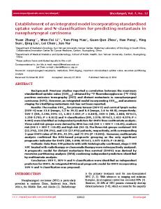
are due to cell mutations caused by normal aging of the human body [105]. Most breast cancers originate in the cells of the lobules, the milk-producing glands, or the ducts, the passageways used to drain milk from the lobules to the nipple. Less commonly, breast cancer originates in the stromal tissue, which contains fatty and fibrous connective tissues of the breast. Figure 2-1 shows a diagram of a typical breast, including the surrounding areas and areas mentioned above where cancer usually begins to form.
Breast profile: A Ducts B Lobules C Dilated section of duct to hold milk D Nipple E Fat F Pectoralis major muscle G Chest wall/rib cage Enlargement A Normal duct cells B Basement membrane C Lumen (center of duct) Figure 2-1. Diagram of a Typical Breast [105]
An understanding of breast cancer biology within the body is critical to the development not only of the tumor model but also of the model for the spread of the disease (metastasis). These disease models will be used to evaluate different screening and treatment policies. The most important anatomical variables are tumor size and the degree of metastasis to other parts of the body. A Gompertzian tumor growth equation [69] will be used to model the size of the initial tumor within the breast. The level of metastasis within the body will also be modeled using a stage progression model that determines the probability of the cancer being in the local, regional, or distant stage given the tumor size. Breast cancer is divided into stages, stages 0, I, II and some cancers in stage III are considered early stage breast
16
cancers. Stage III and stage IV are considered advanced stages of the disease that usually involve the spread of cancer to other parts of the body. Table 2-3 and Table 2-4 show the different stages of breast cancer. Within these stages, there are various assignments for values of T (tumor), N (lymph nodes), and M (distant metastasis). For example, T0 means that there is no evidence of a primary tumor, and TX means that the primary tumor could not be diagnosed. This structured type of system is used to keep track of the state of the disease as it progresses within the human body [105]. Table 2-3. Detailed Stages of Breast Cancer [105] Stage
Definition
Stage 0
Cancer cells remain inside the breast duct, without invasion into normal adjacent breast tissue.
Stage I
Cancer is 2 centimeters or less and is confined to the breast (lymph nodes are clear).
Stage IIA
No tumor can be found in the breast, but cancer cells are found in the auxiliary lymph nodes (the lymph nodes under the arm) OR the tumor measures 2 cm or smaller and has spread to the auxiliary lymph nodes OR the tumor is larger than 2 cm but no larger than 5 cm and has not spread to the auxiliary lymph nodes.
Stage IIB
The tumor is larger than 2 cm but no larger than 5 cm and has spread to the auxiliary lymph nodes OR the tumor is larger than 5 cm but has not spread to the auxiliary lymph nodes.
Stage IIIA
No tumor is found in the breast. Cancer is found in auxiliary lymph nodes that are sticking together or to other structures, or cancer may be found in lymph nodes near the breastbone OR the tumor is any size. Cancer has spread to the auxiliary lymph nodes, which are sticking together or to other structures, or cancer may be found in lymph nodes near the breastbone.
Stage IIIB
The tumor may be any size and has spread to the chest wall and/or skin of the breast AND may have spread to auxiliary lymph nodes that are clumped together or sticking to other structures or cancer may have spread to lymph nodes near the breastbone. Inflammatory breast cancer is considered at least stage IIIB
Stage IIIC
There may either be no sign of cancer in the breast or a tumor may be any size and may have spread to the chest wall and/or the skin of the breast AND the cancer has spread to lymph nodes either above or below the collarbone AND the cancer may have spread to auxiliary lymph nodes or to lymph nodes near the breastbone.
Stage IV
The cancer has spread — or metastasized — to other parts of the body.
Table 2-4. Local, Regional, and Distant Stages of Breast Cancer [105] Stage Local Regional Distant
Definition The cancer is confined within the breast. The lymph nodes, primarily those in the armpit, and possibly those near the collarbone, are involved. The cancer is found in other parts of the body and other organs are involved.
17
There are several risk factors which can affect the possibility of getting breast cancer as well as the outcome of the treatment process. Other than gender, age is the most important risk factor for breast cancer. As a woman ages, the probability of developing some form of breast cancer increases significantly; but it is believed that tumors grow more slowly in older women [21, 27, 28, 59, 73]. Combining this information with the fact that the US population is expected to increase in average age consistently over the next twenty years, the appropriate conclusion is that breast cancer among women in the US will become an increasingly complex problem. In the near future, it will become more important to have screening guidelines for women over the age of 65.
Among the risk factors that have been identified, some are controllable by the person at risk and some are not. Controllable factors include: weight, diet, exercise, smoking, drinking alcohol, exposure to estrogen, recent use of oral contraceptives (birth control pill), stress, and anxiety. Gender, age, family history, personal history, race, exposure to radiation therapy, exposure to estrogen, breast cellular changes, pregnancy and breastfeeding, and exposure to outdated medicine to prevent miscarriages are among the risk factors that cannot be controlled by the person at risk [105]. Some other risk factors that have recently been recognized and are incorporated in this research are body mass index (BMI) and breast density. For the older population, there is an idea that other diseases may actually begin to take precedence over breast cancer. These are referred to as “comorbidities.” Some of the specific diseases in this category include, but are not limited to, the following: hypertension, diabetes, coronary artery disease, congestive heart failure, dementia, and other cancers such as lung or ovarian cancer. Comorbidities for people over a certain age (65, 70, or 75) may substantially reduce the risk of death as a result of breast cancer, thus complicating the decision about how frequently to screen this population.
18
2.2.1.1 Ductal Carcinoma In-Situ (DCIS) DCIS is the most commonly occurring noninvasive breast cancer. As the name indicates, DCIS begins in the milk ducts of the breast. The term in-situ means “in its original place.” Hence, this type of cancer has not spread to any other parts of the breast other than the milk ducts. DCIS is not considered to be life-threatening like invasive cancer; but it needs to be treated, and it increases the risk of being diagnosed with invasive breast cancer later in life. The majority of recurrences happen within 5–10 years of the original diagnosis, and the recurrence rate is about 30% [69]. There are approximately 60,000 cases of DCIS diagnosed each year, and they make up about one in every five new breast cancer cases. This number may seem large, but people are living longer and the risk of cancer increases with increasing age. The increase in screening technology also enables easier and earlier detection this type of cancer, which contributes to the increasing number of DCIS cases [105]. While DCIS is considered an additional risk factor for developing invasive breast cancer later in life, very little is known about the progression of DCIS to invasive cancer. 2.2.2 Screening and Treatment The earlier breast cancer is diagnosed, the greater is the probability of surviving and recovering. Screening is a formal method of examining certain characteristics of the breast, either physically by touch, or by digital medical imaging, in order to determine whether breast cancer or any signs or symptoms related to breast cancer are present at the time of screening. Treatment refers to the process that is begun once it is known that cancer is present, and the stage to which the cancer has progressed within an individual [105]. The treatment process may include surgery, radiation therapy, chemotherapy, hormonal therapy, targeted therapies, and holistic medicines, or some combination of these. In general, there are three types of tests for breast cancer: screening, diagnostic, and monitoring. Screening tests (such as a mammogram) are used for early detection of breast cancer among healthy individuals; diagnostic tests (such as a biopsy) are given to those who are suspected of having breast cancer through symptoms or screening results; and monitoring tests (such as a
19
blood chemistry test) are used to monitor the progress of treatments and therapies as well as to monitor recurring cancers [105]. Table 2-5 outlines the different types of tests that exist.
It is outside the scope of this dissertation to present the details of every test for every category, but the basics of screening and treatment are reviewed. Berg [5] describes tailored supplemental screening for breast cancer, and explains the current state of breast cancer screening as well as the potential future directions.
Table 2-5. Breast Cancer Screening, Diagnostic, and Monitoring Tests Screening Tests
Diagnostic Tests
Monitoring Tests
Physical Exam (Self or Physician)
Biopsy
Blood Cell Counts
Mammography
Breast MRI
Blood Chemistries
Digital Tomosynthesis
Bone Scan
Blood Marker Tests
Ductal Lavage
CAT Scan
Thermograhpy
Chest X-Ray
Molecular Breast Imaging
Ultrasound
Breast MRI
SPoT-Light HER2 CISH
Sonography
Oncotype DX IHC Tests (ImmunoHistoChemistry) FISH Test (Fluorescence In Situ Hybridization)
Physical breast exams and mammography are the most common forms of screening for breast cancer, and mammography is the only screening technique that is currently recommended for population-based screening in the United States [86]. Therefore, mammography and clinical detection (detection of a lump in the breast by physical exam) will be the focus of the modeling efforts in our research. Table 2-6 summarizes current
20
mammography screening guidelines from a variety of sources, including the American Cancer Society (ACS) and American Medical Association (AMA). This table shows that well-defined screening guidelines have not been developed for older woman by any of the public medical authorities.
Table 2-6. Mammography Screening Guidelines [77] Agency
Middle-Aged Recommendation
Medicare/Medicaid Reimbursement
Annual screening mammography for women >40 yrs (if eligible)
American Academy of Family Physicians
Every 1–2 yrs for women 50–69 yrs
American College of Obstetricians and Gynecologists
Every 1–2 yrs for women >50 yrs
American Cancer Society
Yearly for women >40 yrs
American Medical Association
Yearly after 50 yrs
Canadian Task Force on Preventive Health Care
Every 1–2 yrs for women 50–69 yrs
US Preventive Services Task Force
Every 1–2 yrs for women 50–74 yrs
Older Recommendation
For all women after 70 years old, risk factors associated with breast cancer should be explored every 1 – 2 years to discuss breast cancer screening and to address the risks and benefits associated with screening, as well as the individual’s comorbidities, life expectancy, health status and quality of life.
Nine randomized trials have shown that the benefits of mammography screening for breast cancer include a 20% reduction in the mortality rate; however, eight of those trials did not consider the screening of older patients [1, 21]. Of course, this is complicated by the fact that breast cancer in older patients is believed to be less aggressive [21, 27, 28, 73]; and the benefits of screening may be offset by risks from comorbidities that could lead to other causes of death. Upon examination of Table 2-6, it is evident that clear screening guidelines for women over 65 have not yet been developed. These issues form the need for considerable research regarding effective screening strategies for older women, and these strategies could be dependent upon risk factors, chronological age, and a number of other variables.
Biopsy is the most common form of diagnostic test. Once the results of the screening test indicate that the woman may have breast cancer, sample tissue from her breast is taken and analyzed by a specialist in order to determine if cancer is present; and if it is, then the characteristics of the cancer are determined. Several other diagnostic tests are listed in Table
21
2-5, and are generally used in certain instances where a particular type of test has the ability to provide information that cannot be obtained from other diagnostic tests. Blood tests are the standard for monitoring tests; however if the physician believes the cancer has changed stage or spread to other parts of the body including the lymph nodes, then he may order some diagnostic tests in order to confirm or refute his hypothesis about the spread of the cancer [105]. 2.2.4 Past Modeling Approaches In this section, we present an overview of some of the previous modeling approaches to provide the context for the proposed work in the breast cancer screening research area and within the science of simulation. Previous models have made simplifying assumptions that either limit the applicability or validity of the model, or do not focus on the same factors that this research addresses. Previous modeling approaches to breast cancer screening are important because they provide a framework for the research completed in this dissertation. 2.2.4.1 Analytical Models Kirch and Klein [49, 50] develop one of the earlier analytical models for examination schedules of age-dependent diseases, where the objective is to minimize the delay in detection of the disease. The problem is to decide how many examinations should be given in each of the eleven five-year age periods between the ages 25 and 79. In [50], they consider two basic cases, one where the delay until detection is a constant, and one where the delay until detection is a random variable. They also provide a procedure for solving this scheduling optimization problem. In the case of breast cancer, results indicate that nonperiodic schedules will yield and average savings of 2%–3% in the number of exams (delay) prior to detection. The results also showed that a significant improvement in detection time could be achieved if screening were semiannually as opposed to annually. They also suggested that for mass-screening programs, each individual's screening interval should be dependent on age instead of fixed throughout life. This is an early analytical model that contains many simplifying assumptions, and is not particularly flexible or robust.
22
Schwartz [84] develops a Markov model of breast cancer that incorporates the different states of the disease, tumor size progression, and progression to the lymph nodes in order to predict the benefits of screening strategies in terms of life expectancy gain, and probability of detection prior to nodal involvement. The screening strategies compared screening every 10, 5, 3, 2, and 1 year(s). This model assumes test efficiency and rate of disease spread are stationary, does not account for false positives and negatives in the screening process, and assumes the decision maker (the physician) is omniscient and has every piece of necessary information. It also looks at screening from a purely individual perspective, as opposed to allowing some interaction with the surrounding environment. The results of this model suggest that life expectancy and the probability of detection prior to nodal involvement can be improved by screening annually as opposed to less frequent screening. It is also suggested that if the goal is to maximize life expectancy, then screening should be concentrated in the earlier years (40–70 years old). However, if the goal is to minimize the probability of recurrence of the disease, then screening should be concentrated in the later years. Understanding this approach to modeling was beneficial in that it provided a structure for important aspects of breast cancer screening that need to be taken into account in order to have an accurate model. It also provided a basic idea of performance measures used to assess the performance of different screening policies. This model is restricted by Markovian assumptions about progression of the disease.
Ozekici and Pliska’s [70] delayed Markov scheduling model is formulated as a generic scheduling problem that can be solved by dynamic programming, and it is later applied to breast cancer screening. This is one of the first scheduling models to take into account false positives and negatives, and it also allows the time until deterioration begins (i.e., the time until onset of the disease) to be any nonnegative random variable. This model determines the optimal inspection schedule of a system that starts in a “good” state, deteriorates gradually over time with any number of “defective” states, and ends in a “failed” state. The objective is to minimize the sum of the costs associated with the disease, which are subjectively assigned; moreover, the model is sensitive to these costs. Applying this
23
procedure to breast cancer screening yields an optimal policy that involves a sequence of physical and mammographic examinations at ages 50, 55.5, 60, 65, and 75 with some caveats. Some inaccuracies of this model include the assumption of stationary transition probabilities (i.e., disease progression with age is constant), and the fact that once a person has the disease, there is no further diagnostic process. This research is an interesting application of an inspection scheduling problem with false positives and false negatives applied to breast cancer. However, it is a very simple model which a small portion of the problem in breast cancer screening and it also contains a number of simplifying assumptions that this dissertation addresses, such as treatment and survival.
Parmigiani [71] uses Bayesian decision techniques to develop a model that predicts the effect of age on the minimum-cost screening schedule. The model used a non-Markovian stochastic process to represent the progression of the disease; however the model fails to distinguish deaths due to breast cancer from deaths not due to breast cancer. Each factor considered in this model is handled in isolation. An approach which considers the interaction of selected screening factors would clearly be more representative of the real-world system.
Mandelblatt et al. [60] develop a model for breast cancer screening in older women that takes into account two comorbid conditions, hypertension and congestive heart failure. Moreover, this model only considers whether or not screening should be offered to older women, and does not explore the proper screening policies for women in this age group.
Baker [2] develops a model that uses data from a study in the United Kingdom for parameterization; and he uses a continuous-time, non-Markovian stochastic process to model disease progression and screening. There are several limitations of this model, including limited sample data, poor fitting of the data to model parameters, and quite subjective cost values.
24
Lee and Zelen [57] develop a stochastic model that is able to predict breast cancer death and survival rates based on a number of factors such as examination schedules, population age distribution, follow-up time, and survival condition. Maillart and Ivy [59] describe a Markov model of dynamic breast cancer screening policies. They hypothesize that the screening interval at younger ages should be different than the screening interval at older ages and evaluate a large range of policies to develop a set of “efficient policies” measured by a lifetime breast cancer mortality risk metric and by the expected mammogram count. In this scenario, a patient could potentially select a policy based on individual circumstances. One of the authors' main conclusions is that screening should begin early in life and should continue until late in life regardless of the screening intervals adopted, which implies screening older women is beneficial.
The models that have been reviewed to this point are analytical in nature and attempt to provide a closed-form solution to complex problems. One drawback common to all these types of models is their inability to individualize the results. Another drawback is their reliance on many simplifying assumptions. Substantially better screening-and-treatment policies could be achieved by exploiting a methodology that allows results to be applied on an individual level and that allows these results to be a function of the individual risk factors. Simulation has the ability to remedy this problem because of its versatility and the fact that it does not require a closed-form solution to the problem at hand; instead it simulates the system and provides estimates of performance measures of interest over an experimental region containing all feasible solutions to the given problem. Generally, simulation provides a vast amount of insight, as opposed to solving for certain parameters and variables in a very specific mathematical model; moreover, simulation provides a framework of answering a variety of “what if” questions about changes in screening policies. Simulation is ideal for comparing the results of systems when changes are made that could affect the output measures of interest.
25
2.2.4.2 Simulation Models There have been relatively fewer attempts to model the breast cancer screening process using simulation. Michaelson et al. [62] and Fryback et al. [34] both use simulation modeling to answer breast cancer screening questions.
Michaelson et al. [62] develops a simulation methodology to test the ability of different screening intervals to detect breast cancer prior to the spread of the disease, using data on rates of breast cancer growth and metastasis. According to the taxonomy of Section 2.1.1, this is a level-1 model. This is a basic simulation model and does not consider anything that happens after the cancer is diagnosed. The work of Michaelson et al. [62] is a good starting point for modeling the disease in a simulation environment, but it needs to be extended in a number of ways to account for a vast number of risk factors and population variables that are not taken into consideration. The results of the model are fairly straightforward, suggesting that more frequent screening intervals could increase the number of breast cancers identified in the early stage of biological development.
Fryback et al. [34] develop a level-1 DES model of breast cancer in the Wisconsin population. Four interacting processes involved in breast cancer care are modeled: the natural history of the disease, detection of the disease, treatment of the disease, and competing cause mortality. The disease submodel is parameterized using data on breast cancer in the state of Wisconsin, and it provides a good framework for a breast cancer disease model. The model of Frybeck et al. [34] is somewhat simplistic in that it uses several critical factors to come up with a total onset proportion, or proportion of women in the population who get cancer regardless of their individual risk factors; and then it simulates the disease according to the indicated onset. The screening techniques considered are “clinical surfacing,” where the tumor is discovered physically, and mammography. A simple function was used to estimate technological improvements over time. The treatment process is modeled as a process where the women is either cured of the cancer and life proceeds normally, or she dies of breast cancer in the course of treatment. These are the only two possible outcomes of the treatment
26
process in this model. This model is retrospective, whereas our research looks into future and attempts to quantify the potential benefit of screening over the next 25 years. The results of the Wisconsin model indicate that breast cancer mortality for the period 1975–2000 would have been reduced by 38.3% with mammography screening and postcancer therapy compared with the option of not using these techniques during this time period.
2.3 DES Modeling DES is one of the most widely used modeling techniques in operations research. It is important to understand the basic concepts surrounding DES and how it has been used to model health care systems [8, 9]. We begin by providing a brief description of the fundamental concepts behind DES.
DES models progress by scheduling events at discrete points in time, advancing the simulation clock to the time of the event to occur soonest, executing the relevant logic at that event epoch, and then continuing to repeat these steps until reaching the end of the simulation's time horizon. Times between events (such as arrivals or services) are usually stochastic and are represented as random variables. In order to schedule events in the proper chronological order, samples are taken from the appropriate distributions and assigned to the relevant variables in the simulation model logic. The entities that advance through parts of a health care model are typically patients; but this is not always true and other creative modeling techniques can be utilized (for example, the basic entities moving through the various parts of the model might be health care providers or pieces of equipment). Entities (patients) each carry their own patient-specific attributes with them as they move through the model. These attributes can be used to determine the routing of the patient through different wards of a hospital, as well as to keep track of relevant statistics such as waiting time in queues, service times, and many other measurements of interest.
27
The DES approach is typically used for health care systems in which patients join some form of a waiting list for appointments, examination, and treatments [10]. This methodology allows the user to determine the current state of the overall system (in terms of operational efficiency, as well as variable parameters), and to ask “what if” questions to improve the design of the current system, or to design a new system from scratch. Jun et al. [47] provide an extensive survey of applications of discrete event simulation in health care clinics. The authors state that DES can be used to forecast the impact of changes in patient flow, to examine resource needs, or to investigate the complex relationships among the different model variables. These options provide managers with the ability to select and configure system components in order to reconfigure the simulation system and evaluate the simulated performance of the reconfigured system without actually altering the present system in the real world. This mode of using discrete-event simulation is of vital importance in the realm of health care, where it would be highly unethical, for example, to let a breast cancer patient’s cancerous tissue or tumor progress untreated solely for the purpose of gaining insight into its biological behavior. Instead, we define and mathematically model what we know about the disease, and predict the impact of changes to screening practices, medication with certain drugs, and changes in operational procedures and public policy. 2.4 SD Modeling System dynamics modeling is an analytical modeling approach which originated from the general systems theory approach of von Bertalanffy, and the foundations of SD were laid by Jay Forrester at MIT [8]. As stated in the introduction of this proposal, the fundamental principle of SD is that the structure of the system will determine its behavior. The way the separate components of any system are related to each other and affect each other will determine the long-run behavior of the system as a whole. The reason SD modeling is important is because this approach can reveal highly counterintuitive types of emergent behavior by the system at hand, and it is necessary to understand the reasons for such unexpected behaviors [8]. SD provides a framework for solving these types of problems.
28
There are two different sides to SD modeling, the quantitative side and the qualitative side. The qualitative portion involves the construction of a causal loop diagram (CLD), which is a graphic depiction of the manner in which the different components of the system are related. The arrows that relate two components can be signed as either positive or negative. A positive sign indicates that when the level of the state variable for the originating component increases, so does the level of the state variable for the destination component. A negative sign indicates the opposite: when the value of the originating variable increases, the value of the destination component decreases. In essence the signs denote the direction of influence, but do not quantify the magnitude of the influence. The goal of constructing this diagram is to gain a better understanding of the problem through exploring relationships between different variables and the overall structure of the system [51, 91]. Murray [65] defines an SD model that predicts the demand for screening for colorectal cancer. The causal loop diagram from Murray [65] is given in Figure 2-2. The colorectal cancer screening process contains many of the same elements as the breast cancer screening process. That is, the structure of the two systems is made up of very similar components: The role of the patient, the role of the physician, and the role of the health care delivery system.
29
Figure 2-2. Casual Loop Diagram of Colon Cancer Care System [65]
For causal loop diagrams, balancing loops are defined as loops that retain the status quo of the system and keep it operating in steady state. Note carefully that in a CLD, the term loop is synonymous with the graph-theoretic terms circuit and cycle because a CLD is a directed graph [26]. In a CLD, balancing loops can be identified by the use of an odd number of negative signs in given loop. On the other hand, reinforcing loops, or vicious circles,
30
contain an even number of negative signs and are loops where the system can effectively spiral out of control. The identification of these two types of loops is in itself a valuable learning process. A major benefit of this type of approach is that it often identifies unintended consequences of changes in system variables [8]. The quantitative portion of the SD model development process requires the modeler to convert the causal loop diagram from the qualitative part of the modeling process to a stock-flow diagram. The best way to think of this type of diagram is as a system of water reservoirs connected by pipes and governing valves. The water that flows around the system is analogous to entities in a DES model, except that the aggregation of all relevant entities is represented as a continuous quantity (state variable or stock); thus such a quantity can represent an amount of money, the size of a population or subpopulation, the mass of an inventory of raw material, or the number of items in a finished-goods inventory [8]. The influences in the causal loop diagram need to be converted to mathematical relationships that relate component X to component Y. Determining the form of these mathematical relationships is one of the goals of this research. Figure 2-3 is from a model of hospital admissions [8], and it provides a graphical depiction of a casual loop diagram and the stockflow diagram that was generated from it.
31
Figure 2-3. Casual Loop and Stock-Flow Diagrams from Hospital Admissions Model [8]
32
Models are parameterized by defining the initial values of the governing rates, and initial levels of the stocks. After these mathematical relationships are defined, the model can be run and the values of variables over time and under different conditions can be analyzed. It should be noted that SD models are typically deterministic in nature, and do not involve any stochastic components or samples of random variables. Time is handled by a discretization process where the time-step, Δt, is chosen so that all rates are constant over the short period of time Δt. SD modeling is suited for health care systems because it has the ability to model complex systems whose boundaries extend into other organizations. In health care, it is very rare that a system of interest involves well-defined boundaries and does not interact with the surrounding environment. This is compounded by the fact that health care has multiple stakeholders who have different perspectives regarding different aspects of system operation. In DES, an overwhelming amount of expert opinion is needed to define model variables and parameters and the relationships between these quantities, because the necessary data is often limited or not available at all. Data requirements for SD models are much less since the view of the model is more aggregated and at a higher level, but the required data are harder to get. Another key advantage of SD models is that they typically run very fast, while on the other hand, execution time is a major concern with of DES models.
Similarly, Dangerfield [22] describes the emergence of the use of SD modeling in the European health care arena. The paper describes the origins and methodology of system dynamics and relates it to health care, similar to Brailsford [8]. One of the most important conclusions drawn by the authors of both these papers is that there are a relatively few papers that utilize SD modeling, and that the health care arena has an immense potential to benefit from this type of modeling methodology. Brailsford [8] states “there is a clear potential for system dynamics modeling to be employed in support of health care policy.”
33
2.5 Integration of DES and SD Several studies have investigated the differences between DES and SD [8, 11, 19, 63, 89, 93, 96, 97, 103], some on a theoretical and methodological level, and some from the user’s perspective. It is important to understand all the underlying differences from a variety of perspectives in order to assess the possible benefit of combining the two methodologies. The DES methodology is based on the premise that the randomness inherent in the system as well as the system structure will determine its long-run behavior, and DES is usually used to model systems in a high level of detail. SD modeling on the other hand, is based on the idea that the structure of system will determine its long-run behavior. Based on an extensive review of the literature and lengthy discussions with faculty specializing in the fields of health care and simulation, we have great reason to expect that combining the DES and SD techniques could be greatly beneficial in gaining insight into complex systems, such as breast cancer screening. The idea being that overall system dynamics can be modeled using SD modeling techniques, and other aspects which occur at discrete points in time can be modeled using DES. Structure and randomness both play a vital role in the long-run behavior of the system, and the integrated screening model combines these two methodologies into a single approach with the ability to capture the dynamics of high-level system strategy (health care delivery and government involvement), as well as low-level randomness and structure involved in the screening process for individual women. We assert that there is the potential for significant benefits to combining these two modeling approaches, and relevant literature on the subject largely supports this conclusion.
Morecraft and Robinson [63] develop an SD model and a DES model of the same system, a simple fishery. The process of building both the SD and DES models is described in detail, although the models themselves are actually very simple. The importance of this work lies in the conclusions reached after developing both types of models, which include the following:
34
1. “Overall it is apparent that while SD illuminates ‘deterministic complexity,' DES illuminates ‘constrained randomness.' Either or both may be important in understanding and explaining puzzling dynamics. SD and DES should therefore not be seen as opposing modeling approaches, but as complementary.”
2. “The tragedy is that too few researchers realize that both deterministic [SD] and stochastic [DES] models have important roles to play in the analysis of any particular system. Slavish approach to one particular approach can lead to disaster. ... So pursuing both approaches simultaneously ensures that we do not become trapped by either deterministic fantasy or unnecessary mathematical detail.”
Table 2-7 and Table 2-8 [63] provide insights into technical and conceptual differences between the two approaches. These tables reinforce the idea of combining both approaches. These conclusions are in direct support of the concepts behind this research. Table 2-7. Technical Differences between DES and SD [63] Issue
Discrete Event Simulation
1
Systems (such as health care) can be viewed as networks of queues and activities.
2
3
4 5 6
Objects in a system are distinct individuals (such as patients in a hospital), each possessing characteristics that determine what happens to that individual. Activity durations are sampled for each individual from probability distributions and the modeler has almost unlimited flexibility in the choice of these functions and can easily specify nonexponential dwelling times. State changes occur at discrete points of time. Models are by definition stochastic in nature. Models are simulated in unequal times steps, when “something happens.”
System Dynamics Systems (such as health care) can be viewed as a series of stocks and flows. Entities (such as patients) are treated like molecules of a fluid, flowing through reservoirs or tanks connected by pipes. The time spent in each reservoir is modeled as a delay with limited flexibility to specify a dwelling time other than exponential. State changes are continuous. Models are deterministic. Models are simulated in finely sliced time steps of equal duration.
35
Table 2-8. Conceptual Differences between DES and SD [63] Issue
Discrete Event Simulation
System Dynamics
Perspective
Analytic, emphasis on detail complexity.
Holistic; emphasis on dynamic complexity.
Resolution of Models
Individual entities, attributes, decisions, and events.
Homogenized entities, continuous policy pressures and emergent behavior.
Data Sources
Primarily numerical with some judgmental elements.
Broadly drawn.
Problems Studies
Operational.
Strategic.
Model Elements
Physical, tangible, and some informational.
Agents Represented in Models As…
Decision Makers.
Clients Find the Model…
Opaque/dark grey box, nevertheless convincing.
Physical, tangible, judgmental and information links. Boundedly rational policy makers. Transparent/fuzzy glass box, nevertheless compelling.
Model Outputs
Point predictions and detailed performance measures across a range of parameters, decision rules, and scenarios.
Understanding of structural source of behavior modes, location of key performance measures and effective policy levers.
Tako and Robinson [96] provide an empirical study comparing the DES and SD simulation approaches, and the results are summarized in Table 2-9. The uniqueness in this study is that it allows one group of users to become familiar with and analyze the output of a DES model and an SD model of the same system, the prison system in the United Kingdom, where the populations of the prisons are growing rapidly and a major problem seems to be people who reoffend. Before conducting the experiment, the authors review the relevant literature and compare DES and SD in a number of categories, including: model understanding, complexity, validity, usefulness, and results. The authors conclude that users would find significant differences in the two modeling approaches. The results of the study are actually quite interesting. Due to the limited amount of time available, users could not be exposed to both types of models.
36
Table 2-9. Comparison of DES and SD Model Literature [96] Methodology Model Understanding Understanding (parts of) the Model
DES
SD
The client does not understand the underlying mechanics. Animation and graphic tools help model understanding.
Models (links and flows) are transparent to the client. No animation. Visual display of model aids model understanding.
Level of Detail
Emphasis on detail complexity.
Feedback
Feedback is not explicit.
Emphasis on dynamic complexity. Feedback effects are clear to the client.
Animation Complexity
Model Validity Credibility
Both models are perceived as representative, provide realistic outputs and create confidence in decision-making.
Model Usefulness Learning Tool Strategic Thinking Communication Tool
Used less as learning tools.
“Learning laboratories” that enhance users learning.
Mostly used in solving Aid strategic thinking. operational/tactical issues. Both models are seen as good communication tools and facilitate communication with the client.
Model Results Nature of Results
Interpretation of Results
Results Observation
Provides statistically valid estimates of systems performance. Results aid in instrumental learning. More difficult, requires users to have a statistical background. Randomness/variation of results is explicit.
Results provide a full picture of the system. Results aid in conceptual learning. Outputs are easily interpreted, little or no statistical analysis is required. Generally deterministic results, which convey casual relationships between variables.
Each group of users was only subjected to one of the two types of modeling, took for granted the features of the model they were given to work with, and were not really able to provide deep insights into the differences in the way users perceive the two simulation modeling approaches. The results of the comparison experiment are given in Table 2-10. Slight differences were found in terms of interpretation and importance of animation, but no real significant differences were identified in the study. This suggests that users are comfortable with both modeling approaches; therefore, the integration of the two approaches might easily 37
be accepted by typical users. A combined model has the potential to bring the strengths of the two separate concepts together, but this will surely create some unexpected dilemmas about which methodology should prevail when modeling certain system components. Table 2-10. Comparison Between DES and SD Model Experiments and Results [96] Model Use Model Understanding Understanding (parts of) the Model
DES
Factors That Helped Understanding Complexity
Animation the most important factor.
Level of Detail Feedback
SD
No differences in user's opinions. Accompanying model descriptions most important.
Similar in level of perceived detail. Feedback effects more explicit to DES model users..
—
Model Validity Representative of Real Problem Realistic Outputs Confidence in Outputs
SD model just more representative as compared to DES model. Outputs are perceived to be similarly realistic. —
Similar level of confidence in model outputs.
Model Usefulness Learning
Similar level of learning achieved from using the models.
Strategic Thinking
Same level of perceived strategic thinking involved.
Communication of Ideas Model Results Instrumental/Conceptual Learning Interpretation of Results Attitude When Interpreting Results
Same level of communication perceived to have taken place.
Both DES and SD aid instrumental learning. SD aids in conceptual learning to a higher extent. DES models were more difficult to interpret.
-
No differences in user's attitude.
38
Sweester [93] compares the two modeling approaches from the perspective of evaluating businesses’ decision making, and the purpose is to identify the strengths and weaknesses of both approaches. Another visual representation of the differences between the two approaches is shown in Figure 2-4, which presents a typical diagram of production process from both the SD and DES perspectives.
39
Figure 2-4. DES Model (Top) and SD Model (Bottom) of Same Production Process [93]
40
The study concludes SD is suited to modeling problems with continuous variables whose interactions significantly affect the behavior of the system, while DES is used to develop a statistically valid estimates of system performance. What the paper fails to explore is the idea that the system could contain continuous variables whose feedback significantly affects the behavior of the system, and there could be a need to develop a statistically valid estimate of system performance. The relevant literature clearly reveals that many aspects of breast cancer screening present both of these characteristics and that the system is suitable for an integrated model.
Tako and Robinson [96] fulfill one of their goals for future research from [97] by observing users as they build SD and DES models of the same UK prison system, and noting the differences in model development from a user standpoint. Seven specific topics were observed: problem structuring, conceptual modeling, data inputs, model coding, validation and verification, results and experimentation, and implementation. The results of the study suggest that all modelers switch continuously between the seven topics, but DES modelers seem to follow a more linear progression than SD modelers. Model coding is the central topic for DES modelers, while conceptual modeling is primarily followed by model coding for SD modelers. However, the major finding was that neither approach revealed users that followed these seven topics in any particular order. Other findings contribute to the framework for integrating the two approaches, such as “most modelers did not consider alternative modeling approaches before modeling. This conveys that modelers use the simulation approach they are more familiar with rather than the approach that is most appropriate to the situation.” It is reasonable to believe certain systems could benefit from a combined approach.
Brailsford et al. [11] compares the two approaches in the modeling of an emergency department (ED) and suggests that the relative strengths and limitations of both approaches are in fact complementary. “We argue that the integrated simulation models can assist in dealing with the efficiency issues within the ED walls, but can also provide a better understanding of system-wide factors beyond the immediate control of ED managers.” They
41
describe three potential benefits of the integrated approach: inward and outward looking, analytical insights at all levels of the system, and manager-friendly models. The conclusion of the paper states: “We believe there is much to be gained by integrating these two approaches into a unified framework for both conceptual and implementation purposes. ... We recommend future research into conceptual and implementation issues of this area.” This is further support for the integration of these two approaches.
Chahal and Eldabi [19] explore reasons that DES and SD could potentially be combined and predict some potential benefits of this type of model. This paper provides a remarkably clear and compelling argument for why DES and SD should be combined. The benefits and reasons are nearly identical to those discussed in [11]. The authors suggest using the outputs of a DES model as the inputs of an SD model, and vice versa; and they also admit that they have not developed a hierarchical structure to specify the way the two models would interact. There may be some merit to this type of approach, but there seems to be much greater value in integrating these two approaches so that elements of both types of simulation models can be represented simultaneously. This provides evidence of the fact that a gap exists in simulation methodology that has not yet been addressed by researchers.
Ventkatesawaran [103] develops an SD model of a production planning system and uses the output from that model as the input to a DES model of the shop floor. This model is a first-step to combining these two approaches, but these two models are built in different software packages and are not integrated together in a single model. This dissertation is a direct extension of this work and seeks to integrate these two approaches into a single model where both models run concurrently and are linked.
To the best of our knowledge, a simulation methodology which integrates DES and SD modeling approaches into a single model has not yet been developed. The problem statement and research questions addressed are presented in the final sections of this chapter.
42
2.6 Research Motivation In this section we elaborate the motivation for the proposed research beyond the brief discussion given in Chapter 1. By enumerating the main problems in breast cancer screening and in simulations of the breast cancer screening-and-treatment process, we also identify the main research questions to be addressed in the dissertation.
2.6.1 The Problems in Breast Cancer Screening The problem in breast cancer screening has been introduced earlier sections, but will be explored in more detail in this section. The aging U.S. population, the lack of screening guidelines for older women, and the prominence of breast cancer among women in the United States are three of the major factors that contribute to the need for research in breast cancer screening [1, 28, 42, 58, 77]. With regard to breast cancer screening, there is little guidance for older patients [42, 45, 77, 86].
Currently, there are no specific guidelines for women 70 and older (Table 2-6), nor are there established clinical guidelines for the age at which screening for breast cancer should be discontinued. The vagueness of the current guidelines leaves much to be desired in terms of an effective decision making process for the older patient and her physician. The major goals of the simulation model are to determine the most effective screening policy for women over 65, which could be dependent on age or specific individual risk factors. The appropriate age at which to stop screening is also of interest. The Carolina Mammography Registry (CMR), the Surveillance, Epidemiology and End Results (SEER) Program (www.seer.cancer.gov), the Breast Cancer Surveillance Consortium (BCSC), and relevant literature provide reliable sources of information that are used for model parameterization and validation. Medicare claims data assist in determining cost-of-care estimates, since most women over 65 have Medicare as their primary insurance.
43
The majority of the studies in this area have ignored factors that will be considered in this research, do not focus on older patients, or contain assumptions that may have become outdated with technological advances. Previous studies have not focused on policy evaluation and development, whereas that will be the primary objective of this research. In addition, the American Cancer Society has stated there is no reason to believe there will be more randomized clinical trials for breast cancer screening in the near future [1, 21, 45, 86]. Upcoming clinical trials will be pharmaceutical clinical trials as opposed to clinical trials for breast cancer screening. Maillart and Ivy [59] provide motivation for solving this problem:
Considerable controversy remains regarding how frequently to recommend screening and at what age to stop screening older women. There is considerable evidence that physicians have difficulty differentiating the highly heterogeneous older patient population. Given that women over the age of 65 will become the most prevalent patient cohort in the breast cancer population, it is imperative that research into the optimal screening strategies for this patient population be performed.
Next, we state the problems from a simulation modeling perspective.
2.6.2 The Problems in Simulation Modeling Jun et al. [47] describes specifically how discrete event simulation has been used to optimize patient flow and allocation of scarce resources with health care systems. The results of the survey show that there is a lack of papers that address integrated systems. Another conclusion is that this type of modeling has been made possible by recent advances in software. This is important, because the tools to proceed with this integrated type of model have only recently become available. Arena 13.9 [48] is an extremely versatile simulation program that has the ability to model discrete and continuous variables simultaneously, as well as use arbitrarily complex expressions in routing entities through the model. This software will be used in the development of both the natural history simulation model and the integrated screening model. The fact that simulation software that has the ability to handle
44
this type of integrated system is already available is a result of advancing technology. Arena also has the ability to interface with Visual Basic and C++ programming languages.
Several problems arise when the concept of integrating these two approaches is pursued, including:
1. What components of the overall breast cancer screening-and-treatment system should be modeled using the DES approach and which should be modeled using SD? 2. Why and how does the combining these two approaches result in a model which more accurately depicts the overall system, and how can this benefit be quantified? 3. What specific links between the components of the DES submodel and the SD submodel should be present for the most realistic representation of the system? 4. What information needs will be shared by the two approaches as the simulation runs both simultaneously? 5. What are the inputs for the SD components and for the DES components of the system? a. Are they related? b. Will choosing whether a component is to be modeled using SD or DES be based upon the available data to model that system component? 6. What will the outputs of the system be that provide the analyst with better insights compared with developing and analyzing these two types of models separately? 7. What types of “prefabricated” modeling tools and components should be developed to facilitate rapid building of simulation models for design and evaluation of breast cancer screening policies? a. Submodels of the process-interaction part of the overall model representing operations or flows of people, information, etc., that occur in essentially similar form at numerous points in the overall model; and
45
b. New output-analysis procedures that have been adapted to the specific requirements of simulation-based evaluation of breast cancer screening policies.
Our research addresses these questions. However, capturing the big picture and the small details of a system at the same time is intrinsically difficult, and can pose unanticipated challenges during the development of the integrated screening model.
2.6.3 Research Questions Addressed Along with the problems and research questions already stated, this research seeks to address the following questions:
1. How does using a combined DES/SD model help to capture the actual behavior of the breast cancer screening process? 2. What aspects of the breast cancer screening process should be modeled with DES and what aspects should be modeled with SD? 3. How will the SD and DES submodels be linked? 4. How can we measure the risk of having a cancer detected based on specific breast cancer risk factors? 5. How can we attain a database of women with appropriate breast cancer risk factors to sample from so that we can determine incidence on an individual basis? 6. How can we model the disease within the body? 7. How can we determine the stage of breast cancer at diagnosis? 8. What performance measures typically define the effectiveness of a breast cancer screening policy? 9. How can the effect of policy changes on the performance of breast cancer screening best be measured?
46
10. Which screening policy is most effective for older US women in terms of the performance measures referenced in Question 8? 11. How can the Tejada model be validated using historical data? 12. How can the best screening policy be identified from the (large) set of feasible screening policies? 13. How can we honestly represent the randomness and intrinsic uncertainty in the simulation-generated estimates of expected system performance?
These questions characterize the key goals of our research. This dissertation will be the first step of many in combining these two simulation methodologies in the health care modeling arena. To this point, we have provided background on simulation modeling methodology, health care, and specifically breast cancer. The following three chapters review the models used to address the problems and research questions we have just discussed. We begin with our natural history simulation model.
47
CHAPTER 3: THE NATURAL HISTORY SIMULATION MODEL This chapter focuses on the detailed development and results of the natural history simulation model. As stated previously, this model is made up of a number of interacting submodels, namely: (a) the cancer incidence submodel; (b) the disease progression submodel; (c) the survival and mortality submodel; and (d) the population growth submodel. The most important input to this model is the sampling database that contains information about breast cancer risk factors from actual women in the designated population of US women 65 and older. All of the submodels and their components are explained in detail in this chapter.
This model establishes a baseline on the benefits of screening and treatment by determining the earliest a cancer could be detected for each woman in the population with perfect annual screening for all women, and it assumes all cancers remain untreated. The time-step for this model is one year, i.e., we simulate all the events for a given year, then we move to the next year and simulate all the events for that year, and this process repeats until we reach the end of the time horizon. This approach to the operation of the natural history model makes the most effective use of the available literature, because the Barlow risk model [3] predicts a woman's risk each year of being diagnosed with breast cancer as a function of her risk factors given that she received a mammogram; and life-tables with breast cancer deaths removed [79] predict the probability of surviving one more year as a function of a woman's current age. To establish a baseline that will later enable us to investigate the results of using the best-case scenario, we assume in the natural history DES model that every woman is screened every year with perfect compliance until one of the following occurs: (a) detection for a woman who develops breast cancer; (b) death from a cause other than breast cancer; or (c) the end of the simulation is reached at the year 2020 for a woman who survives to that time and does not develop breast cancer. However, when a cancer is detected, it is not treated. The cancer is allowed to progress, and it can eventually cause a breast cancer–related death; or a natural death from other causes may occur before death from breast cancer. It is also possible that the woman lives beyond the year 2020. The integrated screening model
48
considers screening at less frequent intervals and as a function of individual risk factors, postdiagnosis (adjuvant) treatment, costs, and survival after diagnosis.
First, the natural history DES model creates the initial population representative of US women at least 65 using a random sampling procedure. The model assigns each woman the attributes (see Table 3-1 for a list of attributes) of a randomly sampled woman in the data set provided by the Breast Cancer Surveillance Consortium (BCSC) [15], and then that woman enters into the disease submodel. The age distribution of the initial simulated population represents that of US women who are at least 65, and was derived from 2000 US census data [43]. Following the creation of the initial population and the assignment of riskfactor attributes for each individual in that population, the risks of breast-cancer detection and natural death are simulated in one-year time-steps until the occurrence of non–breast cancer related death, breast cancer–related death, or the end of the simulation in the year 2020. Each woman in the model can only have breast cancer detected once, and that event is regarded as the first occurrence of breast cancer in her lifetime. The population of US women at least 65 is currently growing rapidly, so it was essential to include a submodel for population growth that adds women who are turning 65 to the current population each year. The initial simulated population size in the initial model year 2001 is 20,582, which is 0.1% of the number of women over 65 in the United States in 2001. This population grows to over 30,000 by 2020. For each of the 10 simulated populations used to initialize an independent replication (run) of the natural history model, 50,000 samples are taken with replacement from the database, which has about 250,000 entries. The natural history DES model was created using Arena 13.9 by Rockwell Software [48].
Figure 3-1 shows the possible transitions among health states for each individual woman in the simulation model. All women who are diagnosed with invasive cancer are followed past 2020 until their death from breast cancer or natural causes. Women who are diagnosed with DCIS are not "cancer free", but they cannot have a death from breast cancer since DCIS cannot progress, the only possible transition from DCIS is to other cause death.
49
Figure 3-1. Health-State Transitions in the Natural History Model
3.1 Cancer Incidence Submodel The cancer incidence submodel consists of the Barlow breast cancer risk model being used in conjunction with each woman's risk factors. Risk factors for each woman were sampled from the BCSC data set at the very beginning of the model. This allows the simulation to estimate the probability of being diagnosed with breast cancer each year given that a mammogram was received in that year (this is the condition imposed by the Barlow risk model). Each year this probability is computed for each woman, and this is the method used for determining which women in the model will get cancer and the year they were
50
diagnosed. Cancer incidence can be defined as the number of women who get cancer over a given time period, typically one year. These raw numbers are usually converted into rates per 100,000 women, which is the format SEER uses to present incidence data that we use for validation. The remainder of this section presents the details of the BCSC data and the Barlow risk model.
3.1.1 BCSC Data A key component of this research is locating a data set containing risk factors similar to those of women in the US population. For example, age, race, body mass index (BMI), breast density, and many other factors are important in determining a woman’s risk of developing breast cancer. We were required to submit a formal research proposal and request for data to the BCSC in order to obtain the individualized data set required for this model, and the BCSC provided a data set containing risk factors for slightly over one million women that has been de-identified to protect the confidentiality of the women, the radiology facilities, and the mammography registries—i.e., the data do not include any dates, origin of the data, patient identifiers, or the assessment (outcome) of the index screening mammogram [15]. Since the data set consists of information from seven mammography registries in different locations across the United States, it is believed to be representative of the women in the US population. Based on the attributes in this data set, the Barlow risk model [3] can be used to calculate an individual woman’s risk of developing invasive breast cancer or DCIS within the next year, given that she received a mammogram. The risk factors used to calculate risk are assumed to stay constant throughout each woman’s lifetime, with the exception of age and menopausal status.
51
The following is a description of the BCSC that has been taken from their web site: The Breast Cancer Surveillance Consortium (BCSC) is a research resource for studies designed to assess the delivery and quality of breast cancer screening and related patient outcomes in the United States. The BCSC is a collaborative network of seven mammography registries with linkages to tumor and/or pathology registries. The network is supported by a central Statistical Coordinating Center. Currently, the Consortium's database contains information on 7,521,000 mammographic examinations, 2,017,869 women, and 86,700 cancer cases (72,800 invasive cancers and 13,800 In Situ). A full description of the history and early work of the BCSC can be found at: http://breastscreening.cancer.gov/espp_report.html. None of the women who participated had a previous diagnosis of breast cancer, but all of them had undergone breast mammography at some point in the previous five years. This limits the application of this research to women who have no prior history of breast cancer. Cancer registry and pathology data were linked by the BCSC to the data on mammography and incidence of invasive breast cancer or DCIS. Table 3-1 contains a list of the breast cancer risk factors for which data was collected during the BCSC study, and the appropriate predictor (independent) variables in the logistic regression equations of the Barlow risk model.
52
Table 3-1. Risk Factor Attributes of Individuals in the BCSC Risk Model Data set Factor #
Factor Symbol
Factor Name
Columns
Factor Coding
0
None
menopaus
1
0 = premenopausal; 1 = postmenopausal or age>=55 ; 9 = unknown
1
x1
agegrp
3–4
1 = 35–39; 2 = 40–44; 3 = 45–49; 4 = 50– 54; 5 = 55–59; 6 = 60–64; 7 = 65–69; 8 = 70–74; 9 = 75–79; 10 = 80–84
2
x2
density
6
BI-RADS breast density codes 1 = Almost entirely fat; 2 = Scattered fibroglandular densities; 3 = Heterogeneously dense; 4 = Extremely dense; 9 = Unknown or different measurement system
3
x3
race
8
1 = white; 2 = Asian/Pacific Islander; 3 = black; 4 = Native American; 5 = other/mixed; 9 = unknown
4
x4
Hispanic
10
0 = no; 1 = yes; 9 = unknown
5
x5
bmi
12
Body mass index: 1 = 10–24.99; 2 = 25– 29.99; 3 = 30–34.99; 4 = 35 or more; 9 = unknown
6
x6
agefirst
14
Age at first birth: 0 = Age < 30; 1 = Age 30 or greater; 2 = Nulliparous; 9 = unknown
7
x7
nrelbc
16
Number of first-degree relatives with breast cancer: 0 = zero; 1= one; 2 = 2 or more; 9 = unknown
8
x8
brstproc
18
Previous breast procedure: 0 = no; 1 = yes; 9 = unknown
9
x9
lastmamm
20
Result of last mammogram before the index mammogram: 0 = negative; 1 = false positive; 9 = unknown
10
x10
surgmeno
22
Surgical menopause: 0 = natural; 1 = surgical; 9 = unknown or not menopausal (menopaus=0 or menopaus=9)
11
x11
hrt
24
Current hormone therapy: 0 = no; 1 = yes; 9 = unknown or not menopausal (menopaus=0 or menopaus=9)
53
3.1.2 The Barlow Risk Model The Barlow breast cancer risk model [3] was developed using the data collected by the BCSC during their six-year study. The risk model consists of logistic regression equations that predict the probability of being diagnosed with breast cancer (invasive or DCIS) in the next year for premenopausal and postmenopausal women, respectively. SAS statistical software was used to conduct the analysis. We verified this model by using SAS code made publicly available by the authors (Appendix A) to reproduce the statistical model and the corresponding results (Appendix B).
The dependent (response) variable is a binary variable for which the values 0 and 1 denote the absence and presence of breast cancer, respectively; and the predictor variables (factors or "covariates") are the categorical independent variables presented in Table 3-1. Note that the BCSC provided data for women ages 35 and older, and we incorporated the possibility of including younger women in the logic of many of the submodels. For premenopausal and postmenopausal women, Barlow [3] formulated separate logistic regression equations to predict the risk of being diagnosed with breast cancer within one year.
Logistic regression [13] is typically used to predict the probability of occurrence of a specific event using the logistic function. There are several reasons for using the logistic function and logistic regression to predict probabilities. The logistic function is given in Equation (3.1). It is easy to see that the function's domain consists of all real numbers, while its range is restricted between zero and one. This form makes logistic regression appealing for predicting probabilities. The logistic function is of the form: = f ( L)
eL 1 + eL
for − ∞ < L < ∞.
(3.1)
54
In logistic regression, the probability of interest, p, is related to the predictor variables using the natural log function. Typically, Equation (3.2) is referred to as the “logit” function in the regression equation:
p L = Logit(p) = ln = β 0 + β1 x1 + β 2 x2 + ... + β n xn . 1− p
(3.2)
where x1 , ... , xn are the independent variables whose values (coded factor levels) are used to estimate the relevant probability p. In order to calculate the predicted probability of the event of interest based on the risk factors, Equation (3.2) must be solved for p. Equation (3.3) shows the results of such algebraic manipulation. Rearranging (3.2), we see: e L − p − pe L = 0 ⇒ p =
eL = f ( L). 1 + eL
(3.3)
Barlow [3] created two separate logistic regression models to predict the risk of being diagnosed with breast cancer within one year. One model is used for premenopausal women, and the other is used for postmenopausal women. Cancer rates suggest that risk patterns are not the same for premenopausal and postmenopausal women, and the risk factors which are significant for developing cancer are quite different for these two groups of women. All eleven of the risk factors in Table 3-1 were tested for significance in both regression models. There are no interaction terms included in order to keep the model simple enough to be used practically, and the authors state that the interaction terms were not significant. In order to verify the models, 75% of the random sample (the training data set) was used to estimate the statistical models; and the prediction was tested in the remaining 25% of the random sample (the validation data set). The concordance value (c-statistic) is used to determine the quality of the prediction for each individual woman. The c-statistic is a value that varies from 0.5 (discriminating power not better than chance) to 1.0 (perfect discriminating power). It can be interpreted as the percent of all possible pairs of cases in which the model assigns a higher probability to a correct case than to an incorrect case. The entire output from the analysis is
55
available in Appendix B. The c-statistic for the Barlow risk model is 0.624, which the authors suggest is acceptable in practice.
It is important to note that a very strict p-value criterion of p < 0.0001 is required for a risk factor to be statistically significant and included in the model. The reason the P-value criterion is so strict is because of the large size of the data set. Small differences could appear significant at the .05 level due to the size of the data set, so a more stringent criterion was chosen by Barlow [3]. For premenopausal women there were only four statistically significant risk factors: age, family history, breast density, and history of breast procedures. The following risk factors were not statistically significant: race (p = 0.40), ethnicity (p = 0.41), age at first birth (p = 0.48), BMI (p = 0.89), and the result of previous mammogram (p= 0.0018). Equation (3.4) is the regression equation for premenopausal women, which was formed using the factors given in Table 3-1. Equation (3.5) is the explicit equation that can be used to calculate the one-year risk when given the values of the regression coefficients and the values of the categorical variables for a specific woman. The logit for premenopausal women can be calculated according to Equation (3.6), noting that the double equals sign is a true/false statement that evaluates to either zero or one.
p LPRE =Logit( pPRE ) =ln PRE =β 0 + β 2 x2 + β3 x3 + β8 x8 + β9 x9 , 1 − pPRE
(3.4)
where the independent variables x2 , x3 , x8 , and x9 are defined in Table 3-1,
( LPRE ) = pPRE f=
exp( LPRE ) 1 , = 1 + exp( LPRE ) 1 + exp(− LPRE )
(3.5)
56
LPRE = −7.4979 + ( AgeGroup = = = 1) * ( 0 ) + ( AgeGroup = 2 ) * ( 0.2046 ) + ( AgeGroup = = = 3) * ( 0.6388 ) + ( AgeGroup = 4 ) * ( 0.8574 ) + ( Previous Procedure = = = 0 ) * ( 0 ) + ( Previous Procedure = 1) * ( 0.3873) + ( Previous Procedure = = = 9 ) * ( −0.1431) + ( First Degree = 0) * ( 0) + ( First Degree = = 1) * ( 0.4306 ) + ( First Degree = = 2 ) * ( 0.7455 )
(3.6)
+ ( First Degree = = = 9 ) * ( −0.1134 ) + ( Breast Density = 1) * ( 0 ) + ( Breast Density = = = 2 ) * ( 0.6818 ) + ( Breast Density = 3) * (1.2067 ) + ( Breast Density = = = 4 ) * (1.3694 ) + ( Breast Density = 9 ) * (1.1966 ) .
For example, consider a premenopausal woman who is age 43 (category 2), has one relative with cancer (category 1), whose breasts are heterogeneously dense (category 3), and who has had no previous breast procedures (category 0). We can calculate her risk of being diagnosed with cancer in the next year. First we must calculate the value of the logit, shown in Equation (3.7), then we can calculate the desired probability, shown in Equation (3.8),
LPRE = −7.4979 + (1) * ( 0.2046 ) + (1) * ( 0 ) + (1) * ( 0.4306 ) + (1) * (1.2067 ) = −5.656, (3.7)
= pPRE
1 1 = = 0.00348. 1 + exp(− LPRE ) 1 + e5.656
(3.8)
For postmenopausal women there were eleven statistically significant risk factors from Table 3.1: age x1 , breast density x2, race x3, ethnicity x4, BMI x5, age at first birth x6, family history x7, history of breast procedures x8, result of last mammogram x9, surgical menopause x10, and hormone replacement therapy x11. Equation (3.9) is the regression equation for postmenopausal women, which was formed using the factors given in Table 3-1. Equation (3.10) is the explicit equation that can be used to calculate the one-year risk given the values of the regression coefficients and the values of the categorical variables for a specific woman. The logit for postmenopausal women can be calculated according to Equation (3.11):
57
p LPOST =Logit( pPOST ) =ln POST =β0 + β1 x1 + β 2 x2 + β3 x3 + β 4 x4 + β5 x5 1 − pPOST + β6 x6 + β7 x7 + β8 x8 + β9 x9 + β10 x10 + β11 x11 ,
= pPOST
f= ( LPOST )
exp( LPOST ) 1 = , 1 + exp( LPOST ) 1 + exp(− LPOST )
(3.9)
(3.10)
58
LPOST = −7.0713 + ( AgeGroup = = = 3) * ( 0 ) + ( AgeGroup = 4 ) * ( 0.283) + ( AgeGroup = = = 5 ) * ( 0.6743) + ( AgeGroup = 6 ) * ( 0.8196 ) + ( AgeGroup = = = 7 ) * ( 0.9034 ) + ( AgeGroup = 8 ) * (1.026 ) + ( AgeGroup = = = 9 ) * (1.1086 ) + ( AgeGroup = 10 ) * (1.2033) + ( Ethnicity = = = 0 ) * ( 0 ) + ( Ethnicity = 1) * ( −0.3072 ) + ( Ethnicity = = = 9 ) * ( −0.0641) + ( Race = 1) * ( 0 ) + ( Race = = = 2 ) * ( −0.2251) + ( Race = 3) * ( 0.0935 ) + ( Race = = = 4 ) * ( −0.6108 ) + ( Race = 5 ) * ( −0.0269 ) + ( Race = = = = 9 ) * ( −0.00037 ) + ( BMI = 1) * ( 0 ) + ( BMI = 2 ) * ( 0.1337 ) + ( BMI = = = = 3) * ( 0.2463) + ( BMI = 4 ) * ( 0.3827 ) + ( BMI = 9 ) * ( 0.0295 ) + ( AAFB = =+ = 0 ) * ( 0 ) ( AAFB = 1) * ( 0.1871) + ( AAFB == 9 ) * ( 0.0203) + ( Previous Procedure = = = 0 ) * ( 0 ) + ( Previous Procedure = 1) * ( 0.262 ) + ( Previous Procedure = = = 9 ) * ( 0.0585 ) + ( First Degree = 0) * ( 0) + ( First Degree = = = 1) * ( 0.2722 ) + ( First Degree = 2 ) * ( 0.5080 ) + ( First Degree = = = 9 ) * ( −0.0488 ) + ( Hormone Therapy = 0) * ( 0) + ( Hormone Therapy = = = 1) * ( 0.171) + ( Hormone Therapy = 9 ) * ( 0.124 ) + ( Surg Meno = = = 0 ) * ( 0 ) + ( Surg Meno = 1) * ( −0.1701) + ( Surg Meno = = = 9 ) * ( −0.0603) + ( Last Mamm = 0) * ( 0)
(3.11)
+ ( Last Mamm = = = 1) * ( 0.5241) + ( Last Mamm = 9 ) * ( 0.1840 ) + ( Breast Density = = = 1) * ( 0 ) + ( Breast Density = 2 ) * ( 0.7377 ) + ( Breast Density = = 3) * (1.0799 ) + ( Breast Density = = 4 ) * (1.1482 ) + ( Breast Density = = 9 ) * (1.0449 ) . The example given for premenopausal women can easily be extended to postmenopausal women; the probability of being diagnosed with cancer at some point within the next year is calculated in the exact same way using the postmenopausal model equations.
The Gail model [35] is another popular breast cancer risk model that is prevalent in breast cancer literature and is used by the National Cancer Institute to educate the public. More information can be found in [35] and at http://www.cancer.gov/bcrisktool/about-
59
tool.aspx#gail. The Gail Model predicts the probability that a woman will be diagnosed with cancer within the next five years. This is different from the Barlow model, which has a oneyear time span. The risk factors used in the Gail model are also different than those used in the Barlow model. The Gail model uses the following risk factors: age, personal history, age at the start of menstruation, age at the first live birth, and the family history of breast cancer. Unfortunately, we were unable to find an up-to-date, readily available data set that contains these attributes for a group of older US women. The Gail also model uses risk factors that were known in 1989. By contrast, the Barlow risk model was published in 2006; and its associated study was started in 1996, when recent information about new relevant risk factors had been discovered. For the reasons discussed, the Barlow model was chosen for use in the natural history simulation model, but it is important to explore and acknowledge other valid models for calculating risk also exist in the literature.
3.1.2.1 DCIS Risk The Barlow risk model provides us with the ability to determine women in the population who would be diagnosed with DCIS given that they received a mammogram. However, it does not provide us with a method of modeling the time-dependent risk of DCIS progressing to invasive breast cancer. According to the data collected during the BCSC study, 20% of breast cancer diagnoses were DCIS and the other 80% were invasive breast cancer. Thus, when the risk model indicates cancer will be diagnosed for a specific woman in the next calendar year, we assume 20% of the cancers are DCIS. After consulting with breast cancer experts such as my cochair Dr. Julie Ivy [59], Stanford professor Dr. Sylvia Plevritis [75], and University of Michigan physician Dr. Kathleen Diehl, we determined that the behavior of DCIS and its progression to invasive cancer are not well understood at this time; and we have chosen to leave that area for future research in hopes that more knowledge about DCIS will be available in the future. Despite not being able to track DCIS progression to invasive cancer, we can estimate incidence in both the natural history model and the integrated screening model. A woman who is diagnosed with DCIS in the natural history model either survives until the end of the simulation or dies from some other cause; but her
60
DCIS cannot progress to invasive cancer in the natural history model so that there is no possibility of breast cancer being the cause of death.
3.2 Disease Progression Submodel The disease progression submodel consists of a Gompertzian tumor growth model [68] that tracks the size of the primary tumor from onset to death, and the Plevritis stage progression model [74] that is used to determine the stage of invasive breast cancer at diagnosis as a function of primary tumor size. The Gompertzian tumor growth model was found in the literature and was validated against three data sets for untreated tumor growth; however, we extended the basic model in several ways that we felt made the model more realistic and better suited to our purposes. The Plevritis model allows us to determine whether the invasive cancer is in the local, regional, or distant stage at the time of diagnosis. The stage of invasive breast cancer at diagnosis has a dramatic effect on the type of treatment and on the patient's quality of life for the rest of her life. Different screening policies will lead to different stage distributions at diagnosis, and this is an important measure of screening effectiveness that we are able to capture in the Tejada model. We are also able to validate the natural history model by comparing the stage distribution resulting from perfect screening to SEER's reported 65-and-older stage distribution, which reflects the actual level of screening in the population. The remainder of this section presents the details of the Gompertzian tumor growth model and the Plevritis stage progression model.
3.2.1 Gompertzian Tumor Growth Model One of the most important pieces of the natural history model is the tumor growth model, which describes the growth of cancer over time within the body. A Gompertz model proposed by Norton [69] of human breast cancer growth is used to describe the size of a single breast cancer tumor at time t by N (t ), the number of malignant cells in the tumor at time t . Other tumor growth models [85, 88] were considered for use in the natural history model, but Norton’s approach was chosen because there are abundant data that suggest breast
61
cancer growth in an individual woman can be accurately represented by a general Gompertz function [61, 69, 72, 76, 92]. Equation (3.12) was derived from Norton [69] and describes tumor growth from some initial tumor size N (t1 ) at time t1 to the size N (t2 ) at some future time t2 ,
N (t2 ) N (t1 ) exp k 1 eb (t2 t1 ) ,
for (t2 t1 ),
(3.12)
where:
N (t1 ) Individual’s tumor size (cells) at the time t1,
N (t2 ) Individual’s tumor size (cells) at the time t2, N () Maximum size of tumor (cells),
N L Tumor size at which breast cancer is considered lethal, b Tumor growth constant,
t t2 t1 Time delay between tumor sizes N (t1 ) and N (t2 ) (months), N () . k ln N (t1 ) Norton assumes N (t1 ) N (tcd ) , the size at clinical (or symptomatic) detection, and
N (t2 ) N L . He was interested in determining the best values of N (tcd ) , N L , and N () ; and he was also interested in determining the distribution of b , the tumor growth rate. Norton uses a data set provided by Bloom [6] that consists of data on 250 women who had untreated breast cancers and were followed in Middlesex Hospital of London between 1805 and 1933. Norton uses an iterative scheme to solve for the following parameter values and distributions when fitting the Gompertzian model to the Bloom data:
N (tcd ) = 4.8 x 10⁹ cells, N L 1012 cells, 62
N () 3.1 x 10¹² cells,
b ~ Lognormal(E[b] = 0.07079, SD[b] = 0.05731). Norton acknowledges that in reality, the values of N (tcd ) , N L , and N () are random variables and not constants. This analysis did not provide information about the size of breast cancer at mammographic detection, which is a parameter that is necessary for our simulation model. Thus, we extended some of the assumptions of this rather simplistic model in a number of ways in order to account for newly acquired information about breast cancer, such that the growth rate is dependent upon age. In the following section, we describe how we extended and updated this tumor growth model for use in the natural history simulation model. It should also be noted that in reality, tumors reach a terminal size; and breast cancer death is caused by metastasis to other organs, which prevents those organs from functioning properly. In this research, we refer to N L as the size at which the tumor becomes lethal because in reality, the cancer will never reach that size, but it represents the size the cancer would be when it becomes lethal if it kept growing and the body could support an ambiguously large tumor.
3.2.1.1 Extensions and Additions to Tumor Growth Model In the relevant literature, some data are given in terms of tumor diameter, and other data is given in terms of number of cells in the tumor. Thus, it is important to be able to relate these two measures. Because most tumor cells are roughly 2.0 x10⁻² mm in diameter [7, 74, 102], it is reasonable to assume that the tumor density, D , has a values of 2.3873 x 10⁸ (cells/cm3) [62]. Therefore at time t, the tumor volume (cm3) is given by Equation (3.13): V (t ) = N (t ) D cm3 .
(3.13)
63
And since each tumor is assumed to be spherical in shape, we see that the tumor diameter (cm) in terms of the number of cells is given by Equation (3.14), and the cell count N (t ) is given in terms of the tumor diameter d (t ) by Equation (3.15): d (t ) = 2 [3 N (t ) (4π D) ] ,
(3.14)
N (t ) = D (4 / 3)π (d (t ) / 2)3 .
(3.15)
1/3
We will exploit the relationships between the tumor-size measures N (t ) , V (t ) , and d (t ) in dealing with estimates of tumor sizes at different phases of growth, and in determining the stage of the cancer at diagnosis.
Another extension to the tumor growth model involved adding variability to the parameters N L and N () since it is known they are not constants. We make the very basic assumption that N L and N () are normally distributed random variables, and that the mean of these normal distributions is the best-fit value provided by Norton (see above). We assume a conservative coefficient of variation of 0.05, which determines the standard deviations of the corresponding normal distributions. Figure 3-2 and Figure 3-3 are graphs of those probability distributions. These assumptions are not great departures from the original model, but we believe they make the tumor growth model a more accurate representation of reality.
64
Figure 3-2. Distribution of Lethal Tumor Size, N L
Figure 3-3. Distribution of Maximum Tumor Size, N ()
65
As previously mentioned, breast cancer is believed to behave less aggresively in older women [21, 27, 28, 60, 73]. Thus, to accuratley capture the behavior of tumor growth in older women, tumor growth rate needs to be age-dependent. Clearly for younger women, the tumor growth rate will generally be larger, leading to faster-growing tumors; and for older women the tumor growth rate will be smaller, leading to slower-growing tumors. In order to make the tumor growth rate a function of age, we vary the lognormal mean and standard deviation based on the original distribution of b given by Norton. We do this as follows: we assign the 25th percentile of the original distribution of b as the expected value of the lognormal distribution of b for a 25-year-old woman; we assign the 75th percentile of the original distribution of b as the expected value of the lognormal distribution of b for a 75year-old woman; and we let the mean of b vary linearly for all ages between 25 and 75. The distribution for women over 75 is the same as for a 75-year-old woman. The coefficient of variation of b is held constant, so the standard deviation of b for each age is directly determined by the mean of b for that age; and the shape of this distribution is the same for each age. Figure 3-4 is a graph of Norton's original lognormal distribution for b (x 1,000 for clarity) and Table 3-2 shows the percentiles of this distribution. Figure 3-5 and Figure 3-6 are graphs of the probability distributions of tumor growth constant b for a 65-and a 75-yearold woman after using the method described above to determine the age dependency. Other approaches for modeling tumor growth as a function of age may be considered in the future.
66
Figure 3-4. Original Distribution of Tumor Growth Constant b x 1,000
Table 3-2. Percentiles for Original Distribution of Tumor Growth Constant b Original Distribution on Tumor Growth Rate b Percentile Value 5 0.01711 10 0.02215 15 0.02336 20 0.03027 25 0.03408 30 0.03791 35 0.04185 40 0.04596 45 0.05032 50 0.05502 55 0.06015 60 0.06586 65 0.07233 70 0.07984 75 0.08882 80 0.10001 85 0.11484 90 0.13668 95 0.17690
67
Figure 3-5. Distribution of Tumor Growth Constant b for a 65-Year-Old Woman x 1,000
Figure 3-6. Distribution of Tumor Growth Constant b for a 75-Year-Old Woman x 1,000
68
The natural history model assumes women are being screened for cancer by mammography every year. However, at some point most women with breast cancer will experience symptoms of the disease or they will detect a lump in the breast; and it is possible that this may happen before or after cancer is detected by mammography. Thus, we need to estimate the size of each cancer at mammographic detection, N (tmd ) , and at clinical detection, N (tcd ) .
The size of the tumor at mammographic detection N (tmd ) in a population being screened yearly will is a direct function of the tumor growth rate for each woman with cancer. If the tumor is growing fast, then it will likely be larger at detection than a tumor that is growing slowly. Duffy [29] provides information on the size of tumors at mammographic detection, and identifies six different categories for tumor diameter (mm) at mammographic and clinical detection: 1–10 mm, 10–15 mm, 15–20 mm, 20–30 mm, 30–50 mm, and 50–75 mm. Thus, we define six different classifications of tumor growth: very fast, fast, slightly fast, slightly slow, slow, and very slow. When a woman's tumor is detected mammographically, her tumor growth constant b is sampled from the appropriate lognormal distribution for an individual of her age as detailed on pp. 66–67. In order to determine which tumors fell in each category, we use selected percentiles of the original distribution for b that was posed by Norton for b . At first glance it might appear that we should use selected percentiles of the age-dependent distribution from which the woman's growth constant b was sampled; however the tumor-growth classifications {very fast, fast, slightly fast, slightly slow, slow, and very slow} apply to the entire population of women with untreated invasive breast cancer instead of a subpopulation of women in a certain age range—and Norton's original lognormal distribution was specifically formulated to describe the former population of all women with untreated invasive breast cancer. If F0 (u ) ≡ Pr {b ≤ u} denotes Norton's original cumulative distribution function (CDF) of the growth constant b for all nonnegative values of u , then, for example, the 10th
69
percentile of Norton's original distribution in F0−1 ( 0.10 ) . In terms of this notation, we used the 10th, 25th, 50th, 75th, and 90th percentiles of Norton's original distribution to define the six growth classifications for each woman's tumor:
Very slow, Slow, Slightly slow, Class = Slightly fast, Fast, Very fast,
if b ≤ F0−1 ( 0.10 ) ,
if F0−1 ( 0.10 ) 50 S < 20 20 ≤ S ≤ 50
S > 50 Distant
Any
Number of Positive Nodes 0 0 0 Any Any 1–3 Nodes Positive 4+ Nodes Positive Any
Stage (Detailed) 1 2A 2B 2A 2B 3A 3B 4
One of the concerns about using the Michelson model for metastasis was the fact that it did not provide a method for determining whether the cancer was in the local or regional stage at diagnosis. If the cancer is not in the distant stage, then the “Total 1+ Nodes” column in Table 3-6 is an estimate of the probability that lymph nodes are involved as a function of tumor size at diagnosis. If lymph nodes are involved, then the cancer is in the regional stage; otherwise the cancer is in the local stage. If it is determined that the cancer is in the regional stage, the number of nodes that are positive can be assigned as previously described, and the detailed stage of cancer can again be determined from Table 3-7. However, the computational inefficiency of the Michaelson model still makes its use infeasible. 3.3 Survival and Mortality Submodel Nearly all of the women who participated in the BCSC study are still alive, so there is no information about death included with the BCSC data. In order to assign a death age for women who do not die of cancer before 2020, we use life-tables provided by Rosenberg [79]
82
in which breast cancer has been removed as a cause of death. An abridged version of the lifetables used in the natural history model is provided in Table 3-8. It should be noted that there is a table like this for every birth year in the period 1900–2000; and although the current version of the natural history model only requires birth years for the period 1907–1955, all birth years were included to facilitate future implementation of an "all-ages model." In the natural history model, each year a woman has a probability of death occurring from causes other than breast cancer, and this probability is assigned according to the aforementioned life-tables. Thus for each woman who does not die from breast cancer, we compute her age at death from other causes; and we store this quantity for use in statistics calculations and for use when the same population is resimulated in the integrated screening model. This approach allows the natural history model to differentiate between breast cancer–related deaths and deaths from other causes, making it possible to calculate life-years saved by using different screening policies, and the number of cancer deaths averted in any given year.
83
Table 3-8. Abridged Life–Tables with Breast Cancer Removed as a Cause of Death [79] Birth Year 1940 1940 1940 1940 1940 1940 1940 1940 1940 1940 1940 1940 1940 1940 1940 1940 1940 1940 1940 1940 1940 … 1940
Age 65 66 67 68 69 70 71 72 73 74 75 76 77 78 79 80 81 82 83 84 85 … 110
Probability of Death from Causes Other Than Breast Cancer 0.01331 0.01475 0.01611 0.01732 0.01844 0.01964 0.02106 0.02266 0.02449 0.02657 0.02897 0.03162 0.03439 0.03725 0.04031 0.04389 0.04806 0.05269 0.05783 0.06358 0.07010 … 1.00
* Life Table data collection stops at age 119. The simulation model uses the assumption that after age 119, the probability of death in the next year remains the same as it was at age 119.
3.4 Population Growth Submodel It is important to account for changes in the size of the screening population over time. To determine the current rate of growth of the population of US women over 65, we obtained US Census data and determined the average growth of the US population for the last ten years [99]. We fitted a linear regression model to the time series of percentage increases in the population of older US women since the year 2000, shown in Figure 3-9. We go back to the year 2000 because we want to capture the recently increasing trend in this
84
population of women, and the end of the year 2000 is the beginning of the simulation "warmup" period. We simulated population growth in the following manner: at the end of the first year, we know the number of women who died in the first year, the number of women still alive, and the expected size of the target population at the end of the next year based on the fitted regression equation. Therefore, we can determine the expected growth of the target population over the next year; and we repeat this procedure every year. The number of women who actually come into the designated population each year is a Poisson distributed random variable with its mean equal to the expected population growth over that year. Figure 3-10 shows 0.1% of the actual size of the 65 and older female population during the period 2001–2010. The graph is presented in this form and with extended axes because later in the chapter to determine if our model accurately represents the data for the period 2001–2010, we plot the same data superimposed on a plot of the sample mean population size for the years 2001–2020 computed across the 10 simulated populations. This is an example of why a "warm-up" period consisting of the years 2001–2010 was chosen, as this greatly assists with model validation.
Figure 3-9. Population Growth for Older US Women Since 2000
85
Figure 3-10. Actual 65 and Older US Female Population Size for the period 2001–2010
3.5 Summary of Model Parameters, Assumptions, and Logic This section summarizes the key assumptions underlying the natural history model as well as the model's main inputs (run parameters). This section also contains a detailed discussion of model logic, including an explanation of how all the individual submodels are used together. We begin by listing the model's run parameters.
3.5.1 Run Parameters 1. We replicate the natural history model for 10 different randomly sampled populations. 2. The initial population size is 20,582 women, which is 0.1% of the actual size of the 65 and older female population at the end of the year 2000.
86
3. The simulation begins in the year 2001 and runs for 20 years, ending in 2020. 4. With the following hardware and software, the average runtime is 1 minute per population, or 10 minutes total: a. 32-bit Arena using 4 GB of RAM; and b. 2.93 GHz Intel Core i7 processor. 5. Each year the model evaluates the risks of death caused by breast cancer and by other causes. 6. Women diagnosed with invasive cancer during the period 2001–2020 are followed until death in "Model Shutdown" to allow computation of QALYs saved in Phase II model. a. Youngest age at 2020 = 65, Oldest Age = 110, and b. Latest Conceivable Year Woman Could be in “Model Shutdown” = 2065.
Most of the statistical and breast cancer–related assumptions were described in detail in previous sections. However, it is also beneficial to list the key assumptions in one place to get an idea of how they all fit together to form the natural history model.
3.5.2 Key Assumptions 1. None of the women in the study population have had a previous diagnosis of breast cancer. Their risk factor attributes (such as family history of breast cancer) are assigned by random sampling from a BCSC data set. 2. None of the cancers in the population are treated. They are allowed to progress until they become lethal, death occurs from other causes, or the model run ends. 3. Annual screening is the most frequent screening interval considered for this group of women. We simulate the best-case detection scenario, which is assumed to occur under annual screening with perfect compliance.
87
4. All women who have not yet been diagnosed with cancer receive a mammogram every year, and no appointments are missed. This best-case scenario theoretically allows us to determine the true history of cancer in the designated population. 5. The designated population grows annually according to a linear model fit to the annual growth percentages observed for that population during the period 2000–2009. 6. There are two ways cancer can be detected: mammography and clinical presentation of symptoms. 7. Out of all older women for whom breast cancer is detectable, 20% are diagnosed with DCIS, and the other 80% are diagnosed with invasive cancer. These percentages are based on BCSC data. 8. If a woman will be diagnosed with breast cancer during the next year and she is scheduled to have a mammogram that year, then the time of diagnosis is uniformly distributed over that year. 9. Every woman diagnosed with invasive cancer is assumed to have a tumor that grows according to the Gompertzian tumor growth model presented in Section 3.3. This tumor growth model is used to determine each woman’s age at mammographic detection, her age at clinical detection, the stage of the cancer at diagnosis, and the age at which the tumor becomes lethal. Section 3.3 includes all detailed assumptions about tumor growth. 10. The Plevritis natural history model of breast cancer staging is used to determine the stage at diagnosis directly from the tumor size at diagnosis. 11. Life-tables from the Social Security Administration are used to determine the projected time at which death would occur from other causes. For women who are afflicted with breast cancer, this determines their death age, unless they survive to the end of the simulation's time horizon. For women who are afflicted with breast cancer, death from other causes could occur before the projected time of death from breast cancer, especially because breast cancer is less aggressive in older women. 12. Women who are not afflicted with cancer will either die from other causes or survive to the end of the simulation. Women who are diagnosed with invasive cancer can
88
either have a breast cancer death from a lethal tumor, or death from other causes before that tumor becomes lethal.
3.5.3 Detailed Discussion of Model Logic All of the aforementioned submodels were created in Arena, and incorporated into a simulation model that we have referred to as the natural history simulation model. The following discussion is a detailed description of the logic contained in the model. First, the model initializes some global variables and arrays used for determining its initial parameter values. A list and description of important global variables including their names, dimension (if array), type, and their initial values is given in Table 3-9. All these initial variable values are written to an external plain-text file so that they can be accessed by the integrated screening model.
Table 3-9 shows that we have accounted for the possibility that in the future this model may be used for younger women. The majority of the model logic would not need to be changed should we decide to investigate the impact of screening and treatment on women of other age groups. The final year of the simulation is 2020, as we felt it would be difficult to predict what breast cancer screening might be like more than ten years from now, and we balanced that with desire to be able to look at breast cancer screening in the future. The initial simulated population size of 20,582 women was chosen for several reasons. Each woman is represented as a separate entity in the simulation model. The number of entities is limited by the memory and speed of the computer; and as the number of entities becomes very large, the simulation run time increases rapidly. The BCSC data set only contains information about 250,000 different combinations of risk factors associated with women in the study who were at least 65, and we wanted to ensure random samples were taken. The different simulated populations of older women can be thought of as a blocking factor in a statistically designed experiment, and we test the screening policies on several different simulated populations of older women so that we are able to factor out the differences between simulated populations
89
and we can more accurately estimate the true mean differences in performance between candidate screening policies. Also, each simulated population grows by about 33% by the final year of the simulation, and then the next logical larger value to choose for an initial population size would be 205,820, 1% of the total population size. All our computational experience indicates that having over 100,000 entities in a simulation model can begin to have a great effect on run time, and it actually is not necessary to sample this many women to get accurate point and CI estimations of mean performance.
90
Table 3-9. Global Variables Initialized at the Beginning of the Natural History Model Variable Name
Type
Description
Initial Value(s)
65OlderModel
Binary(0,1)
0 if ages 35 and older are considered 1 if ages 65 and older are considered
1
IncludeUSPopAge
Binary(0,1)
0 if BCSC database is used to determine age of initial population 1 if 2000 US Census is used to determine age of initial population
1
PopDist65[5]
Real
Given we are only considering women over 65, these conditional percentages represent the percentage of the population that is 65–70, 70–75,75–80, 80–85, and 85+
0
PopDistAllAges[11]
Real
Given we are considering all women over 35, these conditional percentages represent the percent of the population that is 35-40, 40–45,45–50, …, and 85+
0
AllAgesSampleSize
Integer
Total number of women in the BCSC data set
1,007,660
65OlderSampleSize
Integer
Number of women in the BCSC data set who are at least 65 years old
250,509
WriteData
Binary(0,1)
0 if not writing data to files 1 if writing data to files
1
IncludedDistantEvents
Integer(0,1,2)
0 if not considering stage at diagnosis 1 if using the Michaelson metastasis / regional node model 2 if using the Plevritis stage at diagnosis model
2
SimLength
Integer
Length of the simulation in years
25
SimStartYear
Integer
Year the simulation begins
2012
InitialPopSize
Integer
Number of women in the initial population
20,582
CurrentPopSize
Integer
Number of women in currently in the simulation model
InitialPopSize
DeathsThisYear
Integer
Number of deaths that occurred in current model year
0
TotalDeaths
Integer
Total number of deaths at the end of the current model year
0
GrowthIndex
Integer
Value of 1 for the first year and increased by 1 each year; used as x-value in the linear model (y=mx+b) for the population growth percentage (PopGrowthRatePercent)
1
PopGrowthRatePercent
Real
The population growth percentage calculated according to the linear model: PopGrowthRatePercent = (0.165)*(GrowthIndex) – 330.52
0
NeededPopSize
Integer
The expected number of women that would needed to be added to the population for it to grow according to the linear model
0
GrowthThisYear
Integer
The actual population growth for the current year ~ Poisson(NeededPopSize)
0
NumOfMamms
Integer
The number of mammograms given in the 25-year time horizon
0
SignalCouter
Integer
Specifies the signal number for the 25 signals that are used to ensure the data is written to the output file in the proper order
2012
TumorDensityB
Real
The density of breast cancer tumors (Cells/Cm3)
238,732,414.6
91
Following global variable initialization, the initial simulated population is created (still at time 0), and the women in the initial simulated population proceed through their first year of life in the model. After the first year's events occur for each woman in the initial simulated population, time is advanced by one year; women turning 65 during the next year are added to the simulated population; and then we perform the years events for all women in the simulated population. Some of the attributes for each woman are initialized when the corresponding entity is created and inserted into the simulation. We use the term “attribute” to describe each characteristic that is uniquely and specifically assigned to an individual entity; and almost every entity in this model correspond to a woman in the designated population. Table 3-10. Attributes Initialized for the Women When First Created Attribute Name
Type
Description/Initial Values
YearEnteredSim
Integer
InitialAge
Integer
Year the woman entered the simulation model. The woman’s initial age in the simulation model, initialized according to US census data (Table 3-13) for initial population, assumed to be 65 for women who enter the population in later years.
AgeExact
Integer
The woman’s current age in the simulation model, initialized to InitialAge.
PostMenoAge
Real
The age at which each woman enters menopause (needed only if considering younger women) - sampled from a discrete uniform distribution on the range 43–59 [68].
The age distribution of the initial population is based on 2000 US census data [36], which is presented in different forms in Table 3-11, Table 3-12, and Table 3-13. Table 3-11 shows the US Census numbers from 2000 and 2010 by age and gender, and it also shows the percentage change over that ten-year period. Table 3-12 shows the details of the population data from the 2000 census, including the percentage of the population falling into each agegender category. Table 3-13 was derived from the data in Table 3-12, and the two highlighted columns represent the age distribution of the initial population considering only women at least 65, and all women 35 and older. This is another example of an area where we accounted for the fact that this model may be used for younger women in the future. All
92
women who enter the model after the first year are assumed to do so because they are women turning 65 years old; thus, their initial age in the model is 65. The age when menopause occurs is needed only if younger women are considered and is necessary because the risk model used depends on menopausal status. The NIH [54] estimates that 51 is the average age at which menopause occurs, but it could occur at any age in the range from the mid-forties to late-fifties.
Table 3-11. US Population by Age and Sex: 2000 and 2010 [43] 2000
Percent Change, 2000 to 2010
2010
Age
0–4
Male
Female
Male
Female
Male
Female
9,810,733
9,365,065
10,319,427
9,881,935
5.185%
5.519%
5–9
10,523,277
10,026,228
10,389,638
9,959,019
-1.270%
-0.670%
10–14
10,520,197
10,007,875
10,579,862
10,097,332
0.567%
0.894%
15–19
10,391,004
9,828,886
11,303,666
10,736,677
8.783%
9.236%
20–24
9,687,814
9,276,187
11,014,176
10,571,823
13.691%
13.967%
25–29
9,798,760
9,582,576
10,635,591
10,466,258
8.540%
9.222%
30–34
10,321,769
10,188,619
9,996,500
9,965,599
-3.151%
-2.189%
35–39
11,318,696
11,387,968
10,042,022
10,137,620
-11.279%
-10.980%
40–44
11,129,102
11,312,761
10,393,977
10,496,987
-6.605%
-7.211%
45–49
9,889,506
10,202,898
11,209,085
11,499,506
13.343%
12.708%
50–54
8,607,724
8,977,824
10,933,274
11,364,851
27.017%
26.588%
55–59
6,508,729
6,960,508
9,523,648
10,141,157
46.321%
45.696%
60–64
5,136,627
5,668,820
8,077,500
8,740,424
57.253%
54.184%
65–69
4,400,362
5,133,183
5,852,547
6,582,716
33.001%
28.238%
70–74
3,902,912
4,954,529
4,243,972
5,034,194
8.739%
1.608%
75–79
3,044,456
4,371,357
3,182,388
4,135,407
4.531%
-5.398%
80–84
1,834,897
3,110,470
2,294,374
3,448,953
25.041%
10.882%
85+
1,226,998
3,012,589
1,789,679
3,703,754
45.858%
22.943%
93
Table 3-12. US Population by Age and Sex: 2000 Details [43] Age
Male
Percent
Female
Percent
0–4
9,810,733
5–9
10,523,277
3.49%
9,365,065
3.33%
3.74%
10,026,228
3.56%
10–14
10,520,197
3.74%
10,007,875
3.56%
15–19
10,391,004
3.69%
9,828,886
3.49%
20–24
9,687,814
3.44%
9,276,187
3.30%
25–29
9,798,760
3.48%
9,582,576
3.41%
30–34
10,321,769
3.67%
10,188,619
3.62%
35–39
11,318,696
4.02%
11,387,968
4.05%
40–44
11,129,102
3.95%
11,312,761
4.02%
45–49
9,889,506
3.51%
10,202,898
3.63%
50–54
8,607,724
3.06%
8,977,824
3.19%
55–59
6,508,729
2.31%
6,960,508
2.47%
60–64
5,136,627
1.83%
5,668,820
2.01%
65–69
4,400,362
1.56%
5,133,183
1.82%
70–74
3,902,912
1.39%
4,954,529
1.76%
75–79
3,044,456
1.08%
4,371,357
1.55%
80–84
1,834,897
0.65%
3,110,470
1.11%
85+
1,226,998
0.44%
3,012,589
1.07%
Total
138,053,563
49.05%
143,368,343
50.95%
Table 3-13. Initial Age Distribution for Women 35 and Older to Women 65 and Older Conditioned on Minimum Population Age Age
35–39
35–39
0.15180
40–44
40–44
0.15067
0.17764
45–49
45–49
0.13606
0.16041
0.19506
50–54
50–54
0.11957
0.14096
0.17141
0.21295
55–59
55–59
0.09258
0.10915
0.13272
0.16489
0.20950
60–64
60–64
0.07534
0.08882
0.10801
0.13418
0.17048
0.21567
65–69
65–69
0.06822
0.08042
0.09780
0.12150
0.15437
0.19528
0.24897
70–74
0.06597
0.07777
0.09457
0.11749
0.14928
0.18884
0.24077
75–79
0.05810
0.06849
0.08329
0.10347
0.13147
0.16631
0.21204
80–84
0.04160
0.04905
0.05965
0.07410
0.09415
0.11910
0.15185
85+
0.04010
0.04728
0.05750
0.07143
0.09075
0.11481
0.14637
Sum Check
1.00000
1.00000
1.00000
1.00000
1.00000
1.00000
1.00000
Conditional Sum
0.266800
0.2263
0.1861
0.1498
0.1179
0.0932
0.0731
94
After the initial age is assigned, risk factor attributes from women in the BCSC are randomly chosen for each woman in the simulation model. We divided the BCSC data set according to age such that each five-year interval has its own set of risk factors. Each woman in the simulation model is assigned attributes from a randomly sampled woman within her five-year age interval in the corresponding BCSC data set. At this point, each woman in the model proceeds into the submodels for cancer incidence and for survival and mortality, where we determine the risk of being diagnosed with cancer and the risk of death from other causes on a yearly basis. There are four possible outcomes for each woman in any given year: death from other causes, cancer detection, death from breast cancer (postdetection), or no change in health status. The Barlow risk model computes the risk of being diagnosed with cancer given that a mammogram was performed; thus it is assumed that all women are screened every year until they die, a cancer is detected, or the simulation run ends. The lifetables are used to determine the risk of death from other causes each year, which increases as a function of age. If it is determined that a death from other causes would occur, then the corresponding woman is assumed to have died on a random day during the next year, and her death age is recorded, along with the fact that a death from other causes occurred. The woman then departs the simulation model, and all of her attributes are recorded upon her departure. If there is neither a cancer detection nor death during a given year for a particular woman, then she will experience no other events during that year. When all the women in the model have had their risks of cancer detection and death estimated and evaluated, the simulation can advance to the next year. When the simulation moves to the next year, the number of women who died in the previous year is known, and we can estimate the amount by which the population will grow during that year using our linear model of population percentage growth, so we can directly determine the expected number of women who will turn 65 that year and enter the simulated population. To simulate the number of women that will actually turn 65 that year and enter the population, we generate a Poisson random variable with a mean equal to the expected population growth for that year. The specified number of women enter the model, are assigned initial attributes and risk factor attributes, and then join the rest of the simulated population in the cancer incidence and survival and
95
mortality submodels. The next year is simulated in the same manner as the previous year, and this logic is repeated until the end of the simulation time-horizon is reached. We now explain the events that take place when cancer detection occurs, and the simulation model logic that governs the disease progression submodel.
If the Barlow risk model determines that during the current year a woman in the simulation will have a mammogram revealing the presence of cancer (invasive or DCIS), then the time of cancer detection is assumed to be a random day during that year. A brief analysis of the BCSC data shows that 19.58% of cancer diagnoses were DCIS, and all others were invasive breast cancers. Thus, once it is determined that a woman will be diagnosed with cancer during the current year, with probability 0.1958 she will be diagnosed with DCIS; otherwise she will be diagnosed with invasive breast cancer.
As we mentioned previously, little is known about the probability or time it takes for DCIS to progress to invasive breast cancer; thus our model has no method for estimating whether or not a specific case of DCIS will progress to invasive cancer, and a breast cancer death from DCIS is assumed never to occur. Thus, once a woman is diagnosed with DCIS, she continues in the simulation model until she dies from other causes or until the simulation ends. Her annual risk of developing cancer is no longer estimated, because the risk model assumes that women have had no previous cancer diagnoses. We choose to include DCIS incidence, although we could not estimate its progression, because DCIS is becoming more prevalent in the population; and we believe it will be valuable to have an estimate of DCIS incidence as well as invasive cancer incidence. In the integrated screening model we account for DCIS treatment (cost, QALYs, etc.), so DCIS still has an effect on the overall cost of screening and treatment for the designated population. Once physicians have more reliable information on DCIS progression, future work may include adding a submodel that estimates DCIS progression to invasive cancer and incorporating that progression into the natural history simulation model. The remaining 80.42% of women for whom cancer is detected are
96
diagnosed with invasive breast cancer, and we now explain the logic behind the disease progression submodel that governs the progression of invasive cancer.
When a woman is diagnosed with invasive breast cancer, we assume that a primary breast cancer tumor is present; and that this tumor grows from onset to its final size (i.e., the size at which it becomes lethal or its size when the woman dies from other causes) according to the individualized Gompertzian tumor growth model presented in Section 3.3. The first tumor characteristic assigned to a woman with invasive cancer is the tumor growth rate b, which is assigned from a lognormal distribution whose mean and standard deviation are a functions of the woman's age at diagnosis as explained in previous sections. Once the tumor growth rate for the woman is known, we classify her rate of tumor growth into one of six classes in accordance with the percentiles of the original distribution as summarized in Table 3-3. Next, we assigned the size at mammographic detection, which is a random variable whose distribution depends on the growth rate of the tumor (slow-growing tumors have smaller detection sizes, and faster-growing tumors have larger detection sizes). The size at clinical detection is also a random variable that is assigned according to the specified Beta distribution shown in Figure 3-7. If the size at clinical detection is smaller than the size at mammographic detection, then we assume that particular woman would have had her cancer detected before the most recent mammography based on physical symptoms or self-exam, and we count this as a clinical detection instead of a mammographic detection. In these cases, the size and stage of the tumor at diagnosis as well as the age at diagnosis are based on clinical detection and not mammographic detection. We also assign the size at which the tumor becomes lethal according to the aforementioned normal distribution. After assigning the sizes at clinical and mammographic detection, determining the method of detection, and assigning the lethal size, we calculate the following important cancer attributes for the associated woman using the tumor growth equation:
97
1. The cancer onset age (age that the cancer began to grow); 2. The time delay (months) from onset to mammographic detection; 3. The time delay (months) from onset to clinical detection; and 4. The time delay (months) from onset to lethal size.
Other individual cancer characteristics are known for each woman with cancer detected at this point in time; recall that the values from the natural history model are based on perfect annual screening, our assumed baseline (most frequent screening) scenario. Some of the characteristics of interest are:
1. The method of cancer detection (clinical or mammographic); 2. The age of the woman when breast cancer is first detected; 3. The size of the tumor at detection (diameter, volume, and number of cells); 4. The size of the tumor when it becomes clinically symptomatic (diameter, volume, and number of cells); 5. The size that the tumor must reach to have a lethal effect; and 6. The age at which breast cancer will have a lethal effect.
Next, we determine the stage of breast cancer at diagnosis using the Plevritis natural history model of breast cancer stage progression, which estimates the conditional probability that breast cancer is in the local, regional, and distant stages as a function of tumor size, given that breast cancer symptoms have been detected. The units for this model are diameter of the tumor (in mm), and the reason we constructed an effective method for converting between the number of cells in a tumor and the diameter of that tumor now becomes apparent. At this point in the model logic, we know the size of each woman’s tumor at detection (mammographic or clinical), and we use the curves from the Plevritis stage progression model presented in Section 3.4 to estimate the conditional probabilities that the cancer is in the local, regional, and distant stages, respectively. We use this information to
98
determine 95% confidence intervals (CIs) on the stage distribution at diagnosis under perfect annual screening.
Following the determination of the stage at diagnosis, each woman is returned to the survival and mortality submodel in order to determine if she would die of natural causes or the simulation model would end before she would experience death from breast cancer. If a woman with invasive cancer is still alive at the end of the year 2020, then she is deemed a survivor of invasive cancer, although we do compute her cause of death and age at death after 2020. We compute 95% confidence intervals on the outcome of invasive cancer, that is, on the percentage women with invasive cancers surviving through 2020, the percentage of women with invasive cancers that have a breast cancer death before 2020, and the percentage of women with invasive cancer leading that have a death from other causes before 2020. This concludes the discussion of the natural history simulation model logic. The next section presents the results of the natural history model, which include graphs of 95% CIs on incidence rates, prevalence rates, and death rates as well as 95% CIs on some relevant population characteristics.
3.6 Important Results, Discussion, and Validation Considerations There are several important results from the natural history model presented in this section. There are a few different classes of results, including annual 95% CIs on population cancer statistics, 95% CIs on cancer statistics for the population as a whole over the entire model time horizon, 95% CIs on stage distribution, 95% CIs on invasive cancer outcome percentages, and distributions that were inferred from individual observations of certain individual cancer attributes. Many of the results are presented graphically in this section, but the raw data for the confidence intervals and SEER data are available in Appendix C: Raw Data for Graphical Results from the Natural History Model.
99
3.6.1 Annual 95% CI's on Population Cancer Statistics The following is a list of important results for which 95% confidence intervals were computed for each of the years 2001–2020, and their definitions.
1. Cancer Incidence - the expected value of the number of women that are diagnosed with cancer in a population over a certain finite period of time, typically chose to be one year. Usually presented in the form of incidence rates per 100,000 women. 2. Cancer Prevalence - the expected value of the total number of women in a population that have breast cancer in a population regardless of the time of diagnosis. Usually presented in the form of incidence rates per 100,000 women or percentages of the designated population. 3. Deaths - the expected value of the number of cancer deaths, deaths from causes other than breast cancer, and the total number of deaths that occur each year. 4. Population Size - the expected values of the size of the population each year
3.6.1.1 Incidence We begin with cancer incidence rates per 100,000 women, which we divide into invasive cancer incidence rates, DCIS incidence rates, and total incidence rates. Each graph contains the mean, upper and lower limits of a 95% CI about the mean, and SEER data (age adjusted) for comparison and validation purposes. Figure 3-11 is a graph of annual invasive cancer incidence rates, Figure 3-12 is a graph of annual DCIS incidence rates, and Figure 3-13 is a graph of annual total breast cancer incidence rates.
100
Figure 3-11. Annual Invasive Cancer Incidence Rates Per 100,000 Women
Figure 3-12. Annual DICS Incidence Rates Per 100,000 Women
101
Figure 3-13. Annual Total Cancer Incidence Rates Per 100,000 Women These three graphs show that the cancer incidence rate in the natural history model is considerably higher than age-adjusted SEER data over the period 2001–2008 when SEER data were available and the model was in the "warm-up" period. Incidence rates do not require a "warm-up" period because unlike prevalence rates, they are not cumulative statistics. SEER data is representative of the actual level of screening and diagnosis in the population during the period 2001–2008, which is certainly not perfect annual screening. Thus, we would expect that with perfect annual screening, the incidence rates would be considerably higher than what we actually see in the population, which is happening in each of the three graphs. How much higher the incidence rates should be cannot be known, but what is known is that the simulation results should be greater by a significant margin; and our model captures this expected behavior. We claim these are reasonable estimates of mean incidence rates under perfect annual screening during the "warm-up" period and most importantly, in the future. At a minimum, this result does not invalidate the model. We
102
calibrate the SD model, which governs adherence to screening for the population, using the SEER data presented here.
3.6.1.2 Prevalence We move now to cancer prevalence rates per 100,000 women, which we again divide into invasive cancer prevalence rates, DCIS prevalence rates, and total prevalence rates. Each graph again contains the sample mean as well as upper and lower limits of a 95% CI for the true prevalence rates; however, SEER data for prevalence is not presented on an annual basis. SEER prevalence data from a few different studies simply present the prevalence percentages for certain age groups, and we use a recent US study for comparison with our results. Figure 3-14 and Figure 3-15 are graphs of annual invasive cancer prevalence rates and percentages respectively. Figure 3-16 and Figure 3-17 are graphs of DCIS prevalence rates and percentages, respectively. Finally, Figure 3-18 and Figure 3-19 are graphs of total breast cancer prevalence rates and percentages, respectively.
103
Figure 3-14. Phase I Annual Invasive Cancer Prevalence Rates Per 100,000 Women
Figure 3-15. Phase I Annual Invasive Cancer Prevalence Percentages
104
Figure 3-16. Phase I Annual DCIS Prevalence Rates Per 100,000 Women
Figure 3-17. Phase I Annual DCIS Prevalence Percentages
105
Figure 3-18. Phase I Annual Total Prevalence Rates Per 100,000 Women
Figure 3-19. Phase I Annual Total Prevalence Percentages
106
Table 3-14. SEER Prevalence Data by Age from 2000 Study of US Population Age-Adjusted Prevalence Percent (US 2000) Age
Prevalence Rate
65–69 70–74 75–79 80–84 85+
4.62% 4.83% 5.08% 5.34% 4.97%
Prevalence rates require a "warm-up" period before achieving a "representative" behavior pattern in about the year 2012 (the beginning of the future). The reason for this is that we began with a population that was cancer free, so the initial prevalence was zero. However, after several years the prevalence percentage in the simulated population begins to mimic the real population; and it is the "warmed-up" simulation results that are comparable with SEER data. The prevalence percentage of 4.62% for ages 65–69 is plotted in Figure 3-14 and is the primary value we use for comparison because by the end of our model, the majority of the women are between the ages of 65 and 74. Additionally, the prevalence percentage only slightly increases to 4.83% for ages 70–74. We would expect "representative" prevalence from our model to be less than the prevalence given by SEER for a population of women 65 and older for one major reason—we are not treating any of the cancers that are detected. When cancers are treated, the women with those cancers remain alive much longer than if they were left untreated, leading to a greater build up (or prevalence) of breast cancer within the population. Since our natural history model assumes no treatment, women with cancer are dying off at a much faster rate, which in turn leads to lower prevalence rates. Beyond the warm-up period, the total cancer prevalence rate fluctuates between 4.3% and 4.5%, which is slightly less than the values reported by SEER as we would expect. Again, at the very minimum, this does not invalidate our study; and in fact our model results are quite close to SEER data with the slight departure induced by of lack of treatment.
107
3.6.1.3 Deaths Death rates per 100,000 women are divided into breast cancer death rates, death rates from other causes, and total death rates. Each graph contains the sample mean, upper and lower limits of a 95% CI for the theoretical death rate about the mean, and SEER data for comparison and validation purposes. Figure 3-11 is a graph of annual invasive cancer incidence rates, Figure 3-12 is a graph of annual DCIS incidence rates, and Figure 3-13 is a graph of annual total breast cancer incidence rates.
Figure 3-20. Phase I Annual Cancer Death Rates Per 100,000 Women with SEER Data for Comparison
108
Figure 3-21. Phase I Annual Non Breast Cancer Death Rates Per 100,000 Women with SEER Data for Comparison
Figure 3-22. Phase I Annual Total Death Rates Per 100,000 Women 109
Cancer death rates require a "warm-up" period before achieving a "representative" behavior pattern in about the year 2012 similar to prevalence. The reason for this is that we began with a population that was cancer free, so it took several years for cancer to start having lethal effects on the women in the model. Again, after several years the cancer death rate in the simulated population begins to mimic the real population; and it is the "warmedup" simulation results that are useful to compare with SEER age-adjusted data. Since we have perfect annual screening and in addition we are not treating any cancers, we would expect the cancer death rate from the natural history model to be significantly higher than the breast cancer death rate actually occurring in the population. In the first place, we are detecting a greater number of cancers. Secondly and most importantly, we are not treating cancers at all, whereas in the actual US population the majority of breast cancer deaths are treated. Beyond the "warm-up" period, the cancer death rate from the model is about 2.5 times as large as the real cancer death rate. Again, we know the death rate should be greater, but it is impossible to know how much greater it should be. Thus, we feel these are accurate estimates of the mean cancer death rate for the period 2012–2020 under the assumption of perfect annual screening and without treating cancer.
Deaths from causes other than breast cancer also require a "warm-up" period. We would expect the "warmed-up" death rate from causes other than breast cancer to be similar to the SEER age-adjusted death rate. Of course, we only have SEER data for the period 2001–2008, so we shifted this 8 years of data to the last 8 years of the model after it has had a chance to warm up; and then our model tracks the value and the trend remarkably well. This is further evidence that the natural history model is an accurate representation of breast cancer screening amongst older US women.
3.6.1.4 Population Size and Growth As previously stated, the initial population size in 2001 was chosen to be 0.10% of the actual size of the US population of women who were at least 65 at the end of the year 2000, which was obtained from US census data [99]. We chose this value because it allows
110
us to easily compare output from our model with actual data from the population, by either multiplying the output of our simulation model by 1,000 or dividing the actual data from the population by 1,000. In Chapter 1, we asserted that women over 65 would soon become the prevalent breast cancer patient cohort, partially because the older female population is expected to grow rapidly in the near future, and additionally because of their increased risk of developing breast cancer. The age distribution of the initial simulated population was derived from 2000 US Census data, and population growth occurs because women turning 65 enter the simulation model on a yearly basis. Our simulated population size can be compared with the actual 65 and older female population size for the period 2001–2010. Figure 3-23 shows the estimated mean population size from the simulation model alongside the actual population data. The graph shows that the simulated population is tracking the actual population extremely well, which leads us to conclude that our population growth model is a valid model for population growth for the future years 2012–2020. This graph also shows that the population size grows by about 50% over the twenty-year simulation, which is representative of the rapidly growing older population in the United States.
111
Figure 3-23. Phase I Annual Mean Simulated Population Size with Census Data for Comparison
112
3.6.2 95% CIs on Theoretical Cancer Performance Measures for the Entire Population Over Twenty Years This section provides 95% confidence intervals on the expected values of the main cancer performance measures and other population characteristics. The goal of this research is to make recommendations for future breast cancer screening policies, so Table 3-15 contains performance measures for the full twenty years from 2001–2020, for the 11 year "warm-up" period 2001–2011, and for the future period 2012–2020. There are some interesting results in this table that merit a brief discussion.
We reported the number of cancers that were detected by mammography as opposed to clinical detection; and as expected with perfect annual screening, nearly all cancers are detected by mammography before symptoms become present. However, there were still some fast-growing cancers that caused symptoms to become present before mammographic detection. Perfect annual screening over twenty years leads to 470,000 mammograms being administered over this time period; and since the population size is 0.1% of the actual US population, this number can be multiplied by 1,000 to estimate the actual number of mammograms that would be given in the United States if there were perfect annual screening. Equivalent information to that given in Table 3-15 is presented for scenarios in the integrated screening model at the end of Chapter 4.
113
Table 3-15. 95% CIs on Population Characteristics Based on Natural History Model Performance Measure
Years
Mean
HW
LL
UL
2001–2020
2,633.90
43.41
2,590.49
2,677.31
2001–2011 2012–2020
1,344.60 1,289.30
25.99 23.09
1,318.61 1,266.21
1,370.59 1,312.39
Number of DCIS Cancers Detected
2001–2020 2001–2011 2012–2020
641.70 321.30 320.40
18.78 14.36 14.84
622.92 306.94 305.56
660.48 335.66 335.24
Method of Detection - Mammography
2001–2020 2001–2011 2012–2020
2,590.20 1,324.50 1,265.70
42.85 25.63 23.28
2,547.35 1,298.87 1,242.42
2,633.05 1,350.13 1,288.98
2001–2020 2001–2011
43.70 20.10
3.44 2.69
40.26 17.41
47.14 22.79
Number of Invasive Cancers Detected
Method of Detection - Clinical Presentation
2012–2020
23.60
3.11
20.49
26.71
2001–2065 2001–2011 2012–2020 2020–2065
1,348.00 264.00 570.30 513.70
27.00 11.62 11.42 17.76
1,310.00 244.00 546.00 482.00
1,407.00 294.00 593.00 551.00
2001–2065 2001–2011
1,285.90 327.90
33.40 15.29
1,193.00 289.00
1,353.00 352.00
2012–2020
519.70
15.11
490.00
543.00
2020–2065
438.30
12.96
414.00
468.00
2001–2020 2001–2011 2012–2020
468,833.20 235,216.60 233,616.60
1,439.76 606.86 922.14
467,393.44 234,609.74 232,694.46
470,272.96 235,823.46 234,538.74
Number of Mammograms per DCIS Detection
2001–2020 2001–2020
178.09 731.71
3.09 21.38
175.00 710.33
181.18 753.09
Number of Mammograms per Cancer Detection
2001–2020
143.21
2.61
140.60
145.82
Number of Survivors Initial Population Size
2001–2020 2001–2011 2012–2020 2020 2001–2020
21,137.00 11,022.70 10,114.30 28,827.50 20,582.00
78.04 86.32 73.18 147.23 -
21,058.96 10,936.38 10,041.12 28,680.27 20,582.00
21,215.04 11,109.02 10,187.48 28,974.73 20,582.00
Average Population Size
2001–2020
22,529.99
72.07
22,457.92
22,602.06
Final Population Size
2001–2020
28,827.50
147.23
28,680.27
28,974.73
Total Number of Women Simulated
2001–2020
50,799.30
156.58
50,642.72
50,955.88
Number of Cancer Deaths
Number of Non-BC Deaths with Invasive Cancer
Number of Mammograms Given Number of Mammograms per Invasive Detection
Number of Non-BC Deaths
114
3.6.3 95% CIs on Stage Distribution at Diagnosis The stage distribution at diagnosis reveals the percentage of cancers that were detected in the local, regional, and distant stages, respectively. This is a very important statistic, because breast cancer mortality is largely dependent on the stage at diagnosis. According to SEER data, if invasive breast cancer is diagnosed in the local stage, then there is a 20% chance of dying from breast cancer. This increases to 45% if invasive cancer is diagnosed in the regional stage, and to 80% if invasive cancer is diagnosed in the distant stage. Table 3-16 shows 95% CIs on the number of invasive breast cancers diagnosed in each stage and the percentages of invasive breast cancers diagnosed in each stage from the natural history model. Table 3-15 also includes SEER data on the percentages of invasive breast cancer diagnosed in each stage for comparison purposes. Figure 3-25 and Figure 3-27 are pie charts of the stage distribution at diagnosis from the natural history model and from SEER data, respectively. The percentage of cancers diagnosed in the local stage is 20% higher in the natural history model than in SEER data. The comparable percentage of cancers diagnosed in the regional stage was less by 13%, and the comparable percentage diagnosed in the distant stage was less by 4.5%. Clearly cancers are being diagnosed much earlier in the natural history model than they are in the actual population, which is expected with perfect annual screening. As would be expected, most cancers were diagnosed in the local stage, and very few were diagnosed in the regional and distant stages. However, cancers remain untreated in the natural history model; and thus the benefits of these earlier stages at detection cannot be determined until the execution of the integrated screening model, where treatment is considered. We assert that this estimate of the stage distribution at diagnosis from the natural history model is a reasonable estimate of the stage distribution at diagnosis with perfect annual screening. It is impossible to actually know the stage distribution at diagnosis with perfect annual screening, but cancers should be detected much earlier than in the actual population, and the natural history model captures this expected behavior.
115
Table 3-16. Stage Counts and Distribution at Diagnosis: Results of Natural History Model vs SEER Data for Comparison COUNTS Local Regional Distant
Mean 2191.2 410.5 45.3
HW 29.1 6.3 6.5
LL 2162.1 404.2 38.8
UL 2220.3 416.8 51.8
DIST Local Regional Distant
Mean 82.78 15.51 1.71
HW 0.38 0.32 0.25
LL 82.40 15.19 1.46
UL 83.16 15.83 1.96
SEER DIST Local Regional Distant Unstaged
Mean 29.5 44.0 16.3 10.2
Sim Mean 85.027 13.457 1.516 -
Diff 55.5 -30.5 -14.8 -
Figure 3-24. Simulated Stage Distribution at Diagnosis Pie Chart
Figure 3-25. SEER Stage Distribution at Diagnosis Pie Chart
116
3.6.4 95% CIs on Invasive Cancer Outcomes Another result of interest is the distribution of invasive cancer outcomes. There are three potential outcomes for a woman who is diagnosed with breast cancer in the natural history model: breast cancer death, death from a cause other than breast cancer, or survival through the year 2020. Of particular interest is the percentage of women with invasive cancers who die from a cause other than breast cancer. Since there is no treatment in the natural history model, women who are diagnosed with breast cancer and die from a cause other than breast cancer would not benefit from treatment. Ideally, we would like to maximize the percentage of women with invasive cancers who survive through 2020, and minimize the percentage of breast cancers resulting in death. These outcomes from the natural history model are later compared to outcomes from the integrated screening model. Figure 3-26 is pie chart of the distribution of invasive cancer outcomes, and Table 3-17 shows the 95% CI's on those distribution percentages.
Invasive Cancer Outcome BC Death
32.11%
31.70%
Survive Past 2020 Non-BC Death
36.19%
Figure 3-26. Pie Chart of Distribution of Invasive Cancer Outcomes from Natural History Model
117
Table 3-17. 95% CIs on Distribution of Invasive Cancer Outcomes from Natural History Model Performance Measure
Mean
HW
LL
UL
Percentage of Invasive Cancers Leading to BC Death
31.7
0.69
31.0
32.4
Percentage of Women with Invasive Cancers Still Surviving (2020) Percentage of Invasive Cancers Leading to Non BC Death
36.2 32.1
0.66 0.57
35.5 31.5
36.9 32.7
3.6.5 95% CIs on Tumor Growth-Rate Classifications As previously stated, there is evidence that suggests breast cancers grow more slowly in older women than they do in younger women. To account for this, the tumor growth-rate distribution has a mean that decreases with age, leading to slower tumor growth rates than were implied by the original distribution specified by Norton [69]. Tumor growth rates were divided into six classifications: very slow, slow, slightly slow, slightly fast, fast, very fast. Table 3-18 contains 95% CIs on the expected number of tumors in each class and the percentage of tumors in each class. Figure 3-27 is a pie chart for the percentage of tumors in each class. These results show that the majority of tumors are slow growing, and this is expected with an older population. This also implies that the majority of tumors are detected at small sizes with perfect annual screening, and again this is something that is expected. In the next section we use each woman's tumor growth rate to infer the new distribution of tumor growth rates, which is still lognormal but has a smaller mean. Table 3-18. Phase I Tumor Growth Rate Counts and Distribution CI Data After Age Adjustment COUNTS Very Slow Slow Slightly Slow Slightly Fast Fast Very Fast DISTRIBUTION Very Slow Slow Slightly Slow Slightly Fast Fast Very Fast
Mean 1376.5 557.2 419.3 198.2 62.7 20.0 Mean 52.25 21.16 15.92 7.53 2.38 0.76
HW 34.19 12.58 16.26 5.48 7.50 3.92 HW 0.75 0.52 0.54 0.23 0.27 0.15
LL 1342.31 544.62 403.04 192.72 55.20 16.08 LL 51.5038 20.6418 15.3781 7.2978 2.1076 0.6108
UL 1410.69 569.78 435.56 203.68 70.20 23.92 UL 53.0038 21.6818 16.4581 7.7578 2.6476 0.9108
118
Figure 3-27. Phase I Categorical Distribution of Tumor Growth Rate for Women over 65
3.6.6 Distributions Inferred from Individual Observations of Cancer Attributes The StatFit 2.0 statistical distribution fitting software [36] was used to fit probability distributions to cancer attributes that may be of interest to clinicians and other researchers. Each woman from all ten simulated populations who is diagnosed with cancer produces a cancer history; and in addition, certain cancer attributes of interest were written to a separate text file that can easily be loaded into StatFit for analysis. StatFit does have a limit of 8,000 observations, but 8,000 samples is certainly adequate for distribution fitting. Large sample sizes also render p-values meaningless in most cases. The following criteria were used to evaluate the fit of alternative distributions to data: visual inspection of the fitted CDF, visual inspection of the fitted PDF, a P-P plot of the fit, chi-squared and Kolmogrov-Smirinov pvalues, and the ability of the fitted distribution to capture the first four sample moments.
In this section we simply present Table 3-19, which contains the list of cancer attributes we fitted distributions to, the distribution that was the best fit, and the parameters of the best-fitting distribution. For more detailed information about the fits, including graphs of the fitted CDFs and PDFs, the P-P plots, the moments of the sample data and fitted
119
distributions, and the associated goodness-of-fit p-values, see Appendix D: Inferred Fitted Distributions from Natural History Model. StatFit has the capability to fit the following continuous probability distributions: 1. Beta 2. Chi Squared 3. Erlang 4. Exponential 5. Gamma 6. Lognormal 7. Pearson 5 8. Pearson 6 9. Power Function 10. Rayleigh 11. Triangular 12. Uniform 13. Weibull Table 3-19. Phase I Inferred Distributions and Their Parameters Performance Measure
Distribution
Parameters
Time for Tumors to Grow to Minimum Size Detectable by Mammography (months)
Lognormal
µ = 38.6, σ = 29.4, Min (Offset) = 4.0
Time for Tumors to Become Clinically Detectable or Symptomatic (months)
Pearson 6
β = 106.4, p = 2.8, q = 4.7, Min (Offset) = 3.0
Time Until Tumors to Become Lethal (months)
Pearson 6
β = 235.4, p = 2.82, q = 4.81, Min (Offset) = 7.0
Size When Tumor Becomes Detectable by Mammography (mm)
Lognormal
µ = 2.27, σ = 0.639, Min (Offset) = 1.0
Size When Tumor Becomes Clinically Detectable (mm)
BETA
α1 = 2.27, α2 = 0.639, Min (Offset) = 1.0
Cancer Onset Age for Women 65 and Older
Pearson 6
β = 383.7, p = 23.8, q = 248.6, Min (Offset) = 36.0
Tumor Growth Rate for Women 65 and Older
Lognormal
µ = -3.84, σ = 0.75, Min (Offset) = 0.0
120
3.7 Relationship with Integrated Screening Simulation Model As indicated in Chapter 1, the output of the natural history model is used as input to the integrated screening model, and the results from the natural history model can be compared with the results of different screening scenarios simulated in the integrated screening model. The results presented earlier in this section are those we felt might be of most interest to researchers or clinicians, but they are not vital inputs to the integrated screening model. The initial database of women's attributes used in the natural history model contains only women who have not previously been diagnosed with breast cancer and contains no information about each individual's cancer history or future. The natural history model essentially creates cancer histories for each woman who enters the model, and those women who get cancer have their cancer attributes recorded along with their original riskfactor attributes at the end of the simulation. This database is used as the input to the integrated screening model, essentially providing a trace or realization of each woman's entire cancer history. For example, when a woman who has cancer is screened with mammography in the integrated screening model, the size of her primary tumor at the time of the screening is known, and so is probability of detection as a function of tumor size. Thus, we can compute the probability of detection accurately because each woman's tumor size at any point in the model is known. The integrated screening model provides an environment in which the effects of alternative screening policies on individuals can be measured and compared. Chapter 4 details the development of the integrated screening simulation model, with a detailed analysis of the results and screening policy recommendations for older women.
121
CHAPTER 4: INTEGRATED DES/SD SCREENING SIMULATION MODEL This chapter focuses on the detailed development and results of the integrated DES/SD screening simulation model. This model is made up of a number of interacting submodels, namely: a) the screening submodel; b) the treatment submodel; c) the survival and mortality submodel; d) the costing submodel; and e) the population level SD submodel. The most important input to this model is the modified sampling database that is created by the natural history and that now contains information about breast cancer risk factors and entire breast cancer histories from all women in the simulated population. Stochastic details such as screening events, diagnostic procedure decisions, and treatment decisions are modeled using DES; however these stochastic details are subject to the influence of the population-level SD model, which focuses on adherence to screening. Like the natural history model, the time-step for the integrated screening model is one year.
The integrated screening model is unique in that it has elements of all three levels of models discussed in Chapter 2. The level-1 element is the natural history model of the onset and evolution of breast cancer within the sampled population, modeled using DES. The level-2 elements include screening, treatment, and survival; these elements are modeled using DES and represent the stochastic discrete events that take place in the integrated screening model. The level-3 elements are modeled using SD and will include structural elements of the breast cancer screening system that describe the role of the physician, the government, and the overall health care delivery system. Models in the literature typically focus on elements from one of these three levels. The hybrid DES/SD model developed in this research captures the structural behavior of the system through the operation of the SD submodel; and the DES submodel generates the stochastic details that provide additional insight, as opposed to considering the modeling problem from a single perspective.
This integrated simulation model is used to determine the cost-effectiveness of alternative screening policies for older US women, as well as to provide numerous breast cancer related population statistics for each alternative policy. A screening policy consists of
122
a screening interval and an age at which screening should stop. The screening interval and the stopping age can be a function of a woman's individual breast cancer risk factors, her annual risk, or any other individual characteristics captured in the simulation. Each of the 10 randomly sampled populations from the natural history model are simulated with a screening policy in place, and the results of using that screening policy are delivered via in Microsoft Excel. It is important to note that the alternative screening policies tested in this simulation were only in effect for the future years 2012–2020. As we have previously indicated, for the past years 2001–2011, there was no well-defined screening policy for women of this age group. However, population-level breast cancer data from SEER and the BCSC for the majority of those years is available. Thus, we evaluated multiple theoretical screening policies and selected the one that produced results which most closely resembled actual data. Our ability to produce results which resembled actual data with reasonable theoretical screening policies is one piece of evidence that our model is a valid representation of breast cancer screening in the US. With the cancer history of the every woman in each population now known, each woman's tumor size at any given time can be computed along with the probability of detection given that tumor size. Thus, both false positives and false negatives are considered for screening mammograms. If a screening mammogram is positive, whether it is a true positive or false positive, the woman is sent for a diagnostic procedure. False positives are considered for diagnostic procedures but false negatives are not; and if a woman with cancer is sent for a diagnostic procedure (additional imaging), then she will also be sent for additional work-up (biopsy). Additional work-up procedures include open biopsy, core needle biopsies, and FNA; and we assume these exams are perfect. It is then decided whether a woman with cancer will be treated, and her survival is then determined according to whether or not she receives treatment.
If treatment is received, then the number of years until death from cancer given treatment is sampled from a conditional distribution that is a function of the cancer stage (local, regional, or distant) at diagnosis. The cancer's stage at diagnosis is determined by the Plevritis stage progression model [75] just as it is in the natural history model. It is possible
123
for women who are treated to experience death from other causes during or after treatment, and this probability is computed on a yearly basis according to the breast cancer adjusted life-tables. If a woman with cancer who is treated experiences death from other causes in the natural history model, then she is assumed to die at the same time from other causes in the integrated screening model. Women who are not treated are assumed die of cancer or other causes exactly as they did in the natural history model. For women who are treated, it likely that they will live longer than they did in the natural history model, which did not consider treatment; and the difference in the number of years lived are called "life-years saved".
We compute life-years saved and "quality-adjusted life-years (QALYs) saved," which are life years saved discounted by a utility factor that adjusts for the person's quality of life for that year. For example, a woman living with distant cancer will have a lower health utility for that year than a woman living cancer-free or a woman in the local stage of cancer. Nevertheless, screening and treatment costs money; and in the case of women of age 65+ in the United States, it costs society money, as these women are reimbursed for these costs by Medicare, which society pays for in Medicare taxes. The costs can be divided into four categories: screening costs, diagnostic imaging costs, additional work-up costs, and treatment costs. All costs are discounted forwards and backwards to 2012 dollars so that costs from the future and the past are compared on the same basis. The recognized measure of costeffectiveness for population screening policies is cost per QALY saved, that is, how much does society has to spend to save a QALY. While still debated, one method for determining the cost effectiveness of screening policies is by comparing the cost per QALY saved to the Gross Domestic Product (GDP) per capita for the country in which the screening policy is being used [107]. If the cost per QALY saved is less than three times the GDP per capita, then the screening policy is considered cost-effective; and if the cost per QALY saved less than the GDP per capita, then the screening policy is considered very cost-effective [107]. The 2010 GDP per capita in the US is $47,184.47 [106]. Incremental cost per QALY saved is the difference between cost per QALY saved for two policies, and this measure is used to determine if one screening policy is significantly more cost-effective than another. Thus, the
124
major outcome from this model is a screening policy or set of screening policies that minimize some screening-policy performance measure such as cost per QALY saved. In particular, our simulation optimization may seek to minimize the cost of screening, diagnosis, and treatment per QALY saved for cancers detected by mammography in the period 2012–2020. Breast cancer deaths averted by screening is another important performance measure that we may choose to maximize subject to some restrictions on cost.
The remainder of this chapter describes the development of the integrated screening simulation model, including a detailed discussion of all the model assumptions and a description of each of the interacting submodels. We begin with the stochastic details modeled using DES, we describe the population level SD model, and finally we describe how the two models exchange information and affect one another while running simultaneously. Lastly, we present the results of using this model the determine cost-effective screening or life saving policies, provide a detailed discussion of these results, and also discuss verification and validation.
4.1 DES Submodel Detailed individualized components of this simulation are modeled using discreteevent logic, and other population-level components are modeled using system dynamics equations. Individual characteristics, screening appointments, treatment decisions, survival after treatment, costs, and stochastic elements are all modeled using discrete-event logic.
When the integrated screening model is executed, first a user interface is displayed that allows the user to select values for variables using option buttons and drop-down menus. As detailed in Section 4.4, the user interface provides an intuitive method for rapidly assigning values to key parameters the control the operation of the model. The following parameters are initialized each time the model is executed:
125
1. Simulation length = 20 years; 2. Simulation start year = 2001; 3. Initial population size = 20,582 women; 4. Screening interval for 2001–2011 (default value = 1 year); 5. Stopping age of screening for 2001–2011 (default value = Beta distributed with a mode of 75); 6. Screening policy type for 2012–2020 a. Screening interval for all women for 2012–2020; b. Screening intervals for high risk women and low-risk women separately for 2012–2020 (requires user to specify percentage of women that are "high risk") c. Screening Intervals for each woman based on their individual risk factors for 2012–2020; 7. Stopping age of screening for 2012–2020 (default vale = deterministically specified); 8. The level of breast cancer research for 2001–2011 and 2012–2020 separately (default value = 2 (medium)); 9. The Level of breast cancer public advertising for 2001–2011 and 2012–2020 separately (default value = 2 (medium)); 10. The trend in the number of breast cancer screening facilities for 2012–2020 (default value = 0 (constant)); and 11. The trend in the capacity of breast cancer screening facilities for 2012–2020 (default value = 1 (slightly increasing)).
Women (or entities) enter the integrated screening model exactly as they entered the natural history model. At the end of the natural history model, each woman's individual attributes, including those attributes representing her cancer history, were written to a database; and the entities for different women are inserted into the database in the same order that women entered the natural history model (as opposed to the order that they left the
126
model). We sequentially sample women from this database and assign their individual attributes, allowing the correct number of women to enter the population on a yearly basis, exactly as they did in Phase I. Thus, we are resimulating (re-creating) the same 10 randomly sampled populations that were used in the natural history model; but now we have knowledge of each woman's cancer history (or lack thereof), so that we can use the integrated screening model to perform 10 independent replications of the sample path (trajectory) of the selected population over the given time horizon. This approach enables us to compute means and confidence intervals (CIs) for the expected values of all selected performance measures.
Upon entering the integrated screening model, each woman has her individual attributes assigned from the database created by the natural history model, and then she enters the screening portion of the DES submodel. The time-step for discrete events is one year, the same as in the natural history model. The following events occur each and every year for each woman:
1. Age is increased by one. 2. If no comorbid diseases are present, then the probability of developing a comorbid condition within the last year is computed and used to determine health status. 3. If a woman will have a clinical detection in the current year and it will be prior to a mammographic detection, then she has a clinical detection and proceeds to diagnostic exams. 4. The screening submodel determines whether or not the woman should enter by determining if she should have an appointment based on the screening interval and stopping age specified by the screening policy. If she meets the criteria for screening that year (i.e., her age is evenly divisible by the screening interval and is less than the stopping age), then she is screened for breast cancer with mammography, provided she adheres to the screening policy that year. If she is
127
not eligible for screening, then she has no other events in the current year, and she waits for the start of the next year to repeat the process.
The screening submodel enforces the screening policy on an individual basis, samples the probability of adherence to each screening appointment on an individual basis, and determines the type of screening, diagnostic, and work-up exams on an individual basis. False positives and negatives can occur for screening exams, diagnostic exams can have false positive results but not false negative results, and work-up exams such as biopsies are assumed to be perfect. If cancer is present, then the stage at diagnosis is determined according to the Plevritis breast cancer staging model just as in the natural history model.
The treatment submodel is rather simple, as the details of treatment are not currently the focus of this research. Only women who have a breast cancer detected enter the treatment submodel. Through communications with our breast cancer experts, we determined the probability a woman over 65 would be treated for breast cancer given her age and whether or not she has other comorbid diseases. If a woman does not receive treatment, then her death age will be exactly the same as it was in the natural history model, where treatment was not considered; and her cause of death will be the same as it was in the natural history model. If a woman does receive treatment, then the time until death from breast cancer is computed in the survival submodel; and there are different curves for the three different stages of breast cancer, since survival largely depends on the stage of breast cancer at diagnosis. Each year the breast cancer adjusted life-tables are used to compute the probability that death is caused by something other than breast cancer (i.e., it is a non–breast cancer death).
The survival submodel is only entered by a woman who has breast cancer detected and who receives treatment according to the treatment submodel. The survival submodel calculates the ages at which death would be caused by breast cancer or some other cause, and the lower age governs the death age and cause of death. Then utilities from breast cancer
128
literature are used to compute the number of life-years saved as well as the number of QALYs saved.
The costing submodel keeps track of all of the costs within the integrated screening model. There are four major types of costs: the cost of screening exams, the cost of diagnostic exams, the cost of work-up exams, and the cost of treatment. These costs are added up and used to compute the cost-effectiveness of each alternative screening policy.
4.1.1 Screening Submodel If it has been determined that a woman is eligible for screening during a given year according to the prescribed screening policy and that she will adhere to the screening policy during that year, then she enters the screening submodel. Since we have knowledge of each woman's entire cancer history, we know whether or not cancer is present at the time of screening and the size of all tumors at all times. If the woman being screened does not have cancer, then there will not be a cancer detection: although there could be a false positive screening exam and even a false positive diagnostic exam, the work-up biopsy will reveal that she does not have cancer. A false positive screening exam will not necessarily lead to a false positive diagnostic exam, but such an outcome is possible. If the woman being screened does have cancer, then her tumor size at the time of screening is known and is used to determine the probability of detection by screening. If the cancer is detected, then the woman will go onto diagnostic imaging and biopsy, which will confirm the positive diagnosis. If the cancer is not detected (e.g., it is too small, it is missed by radiologists, etc), then no diagnostic exam will be performed, the overall result is counted as a false negative, and the woman will continue to be screened according to the screening policy. With the help of our breast cancer experts, we have designed three types of screening policies that can be used for the period 2012–2020: interval screening, risk-based screening, and factor-based screening. For the period 2001–2011, there was no well-defined screening policy, so we know that a risk-based or factor-based policy was not in effect during that period, and it would be incorrect to use these types of policies for that time period. Thus, when selecting the
129
screening policy that most closely matches SEER data, we only test basic interval screening policies. However, we are mostly interested in how alternative screening policies can impact the future; and using this model, we have the ability to test over 1030 different screening policies, but not all at once.
4.1.1.1 Screening Policies We have constructed three types of screening policies that can be used for the period 2012–2020: interval screening, risk-based screening, and factor-based screening. Interval screening assigns the same screening interval and stopping age to every woman in the population. Risk-based screening allows for different screening intervals for both high-risk and low-risk women. Factor-based screening allows for different screening intervals based on one individual breast cancer risk factor, such as breast density, the number of first degree relatives with breast cancer, or some combination of these factors. These individualized riskbased and factor-based policies may be more cost-effective than simply basing the screening interval on age, a practice for which there is no good basis. For the "model calibration" period 2001–2011, the screening policy is set to the interval screening policy that yields results most closely matching SEER data from that time period. The screening policy changes in 2012 to the prescribed policy, which lasts through 2020.
These three types of screening policies are essentially three different methods of assigning a screening interval to each woman in the population. However, a screening policy is made up of both a screening interval and a stopping age. For our purposes, it is really necessary to consider the different situations presented by screening in the past and screening in the future. In the past, there was no well-defined stopping age for screening, so women stopped screening at different ages. In order to account for this, we have a set of beta distributions for stopping age, all having a minimum of 65 and a maximum of 100, but with modes varying from 70 to 100 in steps of 5. This gives us seven different stopping ages that we can use in combination with different screening intervals and policies for the past. In the future, we actually want to assign a fixed (constant, deterministic) stopping age at which
130
screening is stopped for everyone in the population. For future policies, possible values of the stopping ages range from 70 to 100 in steps of 5. Stopping ages are for everyone in the population, and they are not dependent on risk classification or risk factors. Future work could include allowing stopping ages to be based on individual factors, but this creates another dimension to the optimization and would slow it down considerably.
4.1.1.1.1 Interval Screening Interval screening is the simplest of the types of screening policies. The screening intervals considered are every one, two, three, four, and five years. Whether using deterministic or stochastic stopping ages, there are seven potential values. This leaves us with 35 combinations of screening interval and stopping age, or in other words 35 possible screening policies of this type.
4.1.1.1.2 Risk-Based Screening Risk-based screening was suggested by our breast cancer experts, and we use the Barlow one-year risk to determine each woman's overall risk of being diagnosed with breast cancer. The only risk factor that changes over time for each woman in the model is her age, while all other risk factors remain constant. Thus, we recorded each woman's one-year risk upon entering the natural history model, determined the percentiles of the data given in Table 4-1, and created a histogram of the data given in Figure 4-1. We allowed the definition of high risk to vary, so that it can correspond to the top 5%, 10%, 15%, or 20% of women of age 65+ in terms of one-year risk.
131
Table 4-1. Percentiles for Barlow 1-Year Risk Distribution From Women in the Simulation Model Percentile Data Values 0% 0.00105 5% 0.00276 10% 0.00367 15% 0.00442 20% 0.00495 30% 0.00565 40% 0.00626 50% 0.00689 60% 0.00751 70% 0.00822 80% 0.00909 85% 0.00974 90% 0.01054 95% 0.01187 100% 0.03026
Figure 4-1. Histogram of Barlow 1-Year Risk for Women Entering the Simulation Model
132
Once a definition of high risk is chosen, different screening intervals can be selected for high-risk and low-risk women, with the same options of every one, two, three, four, or five years for each risk classification. In creating optimizations that search through all riskbased policies, we enforce the constraint that the screening interval for low-risk women be greater than or equal to the screening interval for high-risk women, leaving us with 15 combinations of screening intervals. Given that there are 15 combinations of screening intervals, four different definitions of screening, and seven different potential stopping ages, we have 420 risk-based screening policies. An example of a risk-based screening policy is that high-risk women are screened every year, and low-risk women are screened every two years, where by definition high risk corresponds to the top 5% of women of age 65+ in terms of one-year risk; and screening is stopped at the age of 85.
4.1.1.1.3 Factor-Based Screening Factor-based screening is the most versatile and complex type of screening policy that the integrated screening model can handle. There are eleven risk factors used in the Barlow risk model: five of those factors have three levels, three factors have four levels, two factors have five levels, and one factor has six levels, as shown in Table 4-2.
133
Table 4-2. Table of Risk-Factors, Their Levels, and Their Categorical Variables Risk Factor
Categories and Categorical Variables (43 Total) 65-69
70-74
75-79
80+ FreqAge10
Age FreqAge7
FreqAge8
FreqAge9
Non-Hispanic
Hispanic
Unknown
FreqEth0
FreqEth1
FreqEth9
White
Asian
Black
Native American
Other
Unknown
FreqRace1
FreqRace2
FreqRace3
FreqRace4
FreqRace5
FreqRace9
35
Unknown
FreqBMI1
FreqBMI2
FreqBMI3
FreqBMI4
FreqBMI9
35.
(4.31)
The BCSC data yield the following probability distribution of BMI for a randomly sampled individual in the population of women of ages 65+:
172
0.480, if 0.281, if Pr {Value of BMI assigned = j= } 0.140, if 0.099, if
j = 1, j = 2, j = 3, j = 4.
(4.32)
Based on the BCSC data for the distribution of body mass index in the population of older US women, we see that the assigned values of 1, 2, 3, and 4 for the attribute BMI for an individual woman correspond respectively to the following ranges for the individual’s body mass index: [0, 25]; (25, 30]; (30, 35]; and (35, ∞ ).
4.2.5.3 Number of Other Members in Household The number of other members in each woman's household, HouseSize , is assigned upon entering the Phase II model. It is defined in the following way:
0, HouseSize ≡ 1, 2,
if no other members in household, if one other member in household, if two or more other members in household.
(4.33)
For a randomly sampled individual in the population of older US women, HouseSize is assigned according to the following distribution using data from Gierisch [37]:
0.109, if k = 0, Pr { HouseSize for an individual = k= k 1, } 0.527, if = 0.364, if k = 2.
(4.34)
4.2.5.4 Satisfaction with Screening Technician This individual characteristic is assigned each time a woman goes for screening. It is meant to capture each woman's level of satisfaction with her screening technician for that specific appointment. The highest level of satisfaction is one and the lowest level of
173
satisfaction is zero, and after each screening appointment this characteristic is assigned using a uniform distribution between zero and one: ScreenTechSat(t) ~ UNIFORM (0,1).
We use the uniform distribution because the screening technician is typically not the same for a given woman each time she is screened: and according discussions with several women and our experts, there is significant variation in the level of satisfaction with a technician for a given appointment. The screening technician handles women's breasts, and this can be a uncomfortable experience for some women. Some technicians are better about being careful than others. In addition, some women do not like to engage in conversation and others do, and the screening technician may have a hard time judging this leading to a somewhat uncomfortable experience. This factor is an attempt to take into account these types of human interactions that take place during screening exams. 4.2.6 Individual Characteristics Linked Directly to Adherence The individual characteristics that directly determine the probability of adherence to any screening appointment are a function of the Gierisch breast cancer aherence model [37]. These characteristics are functions of both individual characteristics from the DES model and population levels from the SD model. An attribute, "Comorbidity", representing each woman's health status was added to the model, and is a function of age and body mass index. Satisfaction with the screening process is a function of each woman's satisfaction with her individual screening techniican and her staisfaction with the overall screening process. The number of percieved barriers to screening is a function of each woman's house size, health status, and her access to screening. Each woman's intention to adhere to the screening policy is a fucntion of her satisfaction with the screening process, her number of perceived barriers, and the level of public awareness amongst the population. Lastly, the number of times a woman was adherent to the screening policy out of the last two screening opportunities contributes to the regression equation.
174
4.2.6.1 Comorbidity The comorbidity attribute is defined in Equation (4.35) below. When each woman enters the model, there is a probability she already has comorbidities present. If comorbidities are present, then it is assumed that they are not cured and persist for the duration of the woman's life. For women without comorbidities, each year there is some probability that comorbidities will become present. To describe this process, we use a twostate Markov chain as defined by Equation (4.36), with an initial distribution defined by Equation (4.37): Comorbidity(t) = f ( Age(t)+ , BMI + ) ,
0, if Comorbid Conditions Not Present, Comorbidity(t) ≡ 1, if Comorbid Condition(s) Present,
(4.35)
where the 2x2 matrix PCO of one-step transition probabilities with (i, j ) element
Pr {Comorbidity = (t + 1) j | Comorbidity = (t ) i} has the form 0 PCO =
1
0 1 − PCO (t) PCO (t) , 1 0 1
(4.36)
where the variable PCO (t) is given in Table 4-16 as a function of age and body-mass index; and the 2x1 vector πCO of initial probabilities with the j th element Pr {Comorbidity (0) = j} has the form 0 πCO= [1 − π CO
1
π CO ] ,
(4.37)
where the variable π CO is also given in Table 4-16 as a function of age and body-mass index.
175
The relevant state transition probabilities and initial state probability distribution for each woman are a function age and body mass index (BMI).
π CO = f ( InitialAge + , BMI + ) , PCO (t) = f ( Age(t)+ , BMI + ) .
Table 4-16 describes the relationship between age, BMI, and the two relevant probabilities. Piecewise linear functions are used to compute the probabilities not explicitly stated in Table 4-16. Table 4-16. Relationship Between Comorbidity, Age, and BMI Age/BMI BMI = 3 BMI = 1 BMI = 2 BMI = 4 π CO PCO (t) π CO PCO (t) π CO PCO (t) π CO PCO (t) Age 65
.15
0.0100
.20
0.0133
.25
0.0167
.30
0.0200
85
.35
0.0200
.40
0.0267
.45
0.0333
.50
0.0400
95
.45
0.0300
.45
0.0400
.55
0.0500
.60
0.0600
100
-
0.0500
-
0.0667
-
0.0833
-
0.1000
110
-
0.1000
-
0.1333
-
0.1666
-
0.2000
For each woman in the sample, her associated Markov chain governs changes in her attribute Comorbidity(t). Her age will increase over time, but her BMI category will stay the same. The probability Pco (t ) of initially acquiring comorbid conditions at time t increases with age and BMI.
4.2.6.2 Satisfaction with Mammography When a woman enters the model, an initial distribution, πSAT , will be used to determine the each woman's satisfaction with her previous mammographic experiences. This distribution is given in Equation (4.39). The probabilities were derived from data in Gierisch(2010).
176
MammSat(t) = f ( ScreenTechSat(t)+ , ScreenProcSat(t)+ ) ,
0, if somewhat satisfied, dissatisfied, or very dissatisfied, MammSat(t) ≡ 1, if satisfied or very satisfied, 0
1
0
(4.38)
1
πSAT = [1 − π SAT π SAT ] = [0.111 0.889].
(4.39)
After each woman attends her first screening, her probability of being satisfied with the overall screening experience, PSAT (t) , is a function of her experience with her technician on that visit and her satisfaction with the screening process, PSAT (t) = f ( ScreenTechSat(t)+ , ScreenProcSat(t)+ ) .
Each time a woman goes for screening, we sample a value of ScreenTechSat(t) from the uniform distribution on the unit interval [0,1], ScreenTechSat(t) ~ UNIFORM (0,1).
It is important to account for the fact that individual women may be heavily influenced by some factors and slightly influenced by others, and this may vary greatly from woman to woman. To account for this, two personal penalty factors, ScreenTechPen and
ScreenProcPen , are used as weights that describe how much emphasis each individual woman puts on that factor.
ScreenTechPen ~ UNIFORM ( 0.25,1) and ScreenProcPen ~ UNIFORM ( 0.25,1) . Finally, the probability of being satisfied with the overall screening experience for each woman can be computed using Equation (4.40): PSAT (t) = 1 − ( ScreenTechPen ) * (1 − ScreenTechSat(t)) *(0.5) − ( ScreenProcPen ) * (1 − ScreenProcSat(t)) *(0.5),
(4.40)
177
0, with probability 1- PSAT (t), MammSat(t) = 1, with probability PSAT (t).
(4.41)
4.2.6.3 Perceived Number of Barriers to Screening The definition of the number of perceived barriers to screening is given in Equation (4.42). We consider three possible barriers: health status, the number of other members in the household, and the level of access to screening. Each of these is weighted according to a penalty factor to account for the fact that some women may be influenced by some of these factors more than others. NumofBarriers(t) = f ( Comorbidity(t)+ , HouseSize + , Access(t)− ) ,
0, NumofBarriers(t) ≡ 1, 2,
if the woman has 0 perceived barriers, if the woman has 1 perceived barrier, if the woman has 2+ perceived barriers.
(4.42)
Again, in order to account for individual emphasis on certain factors, we introduce three more penalty factors, ComorbidPen , HouseSizePen , and AcessPen . The perceived number of barriers to screening for each woman can be computed according to the Equation (4.43). This is translated into the three states {0, 1, 2} using the established thresholds in Equation (4.44): ComorbidPen ~ UNIFORM ( 0.25,1) , HouseSizePen ~ UNIFORM ( 0.25,1) , AccessPen ~ UNIFORM ( 0.25,1) ,
NumofBarriers(t) = ( ComorbidPen ) * ( Comorbidity(t) == 1) + ( HouseSizePen ) * ( HouseSize == 0 ) (1) + ( HouseSizePen ) * ( HouseSize = = 1) (0.5)
(4.43)
+ ( AccessPen ) * (1 − Access(t)) ,
178
0, = NumofBarriers(t) 1, 2,
0 ≤ NumofBarriers(t) ≤ 0.75, 0.75 < NumofBarriers(t) ≤ 1.75, NumofBarriers(t) > 1.75.
(4.44)
4.2.6.4 Intention to Adhere to Screening Policy: Intention to adhere to the screening policy is defined in Equation (4.45). Upon entering the model, an initial distribution, π INT , will be used to determine the each woman's intention to adhere to the screening policy. This distribution is given below. The probabilities were derived from data in Gierisch [37]. IntentToAdhere(t) = f ( MammSat(t)+ , NumOfBarriers(t)− , PublicAwareness(t)+ ) ,
0, if woman does not intend to adhere, IntentToAdhere(t) ≡ 1, if woman intends to adhere, 0
1
0
(4.45)
1
πINT = [1 − π INT π INT ] = [0.082 0.918].
(4.46)
After each woman attends her first screening, her probability of having the intent to adhere to the screening policy, PINT (t) , is a function of her screening experience satisfaction, her perception of the number of barriers to screening, and public awareness of breast cancer. The intent for each woman is computed according to Equation (4.47): PINT (t) = f ( MammSat(t)+ , NumOfBarriers(t)− , PublicAwareness(t)+ ) , PINT (t) =
MammSat(t) + ( 3 − NumOfBarriers(t)) + PublicAwareness(t) , 5
0, with probability 1- PINT (t), IntentToAdhere(t) = 1, with probability PINT (t).
(4.47)
(4.48)
179
4.2.6.5 Number of Times Adherent to Screening Policy Each individual woman will have an attribute, Last2 Adherence(t) , which designates the number of times that she actually went to screening when the screening policy suggested she should out of the last two opportunities. Possible values are zero, one, or two; and the definition is given in Equation (4.49):
Last 2 Adherence(t) = f ( last 2 times due for screening ) ,
0, Last 2 Adherence(t) ≡ 1, 2,
if she adhered 0 times out of the last 2 opportunities, if she adhered 1 times out of the last 2 opportunities, if she adhered 2 times out of the last 2 opportunities.
(4.49)
4.3 Linkage between DES and SD Submodels There are two fundamental ways in which the DES submodel and the SD submodel interact, through hybrid DES/SD levels and through the Gierisch breast cancer aherence model [37]. The adherence regression model allows us to compute the probability of adhering to each screening appointment specified by the screening policy for each woman in the model. Individual characteristics assigned in the DES submodel are used in the regression model, and some are functions of SD levels; thus the DES and SD submodels are interacting through this adherence regression. In addition, adherence to the screening appointments directly determines demand in the DES submodel, and this value of demand is used to determine the level of congestion at faciltities in the SD submodel. Demand is an example of what we have called "hybrid levels.” These hybrid levels can be functions of other SD levels or individual characteristics from the DES model, or both. They can have an effect on other SD levels, individual characteristics, or both. For example, the level of demand for screening is actually a function of numerous discrete-events, and it is also a function of adherence, which itself is a function of the SD levels. The same is true for the level of adherence.
180
4.3.1 The Gierisch Breast Cancer Adherence Model The Gierisch breast cancer adherence model [37] was developed using the data from the study called Personally Relevant Information about Screening Mammography (PRISM), a health communication intervention study [25]. PRISM was a National Cancer Institute (NCI) funded intervention trial designed to enhance annual mammography adherence. It was conducted from October 2003 to September 2008 as part of the NIH Health Maintenance Consortium. The sampling frame for PRISM was female North Carolina residents, ages 40 to 75 years, who were enrolled in the North Carolina State Health Plan for Teachers and State Employees for two or more years before sampling. According to [37], 80% of the women in this study were 50 years or older. After reviewing relevant literature, we judged that this model presented the best possible match to our situation. We communicated with the author in an attempt to obtain the data and verify the model. Unfortunately the data were obtained as part of an agreement that has since been terminated, so the data are no longer available. However, we did verify that the model produced the given results, and our experts reviewed the results and deeemed them reasonable. Just as with the Barlow Risk Model, there is a dependent (response) variable, and for this model it is a binary variable for which the values 0 and 1 denote non adherence and adherence to annual screening, respectively; and the predictor variables or "covariates" are the categorical independent variables presented in Table 4-17. The level of each of these factors for each woman is dependent on SD levels and individual attributes.
181
Table 4-17. Characteristics of Individuals Directly Affecting Adherence form Gierisch [37] Factor #
Factor
Factor Name
Factor Coding
1
x1
Presence of Comorbidities
0 = No 1 = Yes
2
x2
Satisfaction with Screening Process
0 = Not or Somewhat Satisfied 1 = Mostly or Totally Satisfied
3
x3
Number of Perceived Barriers
0 = 0 Barriers 1 = 1 Barrier 2 = 2+ Barriers
4
x4
Intention to Adhere to Screening Policy
0 = No 1 = Yes
5
x5
Number of Times Adherent to Screening Appointment (of Last 2)
0 = 0 of Last 2 Appointments 1 = 1 of Last 2 Appointments 2 = 2 of Last 2 Appointments
Screening Interval
1 = Annual Screening 2 = Every 2 Years 3 = Every 3 Years 4 = Every 4 Years 5 = Every 5 Years
6
x6
A p-value criterion of p < 0.05 was required for a factor to be statistically significant and included in the model. There were seven statistically significant risk factors: age, health status, satisfaction with the screening process, number of perceived barriers to screening, intention to adhere to screening, and the number times the woman has adherence to the screening policy (of the last two opportunities). The following risk factors were not statistically significant: race, education level, marital status, perceived financial situation, family history of breast cancer, doctor recommendation for mammogram within the last year, attitude about mammography, and perceived risk of getting breast cancer. The levels of age were under fifty and over fifty, so although this factor was significant, its level was constant for our designated population of women of age 65+ and thus was not relevant for our purposes.
182
Equation (4.50) is the regression equation, which was formed using the coefficients given in Table 4-17. Equation (4.51) is the explicit equation that can be used to calculate the probability of nonadherence to a screening appointment when given the values of the regression coefficients and the values of the categorical variables for a specific woman. The probability of adherence can be computed by taking one minus the probability of nonadherence. The logit can be calculated according to Equation (4.52), noting that the double equals sign is a true/false statement that evaluates to either zero or one:
p LAD = Logit( pAD ) = ln AD = β 0 + β1 x1 + β 2 x2 + β3 x3 + β 4 x4 + β5 x5 + β 6 x6 , (4.50) 1 − pAD
= pAD f= ( LAD )
exp( LAD ) 1 = , 1 + exp( LAD ) 1 + exp(− LAD )
(4.51)
LAD = −1.6852 + ( Comorbidity = = = 1) * ( 0.5188 ) + ( Comorbidity = 0) * ( 0) + MammSat = = = = = 0 ) * ( 0.3577 ) + ( MammSat = 1) * ( 0 ) + NumOfBarriers 2 ) * ( 0.392 ) ( ( + ( NumOfBarriers = = = 1) * ( 0.3001) + ( NumOfBarriers = 0) * (0) + IntentToAdhere = = = 0 ) * ( 0.9783) + ( IntentToAdhere = 1) * ( 0 ) (
(4.52)
+ ( Last2Adherence ) * ( −0.1744 ) + ScreenFreq = = 5 ) * ( 0.47 ) ( + ( ScreenFreq = = = 4 ) * ( 0.37156 ) + ( ScreenFreq = 3) * ( 0.26236 ) + ( ScreenFreq = = = 2 ) * ( 0.13976 ) + ( ScreenFreq = 1) * ( 0 ) .
4.3.2 Hybrid Levels Hybrid levels are those which really belong to both the DES submodel logic and the SD submodel logic, and these levels can be accessed by the respective submodels to perform computations whenever they are needed. Demand for screening is a hybrid level, because the DES submodel logic is actually counting the number of women attending screening appointments each year, but this value is shared with the SD submodel when computing the
183
congestion at facilities. In addition, demand is a function of adherence, which is a function of several SD levels. This is very similar to the level of adherence, where the adherence of individual women is contolled in the DES submodel logic, but the likelihood that individual women adhere to the screening policy is a function of several SD levels. The screening interval is assigned using a population-level screening policy; however this screening interval could be different for individual women based on their individual attributes. The population size was actually determined using DES logic in the Phase I natural history model, but it is a population-level characteristic, so that it is also considered to be a hybrid level. The levels of false positives, false negatives, and survival after treatment are in the form of adjustment factors that are determined by the level of screening technology in the SD submodel. These adjustment values are then used in real time to alter the probabilties pertaining to false positives, false negavites, and survival after treatment.
4.4 User Interface The user interface was designed to make alternative screening policies very easy to implement in the integrated screening model. One of the goals of this research was to ensure that this model, or some form of it, could be understood by clinical experts and government policy makers. Because we own the rights to this model, we are able to share its abilities and results with whomever we please. With the appropriate Arena software (or access to it via a web applet of some sort), the user can simply click the run button, and a series of prompts are displayed that allow the user to choose several options related to the screening policies for both the past and future periods. Default values for the period 2001–2011 are suggested to users, and they are encouraged to choose those values unless they have a valid reason not to. In addition to a user interface for screening policies, one of the forms in the user interface allows the user to select values for the SD input variables, such as the trend in the number of screening facilities. Again default values are suggested, but users are allowed to change these values if they want to explore alternative scenarios. These prompts were designed in Microsoft Visual Basic, which has an editor embedded within Arena.
184
Upon clicking the run button the message box in Figure 4-9 appears with the following statement: "Unless you have communicated with the programmer, please select the default values for all prompts which have a default value. This will ensure the correct inputs are used for the past period 2001–2011, and accurate results are obtained for the future period 2012–2020."
Figure 4-9. User Message Box Appearing Before the User Interface Launches
After clicking the OK button, the user is given the form in Figure 4-10, which gives the user the opportunity to choose the screening interval for the period 2001–2011. The default value is one year, and this default value was chosen using our validation technique presented in the next section. The only reason for choosing a different value would be if the user wanted to determine what would have happened from 2001–2011 if a different screening policy had been chosen, but this has limited value since the population starts out cancer free, and it takes five to ten years (depending on the statistic examined) for most of the important performance measures to reach steady-state. If the user closes this box without making a selection, the value of the screening interval defaults to one year.
185
Figure 4-10. User Interface Prompt for Screening Interval during the Period 2001–2011
Following a selection by clicking the OK button (or by closing the prompt), the dialogue box in Figure 4-11 is shown regardless of the previous selection. The user is allowed to choose whether the stopping age is a deterministic value that stops screening for every woman, or is assigned according to a distribution so that each woman has her own individual stopping age. For the period 2001–2011, there was no definitive stopping age, so it makes sense to use a stochastic distribution to assign a stopping age to each woman individually. Thus, stochastic is the default for this prompt, and the user is encouraged to select this value.
186
Figure 4-11. User Interface Prompt for Type of Screening Stopping Age during the Period 2001–2011 If “deterministic” is selected for the type of stopping age, then the prompt in Figure 4-12 will appear, which instructs the user to select a deterministic stopping age. If “stochastic” is chosen as the type of stopping age, then the prompt in Figure 4-13 will appear, which instructs the user to select the mode of a beta distribution for the stopping age with a minimum of 65 and a maximum of 100. The default is a stochastic stopping age with a mode of 75 years, because this was shown to best match the data for that time period (see the next section).
187
Figure 4-12. User Interface Prompt for Deterministic Screening Stopping Age during the Period 2001–2011
Figure 4-13. User Interface Prompt for Stochastic Screening Stopping Age during the Period 2001–2011
188
This concludes the prompts, which govern the screening policy for the period 2001– 2011. The next series of prompts governs the screening policy for the period 2012–2020. First, the user is presented with the prompt in Figure 4-14, which allows the user to select one of three different types of screening policies: interval-based screening, risk-based screening, and factor based screening. The next prompt depends on the type of screening policy selected.
Figure 4-14. User Interface Prompt for the Type of Screening Policy during the Period 2001– 2011 If the user selects “interval screening” as the policy type, then the same screening interval will be used for all women; and the prompt in Figure 4-15 will appear, allowing the user to choose the screening interval. After the user selects the screening interval, the stopping age options will appear.
If the user selects “risk-based screening” as the policy type, then the user is allowed to choose different screening intervals for high-risk and low-risk women. The user also chooses the definition of "high-risk." In the natural history model, we used the Barlow risk model to estimate the one-year risk of being diagnosed with cancer. We recorded individual observations, and then determined the 95th, 90th, 85th, and 80th percentiles of the data, which represent the top 5%, 10%, 15%, and 20% of women in terms of annual risk. Figure 4-16
189
appears first, and prompts the user to select a screening interval for high-risk women and whether high risk corresponds to the top 5%, 10%, 15%, and 20% of women in terms of annual risk. Figure 4-17 then appears and prompts the user to select the screening interval for low-risk women. After the user selects the screening interval for low-risk women, the stopping age options will appear.
If the user selects factor-based screening, then the prompt in Figure 4-18 will appear. The eleven risk factors are listed along the left side, and there is box next to each factor. Selecting the box next to a factor means the screening intervals that the user selects for different levels of that factor will be taken into account when determining the screening interval used for each woman. The user is allowed to select screening intervals of one, two, three, four, or five years for each level of every factor. If the user selects multiple factors, then each woman will be assigned the appropriate screening interval for each factor, and the smallest interval of that group is actually used. This is a rather conservative approach. Another approach could be to average the numbers and round up and or round down, and this may be considered in future work. After the user clicks the OK button, the stopping age options will appear.
Figure 4-15. User Interface Prompt for Screening Interval for the Period 2012–2020
190
Figure 4-16. User Interface Prompt for Screening Interval for High Risk Women for the Period 2012–2020
191
Figure 4-17. User Interface Prompt for Screening Interval for Low Risk Women for the Period 2012–2020
192
Figure 4-18. User Interface Prompt for Factor-Based Screening Intervals for the Period 2012–2020 The prompts for the type of stopping age (deterministic or stochastic) and then the appropriate prompts that follow are the same for the period 2012–2020 as they were for the period 2001–2011. The prompt in Figure 4-19 is shown first, prompting the user to select either a deterministic or stochastic stopping age. A stochastic stopping age was appropriate for the past when there was no actual policy; but for future periods, we would like to set a strict cutoff age at which people are no longer screened. Thus a deterministic stopping age is the default for the period 2012–2020. If the user selects a deterministic stopping age, then the prompt in Figure 4-20 appears and allows the user to select a deterministic stopping age; and if the user selects a stochastic stopping age, then the prompt in Figure 4-21 appears and allows the user to select the mode of a Beta distribution for the stopping age.
193
Figure 4-19. User Interface Prompt for Type of Screening Stopping Age for the Period 20112020
Figure 4-20. User Interface Prompt for Deterministic Screening Stopping Age for the Period 2012–2020
194
Figure 4-21. User Interface Prompt for Stochastic Screening Stopping Age for the Period 2012–2020 After the user selects a stopping age or mode for the stopping age and clicks the OK button, the message box in Figure 4-22 will appear with the following text: "Unless you have communicated with the programmer, please select default values for the Breast Cancer Research and Advertising for the period 2001–2011."
Figure 4-22. User Message Box Appearing Before SD Input Prompt After the user clicks the OK button on the message box, the final prompt in Figure 4-23 appears, allowing the user to select the inputs for the SD submodel. The user is allowed to select the trend in the number of breast cancer screening facilities and the trend in the capacity of those facilities. The user is also allowed to define the level of public advertising and the level of breast cancer research for the periods 2001–2011 and 2012–2020 separately.
195
After the user clicks the OK button on this form, the model executes using the input values selected by the user. Extensive output reports are generated, with results for each of the 10 populations individually, and another report is generated that aggregates those results into 95% confidence intervals.
Figure 4-23. User Interface Prompt for SD Input Levels 4.5 Analysis of Results, Discussion, and Validation Techniques This section presents the results of the validation process for the period 2001–2011, the results from simulation experiments with alternative screening policies for the period 2012–2020, and a discussion of all these results. Optimization was used in several contexts and the details of these optimization problems are also presented in this section. After using optimization to identify the 5 best screening policies for three separate optimizations, we use paired t-tests to identify the policy or group of policies that can be declared statistically better in terms of a given performance measure (possibly subject to some constraints). Although cost per QALY saved is a standard measure of cost-effectiveness, this may not be best
196
measure of effectiveness for breast cancer screening policies. Thus, several optimization routines with different objectives and constraints were run in order to determine the best policies under different circumstances. A detailed discussion of these results and some insight into their meaning is given at the end of the section. In order to make a convincing case that the simulation-generated results for the future period 2012–2020 are a valid representation of what will actually happen, we validate the model output for the period 2001–2011 against SEER breast cancer data available for that time period. Our techniques for validating the model are explained in detail, and compelling evidence toward that the simulation model is a reasonably accurate representation of what actually happened from 2001–2011 is presented. In fact, we make the case several screening policies could have been considered valid for that past time period. Not that while it is not the main goal of this particular research, the model can easily be used to determine what would have happened if alternative screening policies had been used for the period 2001–2011.
4.5.1 Calibration and Validation In order to provide a convincing case that this integrated simulation model is able to accurately predict the results of using alternative screening policies for the future period 2012–2020, we developed a formal method for validating certain performance measures for the period 2001–2009 for which data were available. Since there was no screening policy in place for this period, we cannot impose a policy and then match the results to data by tuning the adherence submodel. Instead, we must impose several polices and then determine which policy best matches the data, and this "historically calibrated" policy will be in effect from 2001 to 2011. Alternative screening policies are tested for the period 2011–2020. We define a screening policy in terms of a screening interval (which could be different for different women based on their individual attributes) and a stopping age. The screening intervals to be compared are screening every one, two, and three years. Since there was no set stopping age for screening during this time period, we do not want to force every woman to stop screening at some specific age. However, we need to have some control over when women stop screening, and we need to be able to vary that stopping age. There may be some women who
197
never go for screening after they turn 65, so the absolute minimum stopping age would be 65. The oldest possible age in the model is 110; however, it is extremely unlikely any woman reaches this age, and so we set the maximum stopping age at 105 years old. Using these values as the upper and lower limits, we fit seven Beta distributions with modes 70, 75, 80, 85, 90, 95, and 100. These beta distributions are defined according to Equation (4.53).
65 + 40* BETA (1.480, 4.360 ) , 65 + 40* BETA ( 2.200,4.600 ) , 65 + 40* BETA ( 3.117,4.529 ) , Stopping Age Distribution = 65 + 40* BETA ( 4.000, 4.000 ) , 65 + 40* BETA ( 4.529,3.117 ) , 65 + 40* BETA ( 4.600, 2.200 ) , 65 + 40* BETA ( 4.360,1.480 ) ,
if mode = 70, if mode = 75,
if mode = 80,
if mode = 85, (4.53) if mode = 90,
if mode = 95,
if mode = 100.
This yields five screening intervals and seven potential distributions for stopping age, and there are a total of 21 combinations of these two inputs, leaving us with 35 potential screening policies that could be used for 2001–2011. We generated full sets of simulated results for all 21 policies, and then we performed our validation technique on selected performance measures for each of these 21 policies. The best policy was selected, and that policy was used for 2001–2011 regardless of the alternative policy in place from 2012–2020.
We have the following situation: the simulation model produces mean values and confidence interval half-lengths (a measure of the precision of the estimated mean value), but the available data only provides mean values, although we know there is some variation in these reported values. Thus, we develop a formal statistical method for comparing the simulation-generated observations to observed values, and we present a reasonable measure of overall closeness of the simulation results to an observed set of real-world observations. We use this overall measure to determine the screening policy that best matches the available
198
data. Ten randomly sampled populations were used to generate mean values and CI halflengths. After experimenting with five simulated populations, we determined that in order to get CI half-lengths of appropriate size, ten simulated populations were necessary. However, as a result, the CI half-lengths for the percentages of women diagnosed in each stage of breast cancer and for the percentage of benign biopsies are very small. Thus, those measures were validated from a practical standpoint by simply declaring whether or not the mean simulation values were within ± 10% of the mean data values. SEER data [66] were used for measures 1–6 below, and literature [39] was used for measure 7. The following categories of performance measures could be used to compare simulation results with observed data:
1. US Female 65+ Population Size (2002–2010) Data were available for the period 2001–2010; but the initial population size in 2001 was set to its the actual value, so this comparison for the start of 2001 would have no meaning. The other nine data points are formally validated against simulation-generated observations using our revised t-statistic method.
2. US Female 65+ Invasive Cancer Incidence Rates (2005–2009) Data were available for the period 2001–2009; however it takes the simulation model until the end of 2004 to "warm up" and produce reasonable values. The five data points for the period 2005–2009 are formally validated against simulation-generated observations using our revised t-statistic method. The volatility in this data make fitting a statistical model to the data very difficult, and testing simulation-generated values against that model would not be a reliable validation technique.
3. US Female 65+ DCIS Incidence Rates (2005–2009) Data were available for the period 2001–2009; however it takes the simulation model until the end of 2004 to "warm up" and produce reasonable values. The five data points for the period 2005–2009 are formally validated against
199
simulation-generated observations using our revised t-statistic method. The volatility in this data make fitting a statistical model to the data very difficult, and testing simulation-generated values against that model would not be a reliable validation technique.
4. US Female 65+ Total Incidence Rates (2005-2009) Data were available for the period 2001–2009; however it takes the simulation model until the end of 2004 to "warm up" and produce reasonable values. The five data points for the period 2005–2009 are formally validated against simulation-generated observations using our revised t-statistic method. The volatility in this data make fitting a statistical model to the data very difficult, and testing simulation-generated values against that model would not be a reliable validation technique.
5. US Female 65+ Cancer Death Rates (2011-2020) Data were available for the period 2001–2009; however it takes the simulation model until the end of 2010 to "warm up" and produce reasonable values for cancer death rates. As expected, there is a longer warm up period needed for cancer death rates than for cancer incidence rates. This leaves us with no data points that can be compared with any meaning. However the observed data was relatively stable, which allowed us to fit a linear model to the data, predict values for the period 2011-2020, and test simulation-generated values against that model. The ten model-predicted points are formally validated against simulation-generated observations using our revised t-statistic method.
6. US Female 65+ Stage Distribution at Diagnosis Unlike the previous five performance criteria, the percentages of women diagnosed in each stage (local, regional, distant) of breast cancer are not available on a yearly basis; instead they are simply overall percentages based
200
on data from the period 2001–2009. Since these values are limited on the range [0,100] because they are percentages, the CI half-lengths generated by the simulation model are very small, on the order of 1%. Thus, this measure was validated from a practical standpoint by simply declaring whether or not the mean simulation values were within ± 10% of the mean data values. There are percentages for local, regional, and distant cancers, so there are three tests for practical significance performed using this data, one for each stage.
7. PPV3 (or Percentage of Benign Biopsies) The percentage of benign biopsies is not available in BCSC data. Thus, we used literature [39] to determine that about 80% of breast biopsies are negative. Since this value is also limited on the range [0,100], the CI halflengths generated by the simulation model are very small, again on the order of 1%. Thus, this measure was validated from a practical standpoint by simply declaring whether or not the mean simulation value is within ± 10% of the value of 80% derived from literature.
Thus, there are 34 performance measures from categories 1–5 above for which statistical analysis is used to determine whether or not a significant difference exists between the data and the simulation-generated observations. The other four performance measures from categories 6 and 7 above were evaluated from a practical standpoint as previously stated. For several scenarios, all 34 statistical tests are passed, and thus our development of a measure of overall closeness. The following section presents the development of our validation techniques.
201
4.5.1.1 Development of Validation Techniques Suppose we have k performance criteria {Yi : i = 1,..., k } that are observed on the real system, and for a given configuration of the simulation model we perform m independent replications so that for the i th performance criteria ( i = 1,..., k ) , we have the independent and identically distributed (IID) simulation generated observations:
{X
: j = 1,..., m} ~ Normal ( µi , σ i 2 )
(4.54)
1 m ∑ X ij m j =1
(4.55)
IID
ij
with sample mean
Xi = and sample variance
(
)
2
1 m = Si ∑ X ij − X i . m − 1 j =1 2
(4.56)
Now the i th performance measure Yi observed on the real system is subject to random variation; however we have no data about the variance, only the mean values. Thus, we assume that Yi ~ Normal ( µi , σ i 2 ) ,
(4.57)
the same distribution as for the i th simulation-generated performance measure on each replication. This assumption may not provide a sufficiently accurate approximation to the variance of each system-performance estimator that is based on real-world data. In future research we will formulate more-accurate approximations to the variances of these point estimators so that we can perform a more-refined validation of the Tejada model versus the system-performance estimators that are based on real-world data. Based on our current variance assumption, we have
202
Var Yi − X i = Var [Yi ] + Var X i =σ i 2 +
σ i2
m +1 =σ i 2 m m
(4.58)
and the sample variance estimate Y − X =( m + 1) S 2 m Var i i i
(4.59)
has a scaled chi-squared distribution with m − 1 degrees of freedom.
A natural measure of discrepancy between Yi and X i that is dimensionless (and thus can be combined with the same measure of discrepancy for other performance criteria) is Yi − X i ti = ( m + 1) Si 2 m
Y − X ) ( m + 1) σ m (Y − X ) ( m + 1) σ m (= = 2
i
i
( m + 1) Si 2 ( m + 1) σ i 2
2
i
i
i
i
Si 2 σ i 2
m m
Z
χ m2 −1
(4.60)
where Z ~ Normal ( 0,1) is independent of χ m2 −1 ~ chi-squared random variable with m − 1 degrees of freedom under the null hypothesis
µi ≡ E [Yi ] H 0 : E X ij =
for i = 1, , k .
(4.61)
It follows that under the null hypothesis, Equation (4.61), the i th performance criterion,
ti =
Yi − X i
( m + 1) Si 2
(4.62)
m
has Students-t distribution with m − 1 degrees of freedom. As an overall measure of the discrepancy between the with k ×1 vector
Y1 Y Y ≡ 2 Yk
(4.63)
203
of responses from the real system and the k ×1 vector
X1 X 2 X≡ X k
(4.64)
of average responses taken over m independent simulation runs, we use the squared length of the k ×1 vector
t1 t t ≡ 2 t k
(4.65)
of standardized discrepancies between the real and simulated responses
2 D =
(Y − X )
2 2 ti ∑ t= ∑= =i 1 =i 1 k
k
i
i
( m + 1) Si 2
2
2 k Y X − m i i = . ∑ + 1 i 1 Si m m=
(4.66)
In vector notation we can write this compactly in a form the closely resembles Hotelling'sT 2 statistic. Let
= V ( m + 1) m diag ( S12 , S 2 2 , , S k 2 ) (4.67)
S12 0 = ( m + 1) m 0
0 0 S2 2 0 ; 0 S k 2
then we see that
(
)
T
(
)
D2 = Y − X V −1 Y − X .
(4.68)
204
We propose choosing the simulation configuration that minimizes D 2 . Then, as an overall validation of the selected simulation configuration, we compute the p-values associated with the revised ti statistics in Equation (4.62), feed the resulting p-values into Φ −1 ( ) , the inverse of the standard normal CDF Φ ( ) to obtain the corresponding Z-values, combine the Z-values in an overall Shapiro-Wilk test [80, 81, 82], and create the corresponding Q-Q plot. Regardless of the final p-value of the overall Shapiro-Wilk test on the Z-values
{Z
i
= Φ −1 ( pi ) :1, k } ,
(4.69)
visual inspection of the associated Q-Q plot is the main tool for validation of the selected simulation model. In terms of Q-Q plots, the more the plotted points resemble a straight line the better. Although this is somewhat subjective, this method of evaluating Q-Q plots to validate simulations has been used in many application contexts [44, 46, 56], and we are confident we can select the simulation model that best matches historical data for the period 2001–2011 using this method.
At first thought, it may seem appropriate to combine the p-values using the Fisher chi-squared test, which involves taking the natural log of each p-value, adding them up, and multiplying by –2. This statistic is chi-squared distributed with n − 1 degrees of freedom where n is the number of t-tests. However, this method is only valid if the p-values are independent. Performing the von Neumann randomness test [4, 53, 54, 90, 104] on the Z i 's from Equation (4.69), we found that that for each scenario, this test was failed with a very small level of significance. Thus, using the Fisher chi-squared test is inappropriate for this particular situation where the p-values are highly correlated.
It should be noted that this method can easily be extended to situations where there are a different number t-tests for each scenario simply dividing the overall effectiveness measure D 2 by the number of t-tests.
205
2 DADJ =
D2 1 k 2 = ∑ ti k k i =1
(4.70)
4.5.1.2 Validation Results Table 4-18 presents the results from implementing our validation technique. For each of the 21 scenarios, we give the number of t-tests passed (of 34), the D 2 value, the number of practical significance tests passed (of 4), and the von Neumann randomness test p-value. We also give the Shapiro-Wilk p-value for the top four policies, and present the Q-Q plots for each of those scenarios.
206
Table 4-18. Results from Validation Technique Interval/ Measure Interval = 1
t-tests Passed # Passed (34)
Value
Rank
Practical Tests (±10%) # Passed (4)
70
32
44.102
3
75
34
26.679
1
80
34
36.013
85
34
90 95 100
VN p-value
SW p-value
p-Value
p-Value
2
7.699E-09
0.2174
3
1.576E-08
0.3030
2
4
1.144E-08
0.5141
47.411
4
4
1.077E-08
29
87.011
18
4
6.653E-09
0.6491 -
29
93.408
19
4
5.690E-09
-
118.876 Value
21 Rank
4 # Passed (4)
4.250E-09 p-Value
-
Interval = 2
26 # Passed (34)
p-Value
70
31
64.607
13
0
7.605E-09
-
75
31
58.995
10
1
9.842E-09
-
80
30
57.736
9
2
7.291E-09
-
85
32
52.924
6
3
9.160E-09
-
90
33
57.182
8
3
1.725E-08
-
95
30
78.783
16
3
7.763E-09
-
100
78.714 Value
15 Rank
3 # Passed (4)
8.854E-09 p-Value
-
Interval = 3
31 # Passed (34)
p-Value
70
26
95.797
20
0
4.850E-09
-
75
30
77.150
14
0
5.641E-09
-
80
31
56.252
7
0
7.302E-09
-
85
30
64.325
12
0
7.575E-09
-
90
30
60.638
11
0
8.777E-09
-
95
33
49.447
5
1
4.453E-08
-
100
29
85.227
17
1
8.619E-09
-
D2
207
Figure 4-24. Q-Q Plot for Annual Screening with Stopping Age Mode of 70
Figure 4-25. Q-Q Plot for Annual Screening with Stopping Age Mode of 75
208
Figure 4-26. Q-Q Plot for Annual Screening with Stopping Age Mode of 80
Figure 4-27. Q-Q Plot for Annual Screening with Stopping Age Mode of 85
209
Upon examination of the D 2 statistic and the associated Q-Q plots, we believe there is clear evidence that annual screening with a beta distributed stopping age with a mode of 75 matches the data the best. It ranks first in the D 2 statistic, and the associated Q-Q plot appears to be the closest to resembling a straight line. This policy also has a Shaprio-Wilk pvalue that is not significant at the 0.05 level. Thus, we select that policy for use in the periods 2001–2011. Our breast cancer experts expected annual screening to match the data best, so this was expected to some degree. In fact, it seems that multiple screening policies could have been deemed "valid" representations of the past, and we feel this is further evidence of the validity of the model. Since none of these policies was actually in place, it makes sense that more than one policy may match the data reasonably well for a model which is accurately capturing the behavior of the actual system. With a valid screening policy in place for the past, we turn our attention to the results of experimenting with alternative screening policies for future periods.
4.5.2 Results and Discussion Each time the model is executed, a plethora of results are generated. A full set of results includes several tables with yearly values for each SD level and SD performance measure, a table with yearly averages for individual characteristics of interest, the percentage of women treated, a table of yearly cancer incidence and death rates, graphs describing the stage distribution at diagnosis, graphs of cost and QALYs saved information by category, and graphs with information about methods of detection. Because the full set of results includes a large number of performance measures, we only present the full set of results for the screening policy which saves the most lives for the period 2012–2020. For other policies which are close (or not statistically different in terms of some selected performance measures), we provide the key performance measures only.
Three different optimization problems were considered, and to each of these problems we applied policies based on interval screening, risk-based screening, and factor-based
210
screening separately. The results across the different policy types were combined in order to determine the best policy or group of policies for each of the three optimization problems. By an optimization problem, we mean the specification of a performance measure to be optimized as well as the specification of constraints on input variables or on other performance measures (outputs). Before we formulate the three optimization problems, we provide a list of key performance measures.
4.5.2.1 Key Performance Measures There are several performance measures of interest when considering the impact of alternative breast cancer screening policies on the designated population as a whole and on the individuals composing that population. The costs of the screening and treatment processes and the years of life saved are important performance measures for a screening policy, and these outputs can be broken down into categories for even more insight. Ultimately, the goal is to minimize breast cancer deaths, so another key performance measure is the number of deaths caused by breast cancer. The following list describes the key performance measures that are generated by the simulation and how each are categorized.
1. Total Cost a. By Past or Future Time Period (2001–2011, 2012–2020) b. By Detection Type (Clinical, Screening) c. By Procedure Type (Screening, Diagnostic, Biopsy, Treatment) 2. Cost/Life-Year Saved a. By Past or Future Time Period (2001–2011, 2012–2020) b. By Detection Type (Clinical, Screening) 3. Life-Years Saved a. By Past or Future Time Period (2001–2011, 2012–2020) b. By Detection Type (Clinical, Screening) 4. Cost/QALY Saved a. By Past or Future Time Period (2001–2011, 2012–2020)
211
b. By Detection Type (Clinical, Screening) 5. QALYs Saved a. By Past or Future Time Period (2001–2011, 2012–2020) b. By Detection Type (Clinical, Screening) 6. Cost of False Positive Exams and Benign Biopsies a. By Past or Future Time Period (2001–2011, 2012–2020) b. By Procedure Type (Screening, Diagnostic, Benign Biopsy) 7. Number of False Negative Exams Where Detection was Possible a. By Past or Future Time Period (2001–2011, 2012–2020) 8. Number of Cancer Deaths a. By Past or Future Time Period (2001–2011, 2012–2020) 9. Stage of Cancer at Diagnosis (Local, Regional, Distant) a. By Past or Future Time Period (2001–2011, 2012–2020) 10. Type of Detection Percentages (Screening and Clinical) a. By Past or Future Time Period (2001–2011, 2012–2020) 11. Benign Biopsy Percentage a. By Past or Future Time Period (2001–2011, 2012–2020)
With the help of our breast cancer experts, we choose five of these performance measures as the most important subset, and we ranked each policy in terms of these five performance measures (with rank one denoting the best policy). The five most important performance measures are:
1. Number of breast cancer deaths during and after the year 2012; 2. Number of QALYs saved by screening during the period 2012–2020; 3. Percentage of cancers diagnosed in the distant stage during the period 2012–2020; 4. Cost/QALY saved by screening during the period 2012–2020; 5. The total cost of false positive exams and benign biopsies during the period 2012– 2020.
212
Preventing deaths from breast cancer is the real objective of screening, and that could be considered the most important performance measure. Similarly, the number of QALYs saved is a measure of the amount of life saved and the quality of that life over the entire population, and this is another important performance measure. The cost-effectiveness of screening is a measure of how much we are paying to save one quality-adjusted year of a given woman's life. The percentage of cancers diagnosed in the distant stage is important because women diagnosed in the distant stage are not likely to live long and their quality of life will be poor. Cost per QALY saved by screening is something that should be considered, but we argue it is not as important as lives saved; and so long as the policy is reasonably cost-effective, the main objective should be to maximize the lives saved by the policy. The total cost of false positives is important, but not as important as lives saved or overall cost-effectiveness. The monetary cost of these exams to the population alone can be seen as a problem, but we must also consider the nonmonetary costs for individual women who endure substantial emotional stress because of a false diagnosis of breast cancer. In addition, the more incisions to a given woman's breasts, the more difficult is it is to identify breast cancers in the future (we did not account for this in our model); and a woman's future adherence to a screening protocol may be adversely affected because of a prior experience where the outcome was a false positive result. For these reasons, we included the total cost of false positive exams and benign biopsies in the top five performance measures; however we argue that this cost is the least important of the main performance measures.
213
4.5.2.2 Factorial Experiment Enumeration and Optimization Routines The following three optimization problems were run for all three different types of screening:
1. Objective: Minimize cost/QALY saved for breast cancers detected by mammography during the period 2012–2020. Constraints: a. All screening intervals (including low and high risk specific intervals) are restricted to 1, 2, 3, 4, or 5 years. b. The stopping age is restricted to 70, 75, 80, 85, 90, 95, or 100. 2. Objective: Minimize breast cancer deaths during the period 2012–2020. Constraints: a. Total cost/QALY saved during the period 2012–2020 cannot exceed $50,000 (~GDP Per Capita). b. All screening intervals (including low-and high-risk specific intervals) are restricted to 1, 2, 3, 4, or 5 years. c. The stopping age is restricted to 70, 75, 80, 85, 90, 95, or 100. 3. Objective: Maximize QALYs saved during the period 2012–2020. Constraints: a. Total cost/QALY saved during the period 2012–2020 cannot exceed $50,000 (~GDP Per Capita). b. All screening intervals (including low-and high-risk specific intervals) are restricted to 1, 2, 3, 4, or 5 years. c. The stopping age is restricted to 70, 75, 80, 85, 90, 95, or 100.
It should be noted that for interval and risk-based screening, we were able to enumerate all possible screening policies and thus conduct a full designed experiment with all input variables at all values for all three optimization problems. However, the number of possible screening policies for factor based screening is over 16 million, and we could not
214
possibly test all possibilities. Thus, we allowed the optimization to run until 5,000 screening policies were tested for each of the three optimizations. While these optimizations are rather simple, extremely complex optimizations could be and may be performed as future work.
After examining the results, we concluded that factor-based screening was unable to compete with risk-based and interval screening in terms of the key performance measures (most likely because we did not find the best group of policies amongst the first 5,000); therefore we focus on the results of risk-based and interval screening. However, the ability to produce results for factor-based screening policies is a key feature of this model and should not be overlooked. The top five policies in terms of the objectives were ranked and placed into tables in order from best to worst. Upon examining the results, we found that optimizations 2 and 3 produced the same set of 5 best policies; and therefore we were left with two sets of 5 best policies—one set for optimization 1, and one set for optimizations 2 and 3.
After reviewing the full sets of results for the best policies from optimization 1 with our breast cancer experts, it quickly became clear that simply minimizing the cost per QALYs saved has detrimental effects on the other performance measures; and our experts stated that those effects could not be tolerated. For example, there was a 15% increase in women diagnosed in the distant stage of breast cancer. This type of shift in the stage distribution was deemed unacceptable. However for the best policy according to optimization 1, the cost-effectiveness of that policy was approximately $29,000 per QALY saved; and this is well below the threshold (GDP per capita) of $50,000 for a cost-effective screening policy. Thus, we decided that a more appropriate method for determining the best screening policy for older US women would be based on optimization problems 2 or 3, wherein an upper bound of $50,000 is imposed on the cost/QALY saved while respectively minimizing deaths due to breast cancer or maximizing QALYs saved.
215
There are trade-offs between these five main performance measures enumerated on p. 208, and no policy will be best in terms of all five measures. After reviewing the key performance measures for the best policies from optimization 3, we determined that only the top two policies merited comparison on a statistical basis. From a practical standpoint, the best policy was superior to the second best in terms of three of the five key performance measures; and, the other two performance measures were the less-important outputs of costeffectiveness and total cost of false positives. It is important to remember that performance measures that are not rates or percentages, like the number of breast cancer deaths or the number of QALYs saved, are for 0.1% of the population; therefore these results should be multiplied by 1,000 in order to scale up to the entire population of women 65 and older. Paired Student t-tests are used to identify the policy that is statistically superior to the others in terms of the most important performance criterion, and we present the full results for our recommended screening policy in Section 4.5.4. We now present tables of key performance measure values for the top five policies from optimizations 1, 2, and 3, along with a discussion of the results and our recommended screening policy based on the results from optimizations 2 and 3. Note that in this dissertation, we only present the mean values of these performance measures over ten randomly sampled populations. However, the means and CI half-lengths for all performance measures can be obtained by contacting the author.
4.5.2.3 Optimization Routine 1 Results The best five screening policies in terms of all five performance measures from optimization 1 are:
1. Annual screening for the top 5% in terms of risk, every four years for everyone else, and screening stops at age 70; 2. Annual screening for the top 10% in terms of risk, every two years for everyone else, and screening stops at age 70; 3. Annual screening stops at age 70; 4. Annual screening stops at age 75; and
216
5. Annual screening stops at age 80. The following series of tables are used to present full sets of results (mean values) for all performance measures of interest. Table 4-19. Optimization 1 Costs by Category for the Period 2012–2020 Policy
Total Cost 2012-2020
Screening Costs 20122020
Diagnostic Costs 20122020
Work-Up Costs 20122020
Treatment Costs 20122020
Screening Detection Costs 20122020
Clinical Detection Costs 20122020
1
$64,625,871
$11,093,776
$1,140,998
$7,097,373
$45,293,725
$40,655,903
$23,969,968
2
$73,456,854
$13,810,013
$1,380,993
$8,439,582
$49,826,266
$54,254,288
$19,202,567
3
$60,168,015
$7,937,157
$872,744
$4,785,568
$46,572,547
$36,021,188
$24,146,827
4
$60,604,416
$8,214,607
$899,738
$4,965,345
$46,524,725
$36,560,556
$24,043,859
5
$58,042,040
$7,212,167
$820,324
$4,342,519
$45,667,030
$32,828,957
$25,213,082
Table 4-20. Optimization 1 Life-Years Saved and Cost Per Life-Year Saved by Category for the Period 2012–2020
$20,875
Cost Per Life-Year Saved Screening 2012-2020 $22,330.23
Cost Per Life-Year Saved Clinical 2012-2020 $21,623
$22,773
$24,752.90
$20,019
1834.1
$18,112
$15,805.43
$18,794
874.2
1612.4
$18,537
$16,224.00
$19,886
1392.5
1409.3
$19,082
$17,604.59
$20,723
Policy
Life-Years Saved 20122020
Life-Years Saved Screening 2012-2020
Life-Years Saved Clinical 2012-2020
Cost Per Life-Year Saved 2012-2020
1
3100.9
1984.0
1116.9
2
3232.3
2262.3
970.0
3
2314.1
479.9
4
2486.6
5
2801.8
Table 4-21. Optimization 1 QALYs Saved and Cost Per QALY Saved by Category for the Period 2012–2020 Policy
QALYs Saved 20122020
QALYs Saved Screening 2012-2020
QALYs Saved Clinical 2012-2020
Cost Per QALY Saved 2012-2020
Cost Per QALY Saved Screening 2012-2020
Cost Per QALY Saved Clinical 2012-2020
1
1695.3
1088.8
606.6
$38,184
$37,410
$39,826
2
1765.9
1238.2
527.7
$41,687
$43,908
$36,795
3
1260.0
264.9
995.1
$33,263
$28,665
$34,642
4
1356.4
483.1
873.3
$33,983
$29,230
$36,715
5
1531.1
768.4
762.7
$34,922
$31,974
$ 38,303
217
Table 4-22. Optimization 1 False Positive Costs by Category for the Period 2012–2020 Policy
False Positive Costs 20122020
False Positive Screening Costs 20122020
False Positive Diagnostic Costs 20122020
Benign WorkUp Costs 2012-2020
False Negative Screening Exams 20122020
1
$8,236,844
$1,076,982
$1,052,020
$6,107,842
360
2
$9,947,794
$1,316,920
$1,280,573
$7,350,301
499
3
$1,301,444
$197,719
$193,360
$910,365
67
4
$2,691,079
$402,405
$391,788
$1,896,887
122
5
$5,457,013
$701,791
$687,035
$4,068,187
199
Table 4-23. Optimization 1 Cancer Deaths, Stage Distribution, and Method of Detection for the Period 2012–2020 Policy
Cancer Deaths 20122065
Stage At Diagnosis Local % 2012-2020
Stage At Diagnosis Regional % 2012-2020
Stage At Diagnosis Distant % 2012-2020
Screening Detection % 20122020
Clinical Detection % 20122020
Benign Biopsy % 20122020
1
522.9
34.7
39.3
26.0
44.1
55.9
86.3
2
513.6
43.2
35.6
21.2
58.2
41.8
87.3
3
538.4
18.0
46.4
35.6
10.0
90.0
51.8
4
537.4
21.6
43.8
34.6
17.1
82.9
68.7
5
532.1
25.5
43.5
31.0
26.3
73.7
82.1
As previously stated, we choose five of these measures as the most important and then ranked each of these five policies according each of the five performance measures. Table 4-24 and Table 4-25 present these measures and their relative ranks. These tables allow one to easily see which policies are best for certain measures, and to make a decision about which policy should be recommended.
Upon reviewing these tables, we can immediately see that the risk-based policies and screening policies with a stopping age of 70 which are best in terms of cost-effectiveness and the cost of false positives are the worst policies in terms of saving life. The magnitude of the shifts in the performance measures that quantify the saving of life are much too large to consider using one of the most cost-effective policies. We now move to the results from the best group of policies identified by optimizations 2 and 3, and we note that two of the
218
policies that were the best in terms of all five measures for optimization 1 (policies 4 and 5) are also in the set of the best of policies for optimizations 2 and 3.
Table 4-24. Top 5 Policies Ranked in Terms of Top 5 Performance Measures for Optimization 1 Part I Policy
Cancer Deaths 2012-2065 Rank
Cancer Deaths 2012-2065
QALY Saved Screening 2012-2020 Rank
QALY Saved Screening 2012-2020
% Distant Stage 2012-2020 Rank
% Distant Stage 2012-2020
1 2 3 4 5
5 4 3 2 1
538.4 537.4 532.1 522.9 513.6
5 4 3 2 1
264.9 483.1 768.4 1088.8 1238.2
5 4 3 2 1
35.6 34.6 31 26 21.2
Table 4-25. Top 5 Policies Ranked in Terms of Top 5 Performance Measures for Optimization 1 Part II Policy
Cost/QALY Saved Screening 2012-2020 Rank
Cost/QALY Saved Screening 2012-2020
Cost of False Positives 2012-2020 Rank
Cost of False Positives 2012-2020
Sum of Ranks
1 2 3 4 5
1 2 3 4 5
$28,665 $29,230 $31,974 $37,410 $43,908
1 2 3 4 5
$1,301,443 $2,691,079 $5,457,012 $8,236,843 $9,947,793
17 16 15 14 13
4.5.2.4 Optimization Routine 2 and 3 Results The best five screening policies in terms of all five performance measures from optimizations 2 and 3 are:
1. Annual screening stops at age 80; 2. Annual screening stops at age 75; 3. Annual screening for top 10% in terms of risk, every two years for everyone else, and screening stops at age 80;
219
4. Annual screening for top 5% in terms of risk, every two years for everyone else, and screening stops at age 80; and 5. Biennial screening stops at age 80. The following series of tables are used to present full sets of results (mean values) for all performance measures of interest. Table 4-26. Optimization 2 Costs by Category for the Period 2012–2020 Policy
Total Cost 2012-2020
Screening Costs 20122020
Diagnostic Costs 2012-2020
Work-Up Costs 20122020
Treatment Costs 20122020
Screening Detection Costs 20122020
Clinical Detection Costs 20122020
1
$64,625,871
$11,093,776
$1,140,998
$7,097,373
$45,293,725
$40,655,903
$23,969,968
2
$73,456,854
$13,810,013
$1,380,993
$8,439,582
$49,826,266
$54,254,288
$19,202,567
3
$41,719,686
$1,821,079
$261,396
$1,749,956
$37,887,255
$7,475,974
$34,243,712
4
$45,985,813
$3,911,390
$466,534
$2,763,279
$38,844,611
$14,099,359
$31,886,454
5
$53,339,837
$7,069,421
$765,449
$4,962,625
$40,542,342
$24,310,164
$29,029,673
Table 4-27. Optimization 2 Life-Years Saved and Cost Per Life-Year Saved by Category for the Period 2012–2020
$20,875
Cost Per Life-Year Saved Screening 2012-2020 $ 24,029
Cost Per Life-Year Saved Clinical 2012-2020 $ 21,623
$22,773
$ 22,724
$ 20,019
1834.1
$18,112
$ 17,643
$ 18,793
874.2
1612.4
$18,536
$ 23,400
$ 19,885
1392.5
1409.3
$19,081
$ 21,173
$ 20,723
Policy
Life-Years Saved 20122020
Life-Years Saved Screening 2012-2020
Life-Years Saved Clinical 2012-2020
Cost Per Life-Year Saved 2012-2020
1
3100.9
1984.0
1116.9
2
3232.3
2262.3
970.0
3
2314.0
479.9
4
2486.6
5
2801.8
220
Table 4-28. Optimization 2 QALYs Saved and Cost Per QALY Saved by Category for the Period 2012–2020 Policy
QALYs Saved 20122020
QALYs Saved Screening 2012-2020
QALYs Saved Clinical 2012-2020
Cost Per QALY Saved 2012-2020
Cost Per QALY Saved Screening 2012-2020
Cost Per QALY Saved Clinical 2012-2020
1
1765.9
1238.2
527.7
$41,687
$43,907
$36,795
2
1695.3
1088.8
606.6
$38,183
$37,409
$39,825
3
1517.1
814.9
702.2
$40,028
$45,086
$34,337
4
1535.1
803.5
731.7
$39,323
$45,053
$ 33,170
5
1538.8
773.4
765.3
$37,892
$42,843
$ 33,160
Table 4-29. Optimization 2 False Positive Costs by Category for the Period 2012–2020 Policy
False Positive Costs 20122020
False Positive Screening Costs 20122020
False Positive Diagnostic Costs 20122020
Benign WorkUp Costs 2012-2020
False Negative Screening Exams 20122020
1
$9,947,793
$1,316,919
$1,280,573
$7,350,301
499
2
$8,236,843
$1,076,982
$1,052,020
$6,107,841
360
3
$5,581,041
$827,949
$808,131
$3,944,960
340
4
$5,361,173
$804,058
$782,947
$3,774,166
320
5
$4,822,807
$747,013
$730,608
$3,345,185
283
Table 4-30. Cancer Deaths, Stage Distribution, and Method of Detection for the Period 2012–2020 Policy
Cancer Deaths 20122065
Stage At Diagnosis Local % 2012-2020
Stage At Diagnosis Regional % 2012-2020
Stage At Diagnosis Distant % 2012-2020
Screening Detection % 20122020
Clinical Detection % 20122020
Benign Biopsy % 20122020
1
513.6
43.2
35.6
21.2
58.2
41.8
87.3
2
522.9
34.7
39.3
26
44.1
55.9
86.3
3
525.7
36.7
38.2
25.1
46.2
53.8
79.6
4
527.5
36.1
38.5
25.4
45.7
54.3
79
5
531.8
33.5
40
26.4
42.5
57.5
77.2
Again we ranked each of these five policies according each of the five performance measures. Table 4-31 and Table 4-32 present these measures and their relative ranks. Upon reviewing these tables, we can see that in terms of cancer deaths, QALYs saved, and percentage of cancers diagnosed in the distant stage, the best screening policy is annual
221
screening with a stopping age of 80. Although this policy ranks third in cost-effectiveness and last in cost of false positives, these are the less important measures; and from a practical standpoint we do not feel the negative impact on life saved cause by using annual screening stopping at age 75 is justified by the increase in cost-effectiveness or by the decrease in cost of false positives. For example, on average there are 9.3 less cancer deaths for annual screening stopping at age 80 as compared with stopping age 75; however, we must remember that since we are only simulating 0.1% of the population, this is actually 9,300 fewer cancer deaths, which we consider practically significant.
Table 4-31. Top 5 Policies Ranked in Terms of Top 5 Performance Measures for Optimization 2 Part I Policy
Cancer Deaths 2012-2065 Rank
Cancer Deaths 2012-2065
QALY Saved Screening 2012-2020 Rank
QALY Saved Screening 2012-2020
% Distant Stage 2012-2020 Rank
% Distant Stage 2012-2020
1 2 3 4 5
1 2 3 4 5
513.6 522.9 525.7 527.5 531.8
1 2 3 4 5
1238.1 1088.8 814.9 803.5 773.4
1 4 2 3 5
21.2 26 25.1 25.4 26.4
Table 4-32. Top 5 Policies Ranked in Terms of Top 5 Performance Measures for Optimization 2 Part II Policy
Cost/QALY Saved Screening 2012-2020 Rank
Cost/QALY Saved Screening 2012-2020
Cost of False Positives 2012-2020 Rank
Cost of False Positives 2012-2020
Sum of Ranks
1 2 3 4 5
3 1 5 4 2
$43,908 $37,410 $45,086 $45,053 $42,843
5 4 3 2 1
$9,947,794 $8,236,844 $5,581,041 $5,361,173 $4,822,807
11 13 16 17 18
222
After selecting annual screening with a stopping age of 80 as the potential best policy, we performed paired-comparison Student t-tests using information from each of the ten randomly sampled populations, and across each of the five major performance measures. Table 4-33 shows the differences across populations for all performance measures, and presents the results of the paired t-tests.
Table 4-33. Results from Paired t-tests on Two Best Policies Differences Between Annual Screening Stopping at 80 vs. Annual Screening Stopping at 75 Population
BC Deaths
QALY Saved
Cost/ QALY Saved
% Distant
False Positive Costs
1 2 3 4 5 6 7 8 9 10 Mean Difference Standard Deviation 95% CI Half-Width 95% CI Lower Limit 95% CI Upper Limit Percentage Difference Statistical Difference? p-value
10.0 -5.0 7.0 -49.0 -33.0 8.0 -8.0 -18.0 -11.0 6.0 -9.3 19.5 13.9 -23.2 4.6 1.8% No 0.165
-36.1 81.3 52.0 265.6 106.3 98.8 -21.6 44.1 133.9 -18.9 70.5 90.2 64.5 6.1 135.0 4.0% Yes 0.035
$12,010 $5,919 $6,902 $3,517 $3,219 $4,295 $7,574 $9,377 $5,846 $6,322 $6,498 $2,693 $1,927 $4,571 $8,425 14.8% Yes 3.225E-05
-6.7 -3.7 -3.4 -6.6 -6.6 -4.3 -3.5 -3.4 -4.3 -5.1 -4.8 1.4 1.0 -5.7 -3.8 22.5% Yes 1.682E-06
$1,761,258 $1,663,328 $1,869,071 $1,785,903 $1,789,784 $1,695,975 $1,752,920 $1,376,040 $1,728,794 $ 1,686,427 $1,710,950 $131,953 $94,393 $1,616,557 $1,805,343 17.2% Yes 1.521E-11
There was not a statistically significant difference in the number of breast cancer deaths, but there was a statistically significant different for all other performance measures. For QALYs saved and the percentage diagnosed in the distant stage, annual screening stopping at age 80 is statistically better. However, for cost-effectiveness and the cost of false positives, annual screening stopping at age 75 is statistically better. As previously discussed,
223
we put more weight on saving lives than on cost-effectiveness and false positives; and in terms of saving lives, we can declare annual screening stopping at age 80 the statistically best policy, and the cost-effectiveness of this policy is under the cost-effectiveness threshold. The only characteristics that could be perceived as negative is the high cost and number of false positive exams and benign biopsies. However, there would not be a significant increase in these parameters over what they were from 2001–2011; and for that period we tolerated a similar number of false positives. Decreasing false positives comes at the cost of saving lives, which we argue is not worth the trade-off.
4.5.3 Full Set of Results for Statistically Best Screening Policy We now present a full set of results for annual screening stopping at age 80, our recommended screening policy. For most measures we included the results for 2001–2011 under the calibrated policy, the results for 2012–2020 under the calibrated policy (if we continued as we did in the past), and the results for 2012–2020 under our recommended best policy. The results are divided up into five sections: population demographics, average values of individual characteristics, SD levels and adherence results, cancer incidence, death, diagnosis, cost, and life-years.
4.5.3.1 Population Demographics Although we sampled from the BCSC database, which we claim is an accurate crosssection of the US population, information about the sampled populations is still of interest. The following four figures show the distribution of race, ethnicity, body-mass index, and breast density.
224
Race Distribution 4.3%
5.0% 1.6% 0.8%
Asian Black Native American
17.5% 70.8%
Other/Mixed Unknown White
Figure 4-28. Distribution of Race for the 10 Sampled Populations
Ethnicity Distribution 22.0%
6.7% Hispanic Non-Hispanic
71.3%
Unknown
Figure 4-29. Distribution of Ethnicity for the 10 Sampled Populations
BMI Distribution 8.3% 14.4% 30.6%
BMI ≤ 25 46.7%
25 < BMI ≤ 30 30 < BMI ≤ 35 BMI ≥ 35
Figure 4-30. Distribution of Body-Mass Index for the 10 Sampled Populations
225
Breast Density Distribution 25.9%
BI-RADS 1
10.0% 39.3%
2.6%
22.2%
BI-RADS 2 BI-RADS 3 BI-RADS 4 Unknown
Figure 4-31. Distribution of Breast Density for the 10 Sampled Populations 4.5.3.2 Average Values of Individual Characteristics There are several individual characteristics that change for each woman over the course of the time horizon and whose average values across the population may be of interest, including: average age, percentage of women with comorbid conditions, satisfaction with the mammography experience, the number of perceived barriers to screening, the intention to adhere to screening, and the number of times adherent out of the last two opportunities. Many of these characteristics affect adherence in the SD submodel, so their average values across the population may be useful to refer back to when examining SD and adherence results. Table 4-34 shows the average values for each year in the model timehorizon. Figure 4-32 and Figure 4-33 are plots of the average values in Table 4-34.
226
Table 4-34. Average Values of Individual Characteristics by Year for Statistically Best Policy Averages of Individual Characteristics Over All Individuals Year
Average Age
Comorbidity Percentage
Satisfaction with Experience
# of Perceived Barriers
Intention To Adhere
Last Two Adherence
Population Size
2001 2002 2003 2004 2005 2006 2007 2008 2009 2010 2011 2012 2013 2014 2015 2016 2017 2018 2019 2020
75.87 75.98 76.04 76.06 76.05 76.02 75.98 75.89 75.78 75.66 75.55 75.43 75.30 75.16 75.03 74.92 74.79 74.66 74.55 74.44
29.91 30.41 30.87 31.16 31.45 31.66 31.83 31.89 31.91 32.02 31.90 31.88 31.86 31.74 31.68 31.61 31.45 31.36 31.28 31.16
0.889 0.828 0.810 0.801 0.795 0.790 0.785 0.780 0.775 0.772 0.766 0.761 0.753 0.746 0.739 0.734 0.733 0.732 0.729 0.727
0.680 0.669 0.644 0.617 0.592 0.569 0.545 0.525 0.506 0.489 0.475 0.464 0.464 0.460 0.462 0.454 0.451 0.446 0.444 0.445
0.850 0.838 0.826 0.822 0.819 0.817 0.814 0.813 0.811 0.809 0.807 0.804 0.797 0.794 0.788 0.787 0.786 0.784 0.784 0.781
1.500 1.532 1.560 1.558 1.557 1.558 1.560 1.561 1.562 1.563 1.563 1.563 1.560 1.559 1.557 1.555 1.555 1.555 1.555 1.552
20582.0 20692.5 20844.9 21031.7 21272.8 21550.2 21835.8 22176.3 22575.2 23016.0 23492.4 24009.6 24573.4 25205.2 25890.4 26606.6 27416.1 28291.8 29252.3 30310.0
227
Average Values Over All Individuals I 2.00 1.80 1.60
Satisfaction with Experience
Average Values
1.40 1.20
# of Percieved Barriers
1.00
Intention To Adhere
0.80 0.60
Last Two Adherence
0.40 0.20 0.00 Year
Figure 4-32. Averages of Individual Characteristics by Year I for Statistically Best Policy
Average Values Over All Individuals II 80 75 70
Average Value
65 60
Average Age
55 50 45
Comorbidity Percentage
40 35 30 25 20 Year
Figure 4-33. Averages of Individual Characteristics by Year II for Statistically Best Policy
228
The results from Figure 4-32 show that average values for the intention to adhere and the number of screening appoints attended of the last two opportunities across the population are relatively constant over time. One thing to note about Figure 4-33 is the decrease in average age over time. More and more women are turning 65 and coming into this population, and thus the number of younger women will tend to exceed the number of older women in the population as time goes on. This also means that a greater percentage of women will be eligible for screening as time goes on, and as a result the demand for screening will increase as more women will be younger than the stopping age for screening.
4.5.3.3 SD Levels and Adherence Results The values of the levels from the SD submodel are important results from this research. These results are typical of SD type simulation models, plots of the levels of interest over the given time-horizon. We present tables and graphs for the SD input levels, intermediate SD levels, primary SD levels, and SD outputs related to adherence to the screening policy.
229
Table 4-35. SD Input Levels by Year for Statistically Best Policy SD Input Levels Year
# of Facilities
Capacity of Facilities
Breast Cancer Research
Breast Cancer Ads
2001 2002 2003 2004 2005 2006 2007 2008 2009 2010 2011 2012 2013 2014 2015 2016 2017 2018 2019 2020
10125 10000 9850 9700 9550 9400 9250 9100 8950 8800 8650 8500 8500 8500 8500 8500 8500 8500 8500 8500
6.125 6.250 6.375 6.500 6.625 6.750 6.875 7.000 7.125 7.250 7.375 7.500 7.600 7.700 7.800 7.900 8.000 8.100 8.200 8.300
2 2 2 2 2 2 2 2 2 2 2 2 2 2 2 2 2 2 2 2
2 2 2 2 2 2 2 2 2 2 2 2 2 2 2 2 2 2 2 2
230
SD Input Levels 11 10 9
# of Facilities / 1000
SD Level Values
8 7
Capacity of Facilities
6 5
Breast Cancer Research
4 3
Breast Cancer Ads
2 1 0
Year
Figure 4-34. Plots of SD Input Levels for Statistically Best Policy
231
Table 4-36. Intermediate SD Levels by Year for Statistically Best Policy Intermediate SD Levels Year
Distance To Facilities
Congestion
Technology
Time to Get Results
Appointment Availability
2001 2002 2003 2004 2005 2006 2007 2008 2009 2010 2011 2012 2013 2014 2015 2016 2017 2018 2019 2020
11.10 11.50 12.01 12.55 13.12 13.73 14.37 15.05 15.77 16.53 17.33 18.16 18.16 18.16 18.16 18.16 18.16 18.16 18.16 18.16
9.47 9.82 9.36 9.08 8.93 8.88 8.93 8.80 8.88 9.07 9.46 9.83 16.04 16.38 16.92 17.74 18.98 20.59 22.53 24.73
0.40 0.42 0.44 0.46 0.48 0.50 0.52 0.54 0.56 0.58 0.60 0.62 0.64 0.66 0.68 0.70 0.72 0.74 0.76 0.78
15.421 15.174 14.805 14.462 14.140 13.832 13.539 13.220 12.932 12.660 12.420 12.174 12.806 12.557 12.337 12.161 12.047 11.989 11.980 12.009
0.905 0.902 0.906 0.909 0.911 0.911 0.911 0.912 0.911 0.909 0.905 0.902 0.840 0.836 0.831 0.823 0.810 0.794 0.775 0.753
232
Intermediate SD Levels I 30
SD Level Values
25
Distance To Facilities
20 Congestion
15 10
Time to Get Results
5 0
Year
Figure 4-35. Plots of Intermediate SD Levels I for Statistically Best Policy
Intermediate SD Levels II 1.00 0.90 0.80
SD Level Values
0.70 0.60
Technology
0.50 0.40
Appointment Availability
0.30 0.20 0.10 0.00
Year
Figure 4-36. Plots of Intermediate SD Levels II for Statistically Best Policy
233
Table 4-37. Primary SD Levels by Year for Statistically Best Policy Year
2001 2002 2003 2004 2005 2006 2007 2008 2009 2010 2011 2012 2013 2014 2015 2016 2017 2018 2019 2020
Primary SD Levels Satisfaction with Public Screening Awareness Process 0.751 0.500 0.746 0.500 0.743 0.500 0.739 0.500 0.733 0.500 0.727 0.500 0.719 0.500 0.712 0.500 0.703 0.500 0.693 0.500 0.681 0.500 0.668 0.500 0.640 0.500 0.642 0.500 0.643 0.500 0.642 0.500 0.639 0.500 0.634 0.500 0.628 0.500 0.620 0.500
Access 0.800 0.788 0.778 0.766 0.752 0.737 0.721 0.705 0.686 0.667 0.645 0.622 0.591 0.589 0.586 0.582 0.576 0.568 0.558 0.547
234
Primary SD Levels 0.90 0.80 Satisfaction with Screening Process
SD Level Values
0.70 0.60 0.50
Public Awareness
0.40 0.30
Access
0.20 0.10 0.00
Year
Figure 4-37. Plots of Primary SD Levels for Statistically Best Policy
235
Table 4-38. SD Population Results SD Population Results Year
# of Mammograms
Appointments Possible
Adherence Percent
Percentage of Population Screened
Percentage of Population Eligible
2001 2002 2003 2004 2005 2006 2007 2008 2009 2010 2011 2012 2013 2014 2015 2016 2017 2018 2019 2020
9846.3 9722.3 9661.4 9681.3 9755.2 9901.4 10023.1 10211.6 10446.0 10720.0 11000.3 13406.9 13785.9 14218.5 14717.3 15305.0 15946.1 16619.4 17302.0 18072.9
12196.6 12137.6 12121.9 12169.9 12279.8 12434.9 12612.9 12864.9 13188.3 13553.7 13920.8 16998.6 17492.6 18109.5 18768.5 19492.0 20332.8 21206.2 22118.3 23100.1
80.73 80.10 79.70 79.55 79.44 79.63 79.47 79.38 79.21 79.09 79.02 78.87 78.81 78.51 78.42 78.52 78.43 78.37 78.22 78.24
47.84 46.98 46.35 46.03 45.86 45.94 45.90 46.05 46.27 46.58 46.82 55.84 56.10 56.41 56.84 57.52 58.16 58.74 59.15 59.63
59.26 58.66 58.15 57.86 57.73 57.70 57.76 58.01 58.42 58.89 59.26 70.80 71.19 71.85 72.49 73.26 74.16 74.96 75.61 76.21
236
Adherence and Population Screening Percentages 85% 80% Adherence Percent
Percentages
75% 70%
Percentage of Population Screened
65% 60%
Percentage of Population Eligible
55% 50% 45%
Year
Figure 4-38. Plots of SD Outputs for Statistically Best Policy Note that adherence trending very slightly downwards from 2001 to 2011, and then starts to trend downwards at a faster rate in 2012. This effect comes from the fact that the number of screening policies is decreasing from 2001 to 2011 and then remains constant for 2012 and beyond; however the demand for screening is increasing for the entire period 2001 to 2020, and especially during the period 2012 to 2020 when the population begins to grow at a faster rate. This increase in demand accompanied by a lack of increase in the number of screening facilities causes congestion to increase and satisfaction with the screening process to decrease.
237
4.5.3.4 Incidence, Deaths, and Stage Distribution, Method of Detection, and Treatment In this section we present the results for cancer incidence, cancer deaths, stage distribution at diagnosis, method of detection, and the percentage of women treated. We present each of these results for both the historically calibrated screening policy and the statistically best screening policy for the years 2012–2020.
Table 4-39. Incidence and Breast Cancer Death Rates for Statistically Best Policy Year
2001 2002 2003 2004 2005 2006 2007 2008 2009 2010 2011 2012 2013 2014 2015 2016 2017 2018 2019 2020 2001-2011 2012-2020 Post 2020
Rates are Per 100,000 Women Breast DCIS Invasive Cancer Incidence Incidence Death Rate Rate Rate 57.3 154.5 0.00 86.0 286.6 5.32 72.0 344.0 15.84 74.2 392.3 20.45 70.0 422.6 34.80 71.9 443.1 52.90 66.8 436.9 64.55 74.4 455.9 65.39 75.8 434.1 78.85 68.7 455.2 82.54 67.3 435.0 87.72 136.6 578.9 93.30 104.2 466.7 85.06 91.3 461.8 105.10 89.3 460.4 94.60 104.1 439.8 105.50 83.9 457.0 107.60 100.1 429.6 83.06 83.4 461.8 98.80 95.0 455.4 101.40 -
Breast Cancer Deaths 0.0 1.1 3.3 4.3 7.4 11.4 14.1 14.5 17.8 19.0 20.6 22.4 20.9 26.5 24.5 28.1 29.5 23.5 28.9 30.7 113.5 235.0 278.6
238
Table 4-40. Incidence and Breast Cancer Death Rates for Historically Calibrated Policy Year
2001 2002 2003 2004 2005 2006 2007 2008 2009 2010 2011 2012 2013 2014 2015 2016 2017 2018 2019 2020 2001-2011 2012-2020 Post 2020
Rates are Per 100,000 Women Breast DCIS Invasive Cancer Incidence Incidence Death Rate Rate Rate 54.4 139.0 0.97 80.7 282.2 3.87 73.9 364.6 11.04 84.6 389.4 23.76 60.6 436.7 39.99 71.5 432.0 55.23 72.8 423.7 60.93 70.4 450.6 66.30 68.2 451.0 78.02 71.3 441.4 78.19 60.9 462.2 77.51 72.5 439.0 94.10 72.1 452.5 101.30 77.4 449.5 96.40 83.8 441.3 97.80 74.8 444.4 105.90 68.6 442.1 109.40 77.4 422.7 101.00 76.2 448.5 90.30 80.8 417.9 102.90 -
Breast Cancer Deaths 0.2 0.8 2.3 5.0 8.5 11.9 13.3 14.7 17.6 18.0 18.2 22.6 24.9 24.3 25.3 28.2 30.0 28.6 26.4 31.2 110.5 241.5 284.0
239
Invasive Cancer Incidence Rate Per 100,000 Women 1000 900 800
Rate
700 600
Rate Per 100,000
500
CI Lower Limit
400
CI Upper Limit
300
SEER
200 100 0
Year
Figure 4-39. Invasive Cancer Incidence Rates for Statistically Best Policy
Figure 4-40. Invasive Cancer Incidence Rates for Historically Calibrated Policy 240
Figure 4-41. DCIS Incidence Rates for Statistically Best Policy
DCIS Incidence Rate Per 100,000 Women 200 175
Rate
150 125
Rate Per 100,000
100
CI Lower Limit
75
CI Upper Limit SEER
50 25 0
Year
Figure 4-42. DCIS Incidence Rates for Historically Calibrated Policy
241
Breast Cancer Death Rate Per 100,000 Women 150 140 130
Rate Per 100,000
120 110 100
CI Lower Limit
Rate
90 80
CI Upper Limit
70 60
SEER Data
50 40 30
SEER Projection
20 10 0
Year
Figure 4-43. Breast Cancer Death Rates for Statistically Best Policy
Figure 4-44. Breast Cancer Rates for Historically Calibrated Policy 242
Figure 4-45. Method of Detection and Benign Biopsy Percentages for Historically Calibrated Policy
Figure 4-46. Method of Detection and Benign Biopsy Percentages for Statistically Best Policy
243
Figure 4-47. Percentage of Women Treated for Breast Cancer
244
Figure 4-48. Stage Distribution for Historically Calibrated Policy
Figure 4-49. Stage Distribution for Statistically Best Policy
245
4.5.3.5 Cost and Life-Years In this section we present the results all of the results related to costs and life-years saved. We graph cost by procedure type, costs by method of detection, costs of false positive exams by procedure type, QALYs saved by method of detection, cost per QALY saved by method of detection, life-years saved by method of detection, and cost per life-year saved by method of detection. We present each of these results for both the historically calibrated screening policy and the statistically best screening policy for the years 2012–2020.
Figure 4-50. Costs by Procedure Type
246
Figure 4-51. Costs by Method of Detection
Figure 4-52. False Positive Costs by Procedure Type
247
Figure 4-53. QALYs Saved
Figure 4-54. Cost Per QALY Saved
248
Figure 4-55. Life-Years Saved
Figure 4-56. Cost Per Life-Year Saved
249
4.5.4 SD Input Experimentation In addition to the experimentation with screening policies, we also conducted a small experiment with the SD input levels in order to demonstrate the effects of changes in the SD input levels on the other SD levels and outputs. There are two SD inputs with three levels, and two other SD inputs with five levels, yielding a total of 225 (32 × 52 ) combinations of SD inputs. Given that SD type results, which are typically yearly values of numerous levels, require a large number graphs in our particular case, we chose to simply demonstrate the ability of changes in the SD inputs to affect what is happening in the model. Adherence is a function of the both SD levels and individual DES attributes, so when changes in the SD input levels affect the adherence, this is evidence that the SD and DES submodels are interacting as they were designed to do. In order to demonstrate this, we choose to take our statistically best screening policy and run best-case and worst-case scenarios with the input levels with that same screening policy in effect. This strategy is a type of blocking which removes the effects of the screening policy, and simply shows the effects of changing the SD input levels on the other SD levels and model adherence related model outputs. What follows is a series of graphs for both best-case and worst-case scenarios for SD input levels, intermediate SD levels, primary SD levels, and adherence outputs. These could be compared with the graphs of these measures for the default levels of the SD inputs given in the previous section.
250
Figure 4-57. SD Input Levels for Worst-Case Scenario and Statistically Best Policy
Figure 4-58. SD Input Levels for Best-Case Scenario and Statistically Best Policy
251
Figure 4-59. Intermediate SD Levels I for Worst-Case Scenario and Statistically Best Policy
Figure 4-60. Intermediate SD Levels I for Best-Case Scenario and Statistically Best Policy
252
Figure 4-61. Intermediate SD Levels II for Worst-Case Scenario and Statistically Best Policy
Figure 4-62. Intermediate SD Levels II for Best-Case Scenario and Statistically Best Policy
253
Figure 4-63. Primary SD Levels for Worst-Case Scenario and Statistically Best Policy
Figure 4-64. Primary SD Levels for Best-Case Scenario and Statistically Best Policy
254
Figure 4-65. SD Outputs for Worst-Case Scenario and Statistically Best Policy
Figure 4-66. SD Outputs for Best-Case Scenario and Statistically Best Policy
255
4.5.5 Discussion We have presented a great number of results from the integrated screening model in this section. The results we presented here were from a limited number of experiments performed with this two-phase simulation modeling methodology. In fact, numerous other experiments with factor-based screening are possible and may yield interesting results. In addition, experiments about how different screening policies for the past years could have led to different results could also be of interest. However, we were only able to present a limited number of results, but these were focused on the performance measures and types of screening policies we felt were most important. We feel these types of experiments demonstrate the power of this simulation methodology and its ability to combine population characteristics with the characteristics of individuals to produce a model that allows policymakers to determine the effects of alternative screening policies and governmental policies towards breast cancer promotion on numerous key performance measures simultaneously.
One major objective of this research is to inform public breast cancer screening policy, and with the ability to evaluate the potential effects of nearly any screening policy or governmental policy on older women undergoing breast cancer screening in our population. For example, you cannot lower the death rate or detect more cancers in the local and regional states without increasing the percentage of benign biopsies and the total cost of false positives, unless technology dramatically improves. The ability to explore these types of trade-offs is a key feature of this model and was a goal of this research from the beginning.
At the outset of this research, we intended to make cost-effectiveness (measured by the cost per QALY saved) the primary measure of effectiveness for a screening policy, but this is a purely theoretical perspective that inadequately reflects several practical considerations. It is important to realize that we are not discussing the number of defective parts or customer satisfaction with a manufactured good, we are dealing with the impact of policy decisions on people's quality of life and ability to share it with their families and friends. The importance of considering this cannot be understated. Therefore, we ultimately
256
choose to make the number of QALYs saved the primary measure of importance, and simply managed cost-effectiveness by exploring policies which were at minimum still considered cost-effective by global disease screening standards.
We also point out that no model is a perfect representation of the real world, and this model is no exception. Should the screening policies suggested here be put into effect, it is likely the realizations of the performance measures would not be equal to the mean values produced by the simulation model. However, simulation is extremely effective at comparing the differences between two scenarios, and this model has the capability to quickly generate a full set of results for any almost any possible screening policy or government policy. At minimum, the effects of the different policies relative to one another provide information about the effects of those policies on individuals. Of course, in an ideal situation, we would screen every woman every year, detect almost every breast cancer early, and be able to treat most women and return them to a state of health and a full life. Unfortunately, that is just not practical, and thus it becomes important to explore alternatives that are effective at saving lives and keeping screening cost-effective for the entire population.
Combining DES and SD submodels into a single model that allows both to run simultaneously and interact with each other was another major goal of this research. As demonstrated in the previous chapter, the integrated DES/SD model provides insights into the effects of alternative screening and governmental policies concerning breast cancer for individual women, which is difficult to achieve if using one methodology alone. For example, if we had chosen to use only DES model logic, then we would have been forced to come up with a method for queuing up over 30,000 entities at different screening facilities; and this effect would have been extremely detrimental to overall runtime and would have rendered optimization of model parameters out of the question. In addition, using only SD model logic would have produced only population-level results, and would not have been able to create complex stochastic interacting submodels nor simulate the lives of individual women, which is what enables us to capture all of the important performance measures
257
simultaneously. Thus, using either one of these methodologies by itself would have limited the ability of the model to achieve its overall goal: to determine the effects of populationlevel policies and decisions and individuals. For this research, we took the approach of creating a complex DES submodel first, and then we incorporated the outside effects of population characteristics by adding and SD submodel which interacted with this DES submodel by altering its equations and logic. Adherence was the focus of the SD submodel, since it is a function of both individual attributes and population characteristics, providing one convenient way for linking DES and SD submodel logic through a logistic regression model for adherence. In addition, we formed several other hybrid SD levels whose values varied over time and altered the DES submodel logic, such as the adjustment for the percentage of false positives based on the increasing level of technology. These types of interactions characterize the exchange of information between the DES and SD submodels, and the results from a combined model are typical DES and SD results, only they are dependent on one another. The benefit of combining these models was well worth the cost of formulating a methodology for doing so. The effects of changing the level of advertising for breast cancer screening or the number of breast cancer screening facilities on the number of lives saved, the congestion at screening facilities, or the cost-effectiveness of screening are revealed quickly upon examining a full set of results from this integrated model. These types of results would not have been possible to obtain using only one of these methodology alone.
258
CHAPTER 5: CONCLUSIONS AND FUTURE WORK The final chapter of this dissertation presents conclusions, contributions to breast cancer screening policy, contributions to simulation modeling methodology, limitations of the model, and potential future work.
5.1 Conclusions The most cost-effective screening policy for US women at least 65 is annual screening until age 70 for women who are in the top 5% in terms of risk, and a one-time screening at age 68 for all others. However, this optimal policy in terms of cost-effectiveness performs poorly in other areas of performance that are relevant to saving life. After reviewing with our experts the results from this policy and other policies with similar costeffectiveness, we realized that simply finding the most cost-effective policy was not equivalent to finding the "best" policy to use for women in this age group. Instead, we examined the following alternate objectives while maintaining a reasonable level of cost effectiveness: (a) maximizing QALYs saved, and (b) minimizing the number of breast cancer deaths. We found that from the perspectives of both practical and statistical significance, annual screening for all women from age 65 until age 80 was a superior policy in terms of saving lives; and we presented a full set of results from using this policy. Nevertheless, some policy makers may not judge these performance measures to be the most important; and one of the key features of this model is its ability to evaluate alternate screening policies in terms of almost any relevant performance measure. Thus, others can review full sets of results generated by this model and make decisions about which policy they consider to be the best, and they will have numerical evidence from a statistically validated simulation model to support their perspective.
Combining the DES and SD simulation modeling methodologies provided a combination of population-level outputs and performance measures for individuals that would have been very difficult to achieve using only one of these methodologies in isolation.
259
The types of results produced give insight into impacts of policy on both individuals and the population as a whole, which was the initial motivation for combining these two types of methodologies. Many researchers and practitioners working at the interface between computer simulation and health care systems engineering have strongly supported the idea of combining these two types of simulation, and in this dissertation research we have taken a step towards creating simulation models that have a broad perspective and take into account effects beyond the boundaries of the system under immediate consideration. We feel that this integrated model demonstrates the values of using these two types of simulations models together, as opposed to only using one perspective when modeling a given system. 5.1.1 Contributions to Breast Cancer Screening Policy The major contributions of this research to breast cancer screening are: (a) the development of the Tejada model, a combined DES/SD simulation tool for the analysis of screening policies and government policies for women at least 65; and (b) comprehensive experimentation with the Tejada model to estimate relevant performance measures for screening policies that have the potential to maximize the number of lives saved, to minimize the number of deaths caused by breast cancer, or to maximize system-wide cost effectiveness. We provided a full set of results for such screening policies by determining the proper screening interval and stopping age for individual patients. The Tejada model can be used by policymakers to inform their decisions about breast cancer screening policy, the number and capacity of breast cancer screening facilities, and the amount of advertising and research for breast cancer. The model has the ability to test individualized policies and determine their performance for the near future.
260
5.1.2 Methodological Contributions to Simulation Modeling The development of the Tejada model has led to several methodological contributions to simulation modeling. The major contributions are listed below.
1. The Tejada model can be regarded as a template or a guide for how future combined DES/SD simulation models may be designed for other application domains. In addition, it provides an approach to modeling a complex disease and the screening and treatment of that disease in a population when several disparate performance measures are of key importance. 2. We have identified key performance characteristics to be estimated for breast cancer screening, and we have formulated appropriate point estimators and confidence interval estimators for the expected values of these performance measures. 3. We have formulated a method for sampling a Pearson IV random variable using a transformation of the standard Beta distribution. See Appendix F for a detailed explanation of this procedure and a proof its validity.
5.2 Limitations As with all mathematical and computer-based models, effective use of the Tejada model requires a thorough understanding of the assumptions on which the model is based and of the resulting limitations on the model's applicability in practice. The key limitations of this model are the following.
1. We considered the population of US women at least 65; thus the model should only be applied to screening policies for women at least 65. 2. We assumed that all risk factors aside from age, including family history of breast cancer and body-mass index, remained constant over time. This may not be true of all risk factors for all women.
261
3. We assumed a Markov chain model for an individual woman's health status; and in doing so we assumed that her health status is a function of only her age and her health status in the previous year, when in fact there may be other key medical and socioeconomic variables on which her health depends. Moreover, the historical values of those variables out to lags greater than one year could have an effect on the probability of changing health states. 4. We assumed the tumor growth is a linear function of age, with no knowledge of the exact functional form for the dependence of tumor size on age. 5. We used expert opinion to determine the probability of being treated as a function of age and health status, and thus these estimates are subjective. 6. We simplified the SD model by using several unitless measures that had a maximum of zero and a minimum of one. We also used simple inputs for breast cancer advertising and research: low, medium, and high. What these levels actually mean may be subject to interpretation; however, the goal of the model is to determine the differences between scenarios, so it is valid to consider the relative difference between a low and high level of breast cancer research. The strategy for causing a shift in those input levels may prove to be complicated, and in the absence of detailed information about the mechanism governing changes in these state variables, we chose to use a minimum-information approach to modeling such changes. 7. The precise functional form of the interdependences by which some SD levels affect other levels was not known for all relationships, so assumptions about these mathematical relationships were made under the guidance of expert opinion when data were not available. While these assumptions are subjective, we are confident that the combination of our experts and available data provided enough information to construct a valid model, and we have mathematical evidence to support the validity our integrated model as a whole. 8. Logistic regression models from the literature were used to represent the following: (a) the probability of being diagnosed with breast cancer given that a screening exam occurred; and (b) the probability of adhering to a given screening appointment.
262
Probabilities (a) and (b) were each expressed as a function of their own set of variables and individual characteristics. It may not really be true that the logistic function is a linear model of the independent variables. In practice, such linear logistic models are analytically convenient, but they may not be adequate in some applications. 9. In the simulation, treatment is simply a treat or no-treat decision; and therefore the estimated impact on survival may not be as accurate as it would if we modeled the specific type of treatment being given and that treatment's impact on survival. However, such detailed survival data were not readily available from the literature or other sources.
5.3 Future Work This modeling methodology as it stands leaves the opportunity for several areas of future work. The most obvious is a version of the model that supports the inclusion of women of all ages and provides the same measures of performance. There is considerable debate about how to screen middle-aged women, and a great deal of literature has focused on those women. Consequently, we choose to inform policy where there was a lack of consensus on the appropriate screening policy as well as a lack of strong evidence supporting what should be done. We could also include a more complicated model of treatment decisions, and a model of survival after treatment that accounts for the type of treatment given. DCIS incidence is accounted for, but our model does not have the ability to account for its progression to invasive cancer, which likely occurs over time with some probability. However, no data or reliable expert opinion was available to create a model for this progression of DCIS. Along similar lines, once a woman was afflicted with cancer, her survival was computed from survival curves and/or life-tables; the possibilities of curing the disease or recurrence of the disease were not explicitly considered. Future work could be to simulate whether or not the cancer was adequately treated using a treatment decision model; and then based on that information, we could simulate the probability of recurrence. A new
263
risk model for recurrent disease would need to be found in the literature or developed using existing data.
Further experimentation with SD model inputs is another possible area of future work. In fact, extending the SD model to include more levels supported by data and creating a less subjective SD model overall could be another potential area of future work. With respect to types of screening policies, we can add as many different screening policies as we want by appropriately extending the model and the user interface. We can also set up a "Web App" to allow others to use this model or at least submit requests for results based on using different screening policies. Essentially anyone could go to a web site that accesses a server with Arena installed, so the user does not need Arena to run the model. The user would be able to interact with an interface that stores the user's inputs and submits them to the model; then after a few minutes, the user would receive an e-mail or hyperlink with the user-selected results from a displayed list. Another alternative to the web application is a regression metamodel that takes the values of inputs and predicts the values of the outputs without actually running the simulation model. However, classic polynomial metamodels are rarely adequate in practice [46], and more flexible parsimonious metamodeling techniques are required. Using neural networks is a popular approach to obtaining more flexibility, but this results in a massively overparameterized model (i.e., a model that is not parsimonious). Perhaps an approach based on wavelets and be made for flexible, parsimonious, and multiresponse simulation metamodeling [53].
264
BIBLIOGRAPHY 1.
Badgwell, B.D., et al. "Mammography Before Diagnosis Among Women Age 80 Years and Older with Breast Cancer." Journal of Clinical Oncology 26, no. 15 (May 2008).
2.
Baker, Rose D. "Use of a Mathematical Model to Evaluate Breast Cancer Screening Policy." Health Care Management Science 1 (1998): 103-113.
3.
Barlow, William E, et al. "Prospective Breast Cancer Risk Prediction Model for Women Undergoing Screening Mammography." Journal of the National Cancer Institute 98, no. 17 (September 2006): 1204-1214.
4.
Bartels, Robert. "The Rank Version of von Neumann's Ratio Test for Randomness." Journal of the American Statistical Association 77, no. 377 (1982): 40-46.
5.
Berg, Wendie A. "Talored Supplemental Screening for Breast Cancer: What Now and What Next?" American Journal of Radiology 192 (February 2009): 390-399.
6.
Bloom, H., M. Richardson, and B. Harries. "Natural History of Untreated Breast Cancer (1804-1933): Comparison of Treated and Untreated Cases According to Histological Grade og Malignancy." British Medical Journal 2 (1962): 213-221.
7.
Boon, M. E., P. A. Trott, H. van Kaam, P. Kurver, A. Leach, and J. P. Baak. "Morphometry and Cytodiagnosis of Breast Legions." Vrichows Archive 396 (1982): 918.
8.
Brailsford, Sally C. "System Dynamics: What's in it for Healthcare Simulation Modelers." Proceedings of the 2008 Winter Simulation Conference, 2008: 1478-1483.
9.
Brailsford, Sally C. "Tutorial: Advances and Challenges in Healthcare Simulation Modeling." Proceedings of the 2007 Winter Simulation Conference, 2007: 1436-1448.
10.
Brailsford, Sally C, and Andrew F Seila. "Opportunities and Challenges in Health Care Simulation." Chap. 1 in Advancing the Frontiers of Simulation: A Festschrift in Honor of George Samuel Fishman, by James R Wilson, Christos Alexopoulos and David Goldsman, 1-37. Springer, 2009.
11.
Brailsford, Sally C, L Churilov, and S-K Liew. "Treating Ailing Emergency Departments with Simulation: an Integrated Perspective." Proceedings of Western Multiconference on Health Sciences Simulation. 2003.
12.
Brailsford, Sally C, V A Lattimer, P Tarnarus, and J A Trunbull. "Emergency and OnDemand Health Care: Modelling a Large Complex System." Journal of the Operational Research Society 55 (2004): 34-42.
265
13.
Brannick, Michael T. Lositic Regression. University of South Florida. June 29, 2007. http://luna.cas.usf.edu/~mbrannic/files/regression/Logistic.html (accessed September 5, 2010).
14.
Breast Cancer Surveillance Consortium (BCSC). "Performance Measures for 1,960,150 Screening Mammography Examinations from 2002 to 2006 by Age." cancer.gov. 2009. http://breastscreening.cancer.gov/data/performance/screening-/2009/perf_age.html (accessed December 7, 2011).
15.
Breast Cancer Surveillance Consortium (BCSC). "Private Individualized Data Set with Risk Factor Attributes of U.S. Women Undergoing Mammography." Data Set, 2010.
16.
Bucci, Michael J., Michael G. Kay, and Donald P. Warsing. "Comparison of MetaHeuristics for Large Scale Facility Location Problems with Economies of Scale." IIE Annual Conference and Expo 2007 - Industrial Engineering's Critical Role in a Flat World. IIE, 2007. 1410-1415
17.
Carter, Christine L., Carol Allen, and Donald E. Henson. "Relation of Tumor Size, Lymph Node Status, and Survival in 24,740 Breast Cancer Cases." Cancer 63 (January 1989): 181-187.
18.
Center for Disease Control and Prevention. Breast Cancer. November 23, 2011. http://www.cdc.gov/cancer/breast/ (accessed March 27, 2010).
19.
Chahal, Kirandeep, and Tillal Eldabi. "Applicability of Hybrid Simulation to Different Modes of Governance in UH Healthcare." Proceedings of the 2008 Winter Simulation Conference, 2008: 1469-1477.
20.
Cramp, Derek G, and Ewart R Carson. "Assessing Health Policy Strategies: A Model Based Approach to Decision Support." Unknown Journal, Unknown Year: 1969-1983.
21.
Crivellari, Dianna, et al. "Breast Cancer in the Elderly." Journal of Clinical Oncology 25, no. 14 (May 2007): 1882-1890.
22.
Dangerfield, B C. "System Dynamics Applications to European Health Care Issues." Journal of the Operational Research Society 50, no. 4 (1999): 345-353.
23.
Davies, R, and Sally C Brailsford. "Screening for Diabetic Retinopathy." In Handbook of OR/MS Applications in Health Care, 493-518. Norwell, Massachusetts : Kluwer, 2004.
24.
Davies, R, P J Roderick, C R Canning, D N Crabbe, and Sally C Brailsford. "Using Simulation Modeling for Evaluating Screening Services for Diabetic Retinopathy." Journal of the Operational Research Society 51 (2000): 476-484.
266
25.
DeFrank, JT, BK Rimer, JM Giersch, JM Bowling, D Farrell, and CS Skinner. "Impact of Mailed and Automated Telephone Reminders on Receipt of Repeat Mammograms; A Radomized Controlled Trial." American Journal of Medicine 36 (2009): 459-467.
26.
Deo, N. Graphy Theory with Application to Engineering and Computer Science. Englewood Cliffs, New Jersey: Prentice Hall, 1974.
27.
Diab, Sami G, Richard M Elledge, and Gary M Clark. "Tumor Characteristics and Clinical Outcome of Elderly Women with Breast Cancer." Journal of the National Cancer Institute 92, no. 7 (2000): 550-556.
28.
Downey, Leona, Robert Livingston, and Allison Stopeck. "Diagnosing and Treating Breast Cancer in Elderly Women: A Call for Improved Understanding." Journal of American Geriatrics Society 55 (2007): 1636-1644.
29.
Duffy, Stephen W., Laszlo Tabar, Bedrich Vitak, and Jane Warwick. "Tumor Size and Breast Cancer Detection: What Might Be the Effect of a Less Sensitive Screening Tool Than Mammography?" The Breast Journal 12, no. 1 (2006): S91–S95.
30.
Eldabi, T, RJ Paul, and T Young. "Simulation Modelling in Healthcare: Reviewing Legacies and Investigating Futures." Journal of the Operational Research Society (Operational Research Society) 58 (2007): 262-270.
31.
Evenden, D, P R Harper, Sally C Brailsford, and V Harindra. "System Dynamics Modelling of Chlamydia Infection for Screening Intervention Planning and Cost Benefit Estimation." IMA Journal of Management Mathematics 16 (2005): 265-279.
32.
Fletcher, Suzanne W, and Joann G Elmore. "Mammographic Screening for Breast Cancer." New England Journal of Medicine 348, no. 17 (2003): 1672-1680.
33.
Fone, David, et al. "Systematic Review of the Use and Value of Computer Simulation Modeling in Population Health and Health Care Delivery." Journal of Public Health Medicine 25, no. 4 (2003): 325-335.
34.
Fryback, Dennis G, Natasha K Stout, Majorie A Rosenberg, Amy Trentham-Dietz, Vipat Kuruchittham, and Patrick L Remington. "The Wisconsin Brest Cancer Epidemiology Model." Journal of the National Cancer Institute Monographs 36 (2006): 37-47.
35.
Gail, Michael H, et al. "Projecting Individualized Probabilities of Developing Breast Cancer for White Females Who Are Being Examined Annually." Journal of the National Cancer Institute 81, no. 24 (1989): 1879-1886.
36.
Geer Mountain Software Corporation. Stat::Fit Distribution Fitting Software. 2012. http://www.geerms.com/ (accessed January 15, 2011). 267
37.
Gierisch, Jennifer M., Jo Anne Earp, Noel T. Brewer, and Barbara K. Rimer. "Longitudinal Predictors of Nonadherence to Maintenance of Mammography." Cancer Epidemiology, Biomarkers & Prevention 19 (March 2010): 1103-1111.
38.
Griffiths, J D, M Price-Lloyd, M Smithies, and J E Williams. "Modelling the Requirement for Supplementary Nurses in an Intensive Care Unit." Journal of the Operational Research Society 56 (2005): 126-133.
39.
Gutwein, Luke G., et al. "Utilization of Minimally Invasive Breast Biopsy for the Evaluation of Suspicious Breast Lesions." The American Journal of Surgery, 2011: 1-6.
40.
Harper, P R, and A K Shahani. "Modelling for the Planning and Management of Bed Capacities in Hostpitals." Journal of the Operational Research Society 53 (2002): 1118.
41.
Hoel, P. G. Introduction to Mathmatical Statistics. 5th Edition. New York: Wiley, 1984.
42.
Holmes, Chris E, and Muss B Hyman. "Diagnosis and Treatment of Breast Cancer in the Elderly." CA Cancer Journal for Clinicians 53, no. 4 (2003): 227-244.
43.
Howden, Linday M., and Julie A. Meyer. "2010 Census Data." Census.gov. May 1, 2011. http://www.census.gov/prod/cen2010/briefs/c2010br-03.pdf (accessed September 2, 2011).
44.
Humphrey, D. G., and James R. Wilson. "A Revised Simplex Search Procedure for Stochastic Simulation Response Surface Optimization." INFORMS Journal on Computing 12, no. 4 (2000): 272-283.
45.
Humphrey, Linda L, Mark Helfand, Benjamin Chan, and Steven H Woolf. "Breast Cancer Screening: A Summary of the Evidence for the U.S. Preventative Services Task Force." Annals of Internal Medicine 137, no. 5-1 (2002): 347-360.
46.
Irizarry, M., A. de los, James R. Wilson, and J. Trevino. "A Flexible Simulation Tool for Manufacturing Cell Design, II: Response Surface Analysis and Case Study." IIE Transactions 33, no. 10 (2001): 837-846.
47.
Jun, J B, S H Jacobson, and J R Swisher. "Application of Discrete-Event Simulation in Health Care Clinics: A Survey." Journal of the Operational Research Society 50 (1999): 109-123.
48.
Kelton, W David, Randall P Sadowski, and David T Sturrock. Simulation with Arena. New York, New Yotk: The McGraw-Hill Companies, 2007.
268
49.
Kirch, R.L. A, and M Klein. "Surveillance Schedules for Medical Examinations." Management Science 20, no. 10 (June 1974): 1403-1409.
50.
Kirch, Roberta L.A., and Morton Klein. "Examination Schedules for Breast Cancer." Cancer 33 (1974): 1444-1450.
51.
Kirkwood, Craig W. "System Dynamics Resources." System Dynamics Methods: A Quick Introduction. Arizona State University. April 4, 1998. http://www.public.asu.edu/~kirkwood/sysdyn/SDIntro/SDIntro.htm (accessed July 5, 2010).
52.
Kuhl, M. E., J. S. Ivy, E. K. Lada, N. M. Steiger, M. A. Wagner, and J. R. Wilson. "Univariate Input Models for Stochastic Simulation." Journal of Simulation 4 (2010): 81-97.
53.
Lada, Emily K., and James R. Wilson. "A Wavelet-Based Spectral Procedure for Steady-State Simulation Analysis." European Journal of Operational Research 174 (2006): 1969-1801.
54.
Lada, Emily K., Natalie M. Steiger, and James R. Wilson. "SBatch: A Spaced Batch Means Procedure for Steady-State Simulation Analysis." Journal of Simulation 2 (2008): 170-185.
55.
Lane, DC, C Monefedlt, and JV Rosenhead. "Looking in the Wrong Place for Healthcare Improvements: A System Dynamics Study of an Accident and Emergency Department." Journal of the Operational Research Society, 2000: 518-531.
56.
Lee, S., James R. Wilson, and M. M. Crawford. "Modeling and Simulation of a Nonhomogeneous Poisson Process Having Cyclical Behavior." Communication in Statistics - Simulation and Computation 20 (1991): 777-809.
57.
Lee, Sandra, and Marvin Zelen. "Early Detection of Disease and Scheduling of Screening Examinations." Statistical Methods in Medical Research 13 (2004): 4433456.
58.
Louwman, W.J., J.C.M Vulto, R.H.A. Verhoeven, G.A.P. Nieuwenhuijzen, J.W.W. Coebergh, and A.C. Voogd. "Clinical Epidemiology of Breast Cancer in the Elderly." European Journal of Cancer 43 (2007): 2242-2252.
59.
Maillart, Lisa M, Julie Simmons Ivy, Scott Ransom, and Kathleen Diehl. "Assessing Dynamic Breast Cancer Screening Policies." Operations Research 56, no. 6 (December 2008): 1411-1427.
269
60.
Mandelblatt, Jeanne S, Mary E Wheat, Mark Monane, Rebecca Moshief, James P Hollenberg, and Jian Tang. "Breast Cancer Screening for Elderly Women with and without Comorbid Conditions." Annals of Internal Medicine 116 (1992): 722-730.
61.
McManus, M J, and C Waelsch. "DNA Synthesis of Benign Human Breast Tumors in Untreated "Nude" Mouse: An In Vivo Model to Study Hormonal Influences on Growth of Human Breast Tissues." Cancer 25 (1980): 2160-2165.
62.
Michaelson, James S, Elkan Halpern, and Daniel B Kopans. "Breast Cancer: Computer Simulation Method for Estimating Optimal Intervals for Screening." Radiology, 1999: 551-560.
63.
Morecroft, John, and Stewart Robinson. "Explaining Puzzling Dynamics: Comparing the Use of System Dynamics and Discrete-Event Simulation." 2004.
64.
Morecroft, John, and Stewart Robinson. "Explaining Puzzling Dynamics: Comparing the Use of System Dynamics and Discrete-Event Simulation." Proceedings of the 23rd International Conference of the System Dynamics Society. 2005. 1-32.
65.
Murray, Talisa Marie. "Examining Intervention Schemes Through Simulation Modeling to Improve Access to Colorectal Cancer Care." Raleigh, 2004.
66.
National Cancer Institute. Cancer Query Systems. 1970-2007. www.seer.cancer.gov (accessed March 21, 2011).
67.
National Cancer Institute. "Costs of Cancer Care." Cancer Trends Progress Report 2009/2010 Update. April 15, 2010.
68.
National Institute of Health. "Menopause Age Page." nih.gov. April 20, 2010. http://www.nia.nih.gov/healthinformation/publications/menopause.htm (accessed December 12, 2009).
69.
Norton, Larry. "A Gompertzian Model of Human Breast Cancer Growth." Cancer Research 48 (December 1988): 7067-7071.
70.
Ozekici, Suleyman, and Stanley R Pliska. "Optimizing Scheduling of Inspections: A Delayed Markov Model with False Positives and Negatives." Operations Research 39, no. 2 (1991): 261-273.
71.
Parmigiani, Giovanni, and Yu Shen. "Optimization of Breast Cancer Screening Modalities." Cancer Epidemiology, Biomarkers, and Prevention, 2003: Working Paper.
72.
Pearlman, A W. "Breast Cancer: Influence of Growth Rate on Prognosis and Treatment Evaluation." Cancer 38 (1976): 1826-1833.
270
73.
Peer, Petronela, Jos van Dijck, Jan Hendriks, Roland Holland, and Andre Verbeek. "Age-Dependent Growth Rate of Primary Breast Cancer." Cancer 71, no. 11 (1993): 3547-3551.
74.
Pesce, C., and R. Colacino. "Morphometry of the Breast Fibroadenoma." Pathology Research Practicum 1986 (1986): 718-720.
75.
Plevritis, Sylvia K, Peter Salzman, Bronislava M Sigal, and Peter W Glynn. "A Natural History Model of Stage Progression Applied to Breast Cancer." Statistics in Medicine 26 (2007): 581-595.
76.
Rae-Venter, B, and L M Reid. "Growth of Breast Cancer Carcinomas in Nude Mice and Subsequent Establishmnet in Tissue Culture." Cancer Research 40 (1980): 95-100.
77.
Resnick, Barbara, and Sandra W McLeskey. "Cancer Screening Across the Age Continuum." The American Journal of Managed Care 14, no. 5 (2008): 267-276.
78.
Roberts, Stephen, Lijun Wang, Robert Klien, Reid Ness, and Robert Dittus. "Development of a Simulation Model of Colorectal Cancer." ACM Transaction on Modeling and Computer Simulation 18, no. 1 (December 2007).
79.
Rosenberg, Marjorie A. "Competeting Risks to Breast Cancer Mortality." Journal of the National Cancer Institute Monographs 36 (2006): 15-25.
80.
Royston, Patrick. "A Toolkit for Testing for Non-Normality in Complete and Censored Samples." The Statistician 42 (1993): 37-43.
81.
Royston, Patrick. "Approximating the Shaprio-Wilk W-test for Non-Normality." Journal of Statistics and Computing 2 (1992): 117-119.
82.
Royston, Patrick. "Corrigendum: A Toolkit for Testing for Non-Normality in Complete and Censored Samples." The Statistician 48, no. 1 (1999): 158.
83.
Sanchez, SM, DM Ferrin, T Ogazon, and JA Sepulveda. "Emerging Issues in Healthcare Simulation." Proccedings of the 2000 Winter Simulation Conference. 2000.
84.
Schwartz, Michael. "A Mathematical Model Used to Analyze Cancer Screening Strategies." Operation Research, 1978: 937-955.
85.
Shumate, Seth D, and Magda El-Shenawee. "Computational Model of Breast Cancer Tumor Growth." IEEE Region 5 Technical Conference. Fayettville: IEEE, 2007. 13-15.
86.
Smith, Robert A, Debbie Saslow, Kimberly Andrews Sawyer, Wylie Burke, and Mary E Costanza. "American Cancer Society Guidelines for Breast Cancer Screening: Update 2003." CA A Cancer Journal for Clinicians 53, no. 3 (2003): 141-169. 271
87.
Social Security Administration. Period Life Tables. USA.gov. April 19, 2010. http://www.ssa.gov/OACT/STATS/table4c6.html (accessed November 2009).
88.
Speer, John F, Victor E Petrosky, Michael W Retsky, and Robert H Wardwell. "A Stochastic Numerical Model of Breast Cancer Growth that Simulates Clinical Data." Cancer Research 44 (September 1984): 4124-4130.
89.
Standridge, Charles R. "A Tutorial On Simulation in Health Care: Applications and Issues." Proceedings of the 1999 Winter Simulation Conference, 1999: 49-55.
90.
Steiger, Natalie M., Emily K. Lada, James R. Wilson, Jeffrey A. Joines, Christos Alexopoulos, and David Goldsman. "ASAP3: A Batch Means Procedure for SteadyState Simulation Analysis." ACM Transactions on Modeling and Computer Simulation 15, no. 1 (January 2005): 39-73.
91.
Sterman, John D. Business Dynamics: Systems Thinking and Modeling for a Complex World. Mcgraw-Hill, 200.
92.
Surborne, A, and L Norton. "Kinetic Concepts in the Treatment of Breast Cancer." Annals of NY Academic Science 698 (1993): 48-62.
93.
Sweester, A. "A Comparison of System Dynamics and Discrete Event Simulation." Proceedings of the 17th International Confernce of the System Dynamics Society. Wellington: System Dynamics Society, 1999.
94.
Tafazzoli, Ali Yazdi. "A Comparison of Screening Methods for Colorectal Cancer." Masters Thesis, Industrial and Systems Engineering, North Carolina State University, Raleigh, 2004.
95.
Tafazzoli, Ali, Stephen Roberts, Robert Klein, Reid Ness, and Robert Dittus. "Probabilistic Cost-Effectiveness Comparison of Screening Strategies for Colorectal Cancer." ACM Transactions on Modeling and Computer Simulation 19, no. 2 (2009).
96.
Tako, A A, and S Robinson. "Comparing Discrete-Event Simulation and System Dynamics: Users Perceptions." Journal of the Operational Research Society, 2008: 117.
97.
Tako, Antuela A, and Stewart Robinson. "Model building in System Dynamics and Discrete-Event Simulation: A Quantitative Comparison." SystemsDynamics.org. 2008.
98.
Tosteson, Anna N.A., et al. "Cost-Effectiveness of Digital Mammography Breast Cancer Screening." Annals of Internal Medicine 148 (2008): 1-10.
272
99.
U.S. Census Bureau. "Estimates Data." Population Estimates. 2009. http://www.census.gov/popest/national/asrh/NC-EST2009-asrh.html (accessed Novermber 11, 2010).
100. U.S. Department of Health and Human Services. Physician Fee Schedule Search. March 2012. http://www.cms.gov/apps/physician-fee-schedule/search/searchcriteria.aspx (accessed May 10, 2011). 101. U.S. Department of Labor. Consumer Price Indexes: CPI Databases. March 2012. http://www.bls.gov/cpi/data.htm (accessed May 7, 2011). 102. Van der Linden, H. C., J. P. Baak, A. W. Smeulders, C. J. Meyer, and J. Lindeman. "Morphology of Breast Cancer: Comparison of the Primary Tumors and the Auxillary Lymph Node Metastases." Pathology Research Practicum 181 (1986): 236-242 103. Venkateswaran, J, and Y J Son. "Hybrid System Dynamic—Discrete Event SimulationBased Architecture for Hierarchical Production Planning." International Journal of Production Research (Taylor & Francis Group) 43, no. 20 (October 2005): 4397-4429. 104. von Neumann, John. "Distribution of the Ratio of the Mean Square Succesive Difference to the Variance." The Annals of Mathematical Statistics 12, no. 4 (1941): 153-162. 105. Weiss, Marissa. Understanding Breast Cancer. November 26, 2008. http://www.breastcancer.org/symptoms/understand_bc/ (accessed March 3, 2009). 106. World Bank, World Development Indicators. GDP Per Capita. December 12, 2011. http://www.google.com/publicdata/explore?ds=d5bncppjof8f9_&ctype=l&strail=false &bcs=d&nselm=h&met_y=ny_gdp_pcap_cd&scale_y=lin&ind_y=false&rdim=countr y&idim=country:USA&ifdim=country&tstart=284756400000&tend=1293080400000 &hl=en&dl=en&q=us+gdp+per+capital (accessed December 23, 2011). 107. World Health Organization (WHO). "The World Health Report 2002 – Reducing Risks, Promoting Healthy Life." Geneva, 2002, 108-109. "Data collection for this work was supported by the National Cancer Institute-funded Breast Cancer Surveillance Consortium co-operative agreement (U01CA63740, U01CA86076, U01CA86082, U01CA63736, U01CA70013, U01CA69976, U01CA63731, U01CA70040). The collection of cancer data used in this study was supported in part by several state public health departments and cancer registries throughout the U.S. For a full description of these sources, please see: http://www.breastscreening.cancer.gov/work/acknowledgement.html. The content is solely the responsibility of the authors and does not necessarily represent the official views of the National Cancer Institute or the National Institutes of Health. We thank the BCSC investigators, participating women, mammography facilities, and radiologists for
273
the data they have provided for this study. A list of the BCSC investigators and procedures for requesting BCSC data for research purposes are provided at: http://breastscreening.cancer.gov/."
274
APPENDICIES
275
Appendix A: SAS Code for Barlow Risk Model data risk; infile 'Risk Model Data set.txt' ; input menopaus agegrp density race Hispanic bmi agefirst nrelbc brstproc lastmamm surgmeno hrt invasive cancer training count ; run; /* The logistic regression models below reproduce the odds ratios and confidence intervals shown in the article. */; title 'Premenopausal Model - Table 3'; proc logistic data=risk descending simple ; class agegrp density race Hispanic bmi agefirst nrelbc brstproc lastmamm / ref=first param=ref; model cancer = agegrp density race Hispanic bmi agefirst nrelbc brstproc lastmamm / nodesignprint NCONCORDBIN=20000; freq count; where (menopaus=0); run; title 'Postmenopausal Model - Table 5'; proc logistic data=risk descending simple ; class agegrp density race Hispanic bmi agefirst nrelbc brstproc lastmamm surgmeno hrt / ref=first param=ref; model cancer = agegrp density race Hispanic bmi agefirst nrelbc brstproc lastmamm surgmeno hrt / nodesignprint NCONCORDBIN=20000; freq count; where (menopaus=1); run;
276
Appendix B: SAS Results from Barlow Risk Model Premenopausal Model - Table 3 18:05 Thursday, September 2, 2010 The LOGISTIC Procedure Model Information Data Set Response Variable Number of Response Levels Frequency Variable Model Optimization Technique Number Number Sum of Sum of
WORK.RISK cancer 2 count binary logit Fisher's scoring
of Observations Read of Observations Used Frequencies Read Frequencies Used
52869 52869 568215 568215
Response Profile Ordered Value
cancer
Total Frequency
1 2
1 0
1726 566489
Probability modeled is cancer=1. Frequency Distribution of Class Variables cancer Class
Value
1
0
Total
agegrp
1 2 3 4
75 539 693 419
36968 238734 193891 96896
37043 239273 194584 97315
density
1 2 3 4 9
19 306 704 251 446
18164 146415 200192 61162 140556
18183 146721 200896 61413 141002
race
1 2 3 4 5 9
1239 98 79 14 28 268
410495 29539 28389 4465 6669 86932
411734 29637 28468 4479 6697 87200
Hispanic
0 1 9
1295 85 346
426303 35205 104981
427598 35290 105327
bmi
1 2 3 4 9
460 212 92 55 907
141956 69264 30911 19455 304903
142416 69476 31003 19510 305810
agefirst
0 1 2 9
470 226 253 777
156454 65288 69796 274951
156924 65514 70049 275728
nrelbc
0 1 2 9
1234 332 18 142
426422 74774 2805 62488
427656 75106 2823 62630
brstproc
0 1 9
1269 378 79
448241 81006 37242
449510 81384 37321
277
lastmamm
0 1 9
1176 47 503
395507 9507 161475
396683 9554 161978
Model Convergence Status Convergence criterion (GCONV=1E-8) satisfied. Model Fit Statistics
Criterion
Intercept Only
Intercept and Covariates
AIC SC -2 Log L
23458.937 23470.187 23456.937
23118.356 23444.614 23060.356
Testing Global Null Hypothesis: BETA=0 Test
Chi-Square
DF
Pr > ChiSq
396.5809 404.6273 391.6477
28 28 28