Review Article
Disorders of Sex Development: Diagnostic Approaches and Management Options— An Islamic Perspective Nasir AM Al Jurayyan
Submitted: 17 Nov 2009 Accepted: 12 Mac 2010
Department of Paediatrics, College of Medicine & King Khalid University Hospital, King Saud University, PO Box 2925, Riyadh 11461, Saudi Arabia
Abstract Ambiguous genitalia, currently defined as disorders of sex development (DSD), are not uncommon in the Muslim community. DSD constitute a complex, major social and medical emergency, as several forms of congenital adrenal hyperplasia can lead to significant salt loss, which may lead to shock if unrecognised and not appropriately treated. To ensure that the affected individual has a high quality of life (a successful outcome), medical practitioners must quickly and correctly assign the individual’s gender and effectively assuage the family’s concerns and anxieties. It is important to review and understand the embryology and physiology of sexual differentiation, and to understand the various aetiological causes of sexual ambiguity. In this review, the diagnostic approach and management of ambiguous genitalia is thoroughly discussed from an Islamic point of view. Keywords: birth defects, case management, diagnosis, genitalia, Islam, sex development disorders
Introduction Ambiguous genitalia, currently defined as disorders of sex development (DSD), constitute a complex, major social and medical emergency. Several forms of congenital adrenal hyperplasia can lead to significant salt loss, which, if unrecognised and not appropriately treated, may lead to shock. To ensure that the affected individual has a high quality of life (a successful outcome), medical practitioners must quickly and correctly assign the individual’s gender and effectively assuage the family’s concerns and anxieties (1–4). When approaching the treatment of any child diagnosed with sexual ambiguity, it is important to first review and understand the embryology and physiology of sexual differentiation. Although the basic developmental events have long been known, the genetic, biochemical, endocrine, and molecular mechanisms are complex and have only been partially elucidated (5–9). The H–Y antigen, a minor male-specific histocompatibility antigen located on the long arm of the Y chromosome, was widely believed to be the primary testis-inducer. However, recent studies have implicated several genes on the short arm of the Y chromosome, including the gene known as sex-determining region Y (SRY), in the development of the testis and in the determination of male gender (10–12). The gene for the anti-
4
Malaysian J Med Sci. Jul-Sep 2011; 18(3): 4-12
www.mjms.usm.my © Penerbit Universiti Sains Malaysia, 2011 For permission, please email:
[email protected]
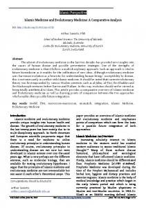
müllerian hormone (AMH) has been elucidated, and a reliable assay for its measurement has been developed (13–15). The gene that codes for the androgen receptor has also been identified, furthering our understanding of complete and partial androgen-resistance syndromes (16). In addition, the genetic marker that results in 5-α-reductase deficiency has recently been identified (17), and this discovery extends our understanding of the actions of testosterone and dihydrotestosterone (DHT). Another important research project shows that the female pathway is not simply a default pathway and that several genes actively regulate female development (18). The processes of sex determination and differentiation proceed in sequence, as shown in Figure 1. Genetically, sex is determined at fertilisation, by the contribution of a Y or an X chromosome from the sperm; this contribution determines the differentiation of the primordial gonads. Thus, the presence or absence of a Y chromosome, with its testis-determining factor, primarily determines the course of sexual differentiation. This differentiation is visible by the 4th week of gestation; the XY karyotype is normally associated with male differentiation, and the XX karyotype is normally associated with female development. Complete female sexual differentiation occurs in the absence of the male determinant, but female differentiation
Review Article | Disorders of sex development: An Islamic perspective is probably regulated by a specific genetic pathway that originates on the long arm of the X chromosome. The presence of a single Y chromosome, even in the presence of more than one X chromosome, is sufficient to cause the development of a testis. Testicular differentiation to produce functional Leydig and Sertoli cells occurs by the 7th week of gestation and is a rapid phenomenon. This rapid differentiation contrasts with the slower development of the ovaries in the 20th week of gestation. At 7th week, the Sertoli cells are actively secreting AMH and Leydig cells are secreting testosterone, and this secretion is controlled by placental human chorionic gonadotrophin (hCG). Thereafter, the foetal pituitary gland controls testicular function in the 2nd and 3rd trimesters. Undifferentiated embryos possess 2 internal duct systems, müllerian (paramesonephric) ducts and wolffian (mesonephric) ducts, but the differentiation of the internal and external genitalia is essentially complete by the end of the 1st trimester. The presence of testosterone and AMH protects the genetically male foetus from inadvertent feminisation (4–19). Locally secreted testosterone promotes the development of the ipsilateral wolffian duct into the epididymis, vas deferens, ejaculatory duct, and seminal vesicle. Testosterone production by 1 testis has no effect on the wolffian duct development on
Y chromosome (testisdetermining factor)
the opposite side. AMH produced by the Sertoli cells causes regression of the ipsilateral müllerian duct. Similar to the local control of the wolffian duct, müllerian duct regression is dependent on the local secretion of AMH and unaffected by AMH secretion from the contralateral testis (4,20,21). Thus, differences in the secretion of testosterone or AMH by the 2 gonads can result in the presence of male internal ducts on 1 side and female structures on the other side. As male internal duct differentiation requires high local concentrations of testosterone, a virilised female with ovaries still has normal female internal structures (4,21). Exposure of female foetuses to androgen in early gestation, before 12th week, can lead to variable degrees of virilisation (Prader’s classification), as shown in Table 1; however, labioscrotal fusion cannot be achieved after 12th week (21,22). The lack of AMH, as well as insensitivity to AMH, is known to yield persistent müllerian duct syndrome. A recent study reports that males with a 46,XY karyotype can be characterised by the presence of Fallopian tubes and a uterus; the external genitalia of these XY individuals are male, and testicular function is otherwise normal (21). Traditionally, the appearance of the external genitalia indicates the appropriate gender assignment. Male development depends
Primordial gonads
Ovary • No testosterone • No AMH
Müllerian ducts development • Fallopian tubes • Uterus • Upper vagina • Normal external female genitalia
Testis
Leydig cells
Sertoli cells
Testosterone
5αreductase
Dihydrotestosterone (DHT)
Anti-müllerian hormone(AMH) Müllerian ducts regression
Wolffian ducts development • Epididymis • Vas deferens • Seminal vesicle
• Penis • Scrotum • Prostate
Figure 1: Simplified model for sexual differentiation and the development of internal and external genitalia. www.mjms.usm.my
5
Malaysian J Med Sci. Jul-Sep 2011; 18(3): 4-12
Table 1: Degree of virilisation of the external genitalia according to Prader’s classification (23) Classification
Characteristics
Type 1 (P-1)
Clitoral hypertrophy
Type 2 (P-2)
Clitoral hypertrophy, urethral and vaginal orifices present, but very near
Type 3 (P-3)
Clitoral hypertrophy, single urogenital orifice, posterior fusion of the labia majora
Type 4 (P-4)
Penile clitoris, perioneoscrotal hypospadias, complete fusion of the labia majora
Type 5 (P-5)
Complete masculinisation (normal-looking male genitalia) but no palpable testes
on adequate testosterone secretion, peripheral metabolism of testosterone to dihydrotestosterone (DHT), and peripheral tissue response to androgens. Male external genitalia differentiate in response to DHT, which is formed in genital skin and other sensitive structures by the metabolism of testosterone by the 5-α-reductase enzyme. The presence of DHT induces the elongation of the genital tubercle, fusion of the genital folds to form the penis, fusion of the labioscrotal folds to form the scrotum, and the formation of the prostate. The response of these structures to DHT requires the presence of a normal intracellular androgen receptor (16,17,23–30). The formation of the male external genitalia is complete by the end of the 1st trimester. After the 1st trimester, further development consists only of growth of the penis and the descent of the testes into the scrotum. Both of these developments depend on the levels of foetal pituitary gonadotrophin (31). In the normal female, the external genitalia do not differentiate into male-specific anatomy; the genital tubercle remains small and becomes the clitoris, and the genital and labioscrotal folds remain unfused to form the labia minora and labia majora, respectively (32).
Diagnostic approach and management
It is important to understand the physiology and embryology of sex-determination and sexual differentiation, but it is also essential to know the different causes of DSD; these causes are shown in Table 2 (1–4,33,37). Most patients are known to have either 46,XX DSD or 46,XY DSD conditions. In 46,XX DSD individuals (previously known as female-pseudohermaphrodites), normal ovaries and internal female organs are present; however, there are variable degrees of 6
www.mjms.usm.my
virilisation of the external genitalia among different individuals. Congenital adrenal hyperplasia, due to a deficiency in the 21α-hydroxylase enzyme or due to deficiencies in the 11β-hydroxylase and 3β-hydroxysteroid dehydrogenase enzymes, constitutes the majority of DSD cases (Figure 2). Congenital adrenal hyperplasia is not an uncommon autosomal recessive condition; it is prevalent in the Arab population due to a high rate of consanguineous matings (38–41). Saedi-Wong et al. (41) reported a high prevalence of congenital adrenal hyperplasia (54.3%) among the Saudi Arabian population. However, 46,XY DSD individuals (previously known as male pseudohermaphrodites) develop normal testes but with incomplete virilisation(undermasculinised external male genitalia, as shown in Figures 3, 4, and 5) due to a variety of causes. Only a small portion of patients have genital ambiguities extensive enough to make the determination of sex-of-rearing difficult. Ovotesticular DSD individuals (previously known as true hermaphrodites) are rare; both testicular and ovarian tissues are present in these individuals. The most common karyotypes in ovotesticular DSD cases is 46,XX, but karyotypes of 46,XY or 46,XX/46,XY can also lead to ovotesticular DSD. The appearance of the internal and external genitalia is variable in individuals with ovotesticular DSD, and the assignment of sex for ovodesticular DSD cases usually depends on the amount of functional testicular tissue. Other disorders of gonadal differentiation, such as pure or mixed-gonadal dysgenesia or testicular regression, should be considered potential causes of ambiguous genitalia, and the presence or absence of these gonadal disorders should be investigated at the time of diagnosis. The diagnosis of ambiguous genitalia can be
Review Article | Disorders of sex development: An Islamic perspective Table 2: Major causes of disorders of sex development (DSD) according to karyotype 46,XX Karyotype 46,XX DSD
Congenital adrenal
Enzyme deficiency
hyperplasia (CAH)
21α-hydroxylase 11β-hydroxylase 3β-hydroxysteroid dehydrogenase
Ovarian/adrenal tumours (mother–child) Exposure to exogenous medication (synthetic progestin preparation) Ovotesticular DSD 46,XY Karyotype 46,XY DSD
Lack of synthesis of
Testicular differentiation
testosterone
pure gonadal dysgenesis absence of Leydig cells or luteinising hormone receptor testicular regression gonadotrophin hormone deficiency Enzyme deficiency in testosterone pathway 20,22-desmolase 17,20-lyase 3β-hydroxysteroid dehydrogenase 17α-ketoreductase
Lack of synthesis of
5α-reductase deficiency
dihydrotestosterone End-organ-unresponsiveness
Partial
(resistance)
Complete
Ovotesticular DSD Multiple or local congenital anomalies Mixed Karyotype Ovotesticular DSD 46,XX/46,XY Mixed gonadal dysgenesis 45,X/46,XY
made only by laparoscopy or by a combination of laparotomy and a histological examination of the gonads (4,42). Ambiguous genitalia in genetic males may be comprised of multiple malformation syndromes, such as Smith–Lemli–Opitz, Vater, and Meckel’s syndromes (43,44). Thus, the presence of other morphogenic anomalies usually indicates a non-endocrine explanation for the abnormal appearance of the genitalia (4). Furthermore, local ano-rectal and
uro-genital anomalies such as epispdius and bladder extrophy are known to be associated with external genital ambiguity (4,43–45). The initial assessment of a DSD patient requires a detailed maternal and family history, as well as a careful physical examination for symptoms including hyperpigmentation and signs of salt-wasting; the initial assessment should also provide documentation of the existing external genitalia (1,3,4,33–37). Proper genital www.mjms.usm.my
7
Malaysian J Med Sci. Jul-Sep 2011; 18(3): 4-12
Figure 2: Ambiguous genitalia in a 46,XX patient known to have congenital adrenal hyperplasia due to 21α-hydroxylase deficiency. Note the complete masculinisation, with normal looking hyperpigmented male genitalia (but no palpable testes).
Figure 3: Ambiguous genitalia in a 46,XY patient known to have congenital adrenal hyperplasia due to 3β-hydroxysteroid dehydrogenase deficiency. Note the pigmented, short, curved phallus, central urogenital slit, and separated labioscrotal testis.
examination at birth and before declaring the sex of a child is always encouraged to prevent the trauma of sex reassignment later in life. Children with ambiguous genitalia should be transferred immediately to a facility where specialised physicians are available. A multidisciplinary team consisting of a paediatric endocrinologist, paediatric surgeon, urologist, plastic surgeon, geneticist, and a psychologist or paediatric psychiatrist should collaborate in managing such a condition. The trauma associated with gender assignment is less when the sex-of-rearing is determined by an expert as soon as possible after birth as opposed to the experience of a gender re-assignment later in life. The issue should be discussed clearly and openly, and parents need to understand immediately that their child’s genitalia are abnormal but can be corrected by surgery, and that the sex-of-rearing will be either a male or female, but this decision requires reasonable time for investigation. The announcement of the baby’s sex should be deferred until supportive data are available. The embryology of sexual differentiation should be reviewed at a level
that allows the parents to understand the issue at hand. Also, parents should understand that virilisation is reflective of the magnitude of androgen stimulation during foetal development, and is not necessarily indicative of the most appropriate sex-of-rearing. Establishing the genetic sex (karyotype) should be the first step, coupled with determining the anatomical status of the internal organs. Further specific hormonal investigations and therapeutic trials need to be undertaken to specify the cause of the anomaly and, hence, the appropriate therapy (3,4,33,46). A paediatric radiologist plays a significant role in elucidating the existing anatomy. Real-time pelvic ultrasonography (US) is not invasive, and this technique remains the modality of choice for such screening; however, US (Figures 6) performed in conjunction with genitography (Figure 7), which reveals further details of the vaginal structures, is far more informative than US alone (47). Magnetic resonance imaging (MRI) is helpful in cases where US and US/genitography examinations are inconclusive (48). Pelvic laparotomy or laparoscopy and gonadal biopsy are rarely needed.
8
www.mjms.usm.my
Review Article | Disorders of sex development: An Islamic perspective
Figure 4: Ambiguous genitalia in a 46,XY patient known to have partial androgen insensitivity. Note the micropenis, urogenital sinus, and labioscrotal folds (the left fold contains a palpable gonad).
Figure
5:
Ambiguous genitalia in a 46,XY patient known to have gonadotrophin hormone deficiency. Note the micropenis, underdeveloped scrotum, and bilateral undescended testes
Figure 6: Ultrasound of the sagittal pelvis showing the bladder (BL) from an anterior view, uterus (UT) from a postero-superior view, and vagina (V) from a postero-inferior view.
Figure 7: A genitogram showing contrast material filling the bladder (BL), vagina (V), and uterus (arrow).
www.mjms.usm.my
9
Malaysian J Med Sci. Jul-Sep 2011; 18(3): 4-12
The current Islamic recommendations put forward by the senior Ulama Council in Saudi Arabia as well as the experiences of local medical practitioners (3,4,46,49) yield a set of very useful general guidelines. These recommendations are translated as follows: 1) A sex-change operation (i.e., converting someone with a completely developed gender to the opposite sex) is totally prohibited, and it is even considered criminal in accordance with the Holy Quran and the Prophet’s sayings. 2) Those who have both male and female organs require further investigation, and if the evidence is more suggestive of a male gender, then it is permissible to treat the individual medically (by hormones or surgery) to eliminate his ambiguity and to raise him as a male. If the evidence is suggestive of a female gender, then it is permissible to treat her medically (by hormones or surgery) to eliminate her ambiguity and to raise her as a female. 3) Physicians must explain the results of medical investigations to the child’s guardians and whether the evidence indicates that the child is male or female so that guardians are well-informed. Therefore, genetically female children (46,XX karyotype) with normal ovaries and internal female organs (uterus, fallopian tubes, and upper vagina), but with some variable degree of virilisation of the external genitalia, should be raised as females; this decision is not only due to the ease of reconstruction of the female genitalia (50–52), but also due to the ability of these females to have high fertility rates and to bear children later in life (53). Any gender-reassignment surgery should generally be performed before 2 years of age, i.e., before the child develops gender-awareness. The techniques used for clitoral recession and vaginoplasty are continually improving. In contrast, a child with a sexual ambiguity and an XY chromosome creates a rather more difficult and challenging problem, not only for the child and his family but also for the medical caretaker. Although familial interactions with the young child during the first months of life are key factors in gender and sexual role-development, it is still difficult to predict the potential for penile growth in relation to future sexual function to determine if the male gender assignment will be satisfactory. Also, it is important to understand that it is more difficult to reconstruct a penis than 10
www.mjms.usm.my
it is to create a vagina. The dominant role of the male gender in the Muslim community should not overrule Islamic Laws; these Laws should not be ignored, and they should be given prime consideration. There may exist a bias concerning the influence of the karyotype, such as the perception that a child with a 46,XY genotypes should be raised as a male. Patients with complete end-organ unresponsiveness due to androgen receptor defects (referred to as androgen insensitivity syndrome or testicular feminisation) lack response to testosterone and, therefore, should be raised as females. It is advisable to remove the gonads at the time of diagnosis rather than wait until puberty, to avoid the adverse effects of testosterone on the neurons and to minimise the risk of the development of gonadoblastoma (4). The availability of oestrogen replacement therapy allows gender reassignment in such cases. However, the use of oestrogen replacement therapy was debated recently, and it was suggested that the removal of gonads should be deferred until puberty to allow for oestrogen formation, as carcinoma in situ is rare and the earliest reported case was at 14 years of age (37). On the other hand, in cases where genetic males demonstrate any appreciable response to exogenous stimulation, a testosterone treatment of short duration might indicate the degree of masculinisation achievable at puberty and facilitate the decision to raise such an individual as a male. In cases of 5-α-reductase deficiency, further virilisation occurs at puberty, along with the development of a male habitus. Many of these individuals will be fertile as adults; therefore, male gender is the appropriate choice for gender assignment, and surgical reconstruction should be performed at 18 months (2,4,21). In testicular regression syndrome, gonadotrophic hormone deficiency, and other defective testosteronesynthesis pathways, a male gender should be assigned, and the appropriate medical therapy should be given. In individuals with ovotesticular DSD (46,XX, 46,XY, or 46,XX/46,XY karyotypes), pure gonadal dysgenesis (46,XX or 46,XY karyotypes), or mixed gonadal dysgenesis (45,X/45,XY), there can be different degrees of internal and external genital formation, depending on the amount of functioning testicular tissue. The sex-of-rearing should be female in most of these cases, and all testicular tissue must be removed at the time of diagnosis because of the risk of virilisation at puberty and the higher incidence of gonadal tumours (4).
Review Article | Disorders of sex development: An Islamic perspective In conclusion, appropriate genetic and psychosocial counselling should be made available to the family and when the patient is old enough to understand, all available information should be progressively disclosed.
Acknowledgement The author would like to thank Ms Loida M Sese for her secretarial assistance.
Correspondence Professor Dr Nasir AM Al Jurayyan MBBS (King Saud University), FRCPC, FAAP Department of Pediatrics College of Medicine & King Khalid University Hospital King Saud University PO Box 2925 Riyadh 11461 Saudi Arabia Tel: +966-1-467-1504 Fax: +966-1-467-1631 Email:
[email protected]
References 1.
Saenger P. Abnormal sex differentiation. J Pediatr. 1984;104(1):1–17.
2.
Rappaport R, Forest MG. Disorders of sexual differentiation. In: Bertrand J, Rappaport R, Sizomenko PC, editors. Paediatric endocrinology: physiology, pathophysiology, and clinical aspects. 2nd ed. London (GB): Williams and Wilkins; 1993. p. 447–470.
3.
Abdullah MA, Katugampola M, al-Habib S, alJurayyan N, al-Samarrai A, al-Nuaim A, et al. Ambiguous genitalia: Medical, socio-cultural and religious factors affecting management in Saudi Arabia. Ann Trop Paediatr. 1991;11(4):343–348.
4.
Couch RM. Disorders of sexual differentiations. Medicine North America. 1987;14:2632–2647.
5.
McLaren A. What makes a man a man? Nature. 1990;346:216–217.
6.
Jost A. Recherches sur la differentiation sexualle de l’embryo de lapin. Arch Anat Micros Morph Exp. 1947;36:271–315.
7.
Whelsons WJ, Russell LB. The Y-chromosome as a bearer of male determining factors in the mouse. Proc Natl Acad Sci USA. 1959;45(4):560–566.
8.
McCarrey JR, Abbott UK. Mechanisms of genetic sex determination, gonadal sex-determination, and germ-cell development in animals. Adv Genet. 1979;20:217–290.
9.
10. Wachtel SS, Ohno S. The immunogenetics of sexual development. Prog Med Genet. 1979;3:109–142. 11. Simpson E, Chandler P, Goulmy E, Disteche CM, Ferguson-Smith MA, Page DC. Separation of the genetic loci for the H-Y antigen and for testis determination on human Y chromosome. Nature. 1987;326(6116):876–878. 12. Capel B. Sex in the 90s: SRY and the switch to the male pathway. Annu Rev Physiol. 1998;60:497–523. 13. Cohen-Haguenauer O, Picard JY, Mattéi MG, Serero S, Nguyen VC, de Tan MF, et al. Mapping of the gene for anti-müllerian hormone to the short arm of human chromosome 19. Cytogenet Cell Genet. 1987;44(1):2–6. 14. Donahoe PK, Care RL, MacLaughlin DT, Epstein J, Fuller AF, Takahashi M, et al. Müllerian-inhibiting substance: Gene structure and mechanism of action of a fetal repressor. Rec Prog Horm Res. 1987;43: 431–467. 15. Rey R. Endocrine, paracrine and cellular regulation of postnatal anti-müllerian hormone secretion by sertoli cells. Trends Endocrinol Metab. 1998;9(7):271–276. 16. Quigley CA, De Bellis A, Marschke KB, el-Awady MK, Wilson EM, French FS. Androgen receptor defects: Historical, clinical and molecular perspectives. Endocr Rev. 1995;16(3):271–321. 17. Wilson JD, Griffin JE, Russell DW. Sterol 5α-reductase 2 deficiency. Endocr Rev. 1993;14(5):577–594. 18. Vainio S, Heikkilä M, Kispert A, Chin N, McMahon AP. Female development in mammals is regulated by Wnt-4 signalling. Nature. 1999;397(6718): 405–409. 19. Josso N. Paediatric applications of anti-müllerian hormone research. 1992 Andrea Prader lecture. Horm Res. 1994;43(6):243–248. 20. Rey R, Picard JY. Embryology and endocrinology of genital development. Baillieres Clin Endocrinol Metab. 1998;12(1):17–33. 21. Saenger PH. Physiology of sexual determination and differentiation. In: Brook CGD, Hindmarsh PC, editors. Clinical paediatric endocrinology. 4th ed. Oxford (GB): Blackwell Scientific Publisher; 2001. p. 60–76. 22. Mann DR, Akinbarni MA, Gould KG, Paul K, Wallen K. Sexual maturation in male rhesus monkeys: Importance of neonatal testosterone exposure and social rank. J Endocrinol. 1998;156(3):493–501. 23. Prader A. Der Genital befund beim pseudohermaphroditism feminus des kongenitalen adrenogenitalen syndromes. Helv Paed Acta. 1954;9:231–247. 24. Wilson JD, Griffin JE, Leshin M, George FW. Role of gonadal hormones in development of sexual phenotypes. Hum Genet. 1981;58(1):78–84.
Jost A, Vigier B, Prepin J, Perchellet JP. Studies on sex differentiation in mammals. Rec Prog Horm Res. 1973;29:1–41.
www.mjms.usm.my
11
Malaysian J Med Sci. Jul-Sep 2011; 18(3): 4-12
25. Griffin KD, Wilson JD. The androgen resistance syndromes: 5α-reductase deficiency, testicular feminization, and related disorders. In: Scriver CR, Beaudet AL, Sly WS, Valle D, Childs B, Kinzler KW, et al., editors. The metabolic and molecular bases of inherited disease. 6th ed. New York (US): McGrawHill; 1989. p. 1919–1944. 26. Russell DW, Wilson JD. Steroid 5-reductase: Two genes, two enzymes. Annu Rev Biochem. 1994;63: 25–26. 27. McPhaul MJ, Marcelli M, Zoppi S, Griffin JE, Wilson JD. Genetic basis of endocrine disease. 4. The spectrum of mutations in the androgen receptor gene that causes androgen resistance. J Clin Endocrinol Metab. 1993;76(1):17–23. 28. Sinnecker GHG, Hiort O, Nitsche EM, Holterhus PM, Kruse K. Functional assessment and clinical classification of androgen sensitivity in patients with mutations of the androgen receptor gene. German Collaborative Intersex Study Group. Eur J Pediatr. 1997;156(1):7–14. 29. Brinkmann AO, Blok LJ, de Ruiter PE, Doesburg P, Steketee K, Berrevoets CA, et al. Mechanisms of androgen receptor activation and function. J Steroid Biochem Mol Biol. 1999;69(1–6):307–313. 30. Holterhus PM, Wiebel J, Sinnecker GHG, Brüggenwirth HT, Sippell WG, Brinkmann AO, et al. Clinical and molecular spectrum of somatic mosaicism in androgen insensitivity syndrome. Pediatr Res. 1999;46(6):684–690.
39. Salman H, Abanamy A, Ghassan B, Khalil M. Congenital adrenal hyperplasia. Ann Saudi Med. 1991;11(1):9–14. 40. Al-Jurayyan NAM, Al-Herbish AS, Abo Bakr AM, Al-Rabeeah AA, Al-Samarrai AI, Jawad AJ, et al. Congenital adrenal hyperplasia in a referral hospital in Saudi Arabia: Epidemiology, pattern and clinical presentation. Ann Saudi Med. 1995;15(5):447–450. 41. Saedi-Wong S, Al-Frayh AR, Wong HYM. Socioeconomic epidemiology of consanguineous matings in Saudi Arabian population. J Asian Afr Stu. 1989;24:247–52. 42. Van Niekerk WA. True hermaphroditism. In: Josso N, editor. The intersex child. London (GB): Karger; 1981. p. 80–99. 43. Dincsoy MY, Salih MAM, al-Jurayyan N, al Saadi M, Patel PJ. Multiple congenital malformations in two sibs reminiscent of hydrolethalus and pseudotrisomy 13 syndromes, new syndromes. Am J Med Genetics. 1995;56(3):317–21. 44. Verloes A, Ayme S, Gambarelli D, Gonzales M, Le Merrer M, Mulliez N, et al. Holoprosencephalypolydactly (‘pseudotrisomy 13’) syndrome: A syndrome with features of hydrolethalus and Smith– Lemli–Opits syndromes. A collaborative multi-center study. J Med Genet. 1991;28(5):297–303. 45. Rimoin DL, Schimke RN. Genetic disorders of the endocrine glands. St Louis (US): C.V. Mosby Co.; 1971.
31. Lovinger RD, Kaplan SL, Grumbach MM. Congenital hypopituitarism associated with neonatal hypoglycemia and microphallus: four cases secondary to hypothalamic hormone deficiencies. J Pediatr. 1975;87(6 Pt 2):S1171–S1181.
46. Al Herbish AS, Al Jurayyan NA, Abo Bakr AM, Abdullah MA, Al Hussain M, Al Rabeah AA, et al. Sex-reassignment: A challenging problem—Current medical and Islamic guidelines. Ann Saudi Med. 1996;16(1):12–15.
32. Pryse-Davies J, Dewhurst CJ. The development of the ovary and uterus in the foetus, newborn and infant: A morphological and enzyme histochemical study. J Pathol. 1971;103(1):5–25.
47. Al Jurayyan NA, Patel PJ, al Herbish AS, Abdullah MA, Abo-Bakr AM, al Rabeeah AA, et al. Ambiguous genitalia: Comparative role of pelvic ultrasonography and genitography. Ann Trop Pediatr. 1995;15(3): 203–207.
33. Pagon RA. Diagnostic approach to the newborn with ambiguous genitalia. Pediatr Clin N Am. 1987;34(4):1019–1031. 34. Jini M, Sen S, Chacko J, Zachariah N, Raghurpathy P, Mammen KE. Gender assignment in male pseudohermaphroditism: An Indian prospective. Pediatr Surg Int. 1993;8(6):500–501. 35. Hughes IA. Disorders of sex development: A new definition and classification. Best Pract Res Clin Endocrinol Metab. 2008;22(1):119–134. 36. Hughes IA, Nihoul-Fekete C, Thomas B, CohenKettenis PT. Consequences of the ESPE/LWPES guidelines for diagnosis and treatment of disorders of sex development. Best Pract Res Clin Endocrinol Metab. 2007;21(3):351–365. 37. Hughes IA, Houk C, Ahmed SF, Lee PA. Consensus statement on management of intersex disorders. J Pediatr Urol. 2006;2(3):148–162. 38. Lubani MM, Isaa ARA, Bushnag R, al-Saleh QA, Dudin KI, Reavey PC, et al. Prevalence of congenital adrenal hyperplasia in Kuwait. Eur J Pediatr. 1990;149(6):391–392.
12
www.mjms.usm.my
48. Biswas K, Kapoor A, Karak AK, Kriplani A, Gupta DK, Kucheria K, et al. Imaging in intersex disorders. J Pediatr Endo Met. 2004;17(6):841–845. 49. Consensus statement on intersex issues: No. 176. Saudi Arabia: The Senior Ulama Council; 1992. 50. Lee PA. Should we change our approach to ambiguous genitalia. Endocrinologist. 2001;11(2):118–123. 51. Dukett JW, Baskin LS. Genitoplasty for intersex anomalies. Eur J Pediatr. 1993;152(Suppl 2): S580–S584. 52. Coran AG, Polley TZ. Surgical management of ambiguous genitalia in the infant and child. J Pediatr Surg. 1991;26(7):812–820. 53. Mulaikal RM, Migeon CJ, Rock JA. Fertilizing rates in female patients with congenital adrenal hyperplasia due to 21-hydroxylase deficiency. N Eng J Med. 1987;316:178–82.