413
An optimized enumeration method for sorbitolfermenting Bifidobacteria in water samples Sharon C. Long, Catalina Arango P., and Jeanine D. Plummer
Abstract: With increased focus on watershed protection under the Surface Water Treatment Rule, indicators that discriminate among sources of microbial inputs (microbial source tracking) are needed to supplement the quantitative information provided by total and fecal coliform measurements for drinking water monitoring. Bifidobacteria are found in the digestive tract and feces of humans and other animals, and also in sewage. Sorbitol is a food additive used exclusively in food intended for human consumption. Therefore, the presence of sorbitol-fermenting Bifidobacteria in environmental waters can be indicative of sources of human fecal contamination. A series of media were evaluated using ATCC cultures of B. breve and B. adolescentis, feces from different animals, and domestic wastewater samples. The media evaluated were Human Bifid Sorbitol agar (HBSA), modified Human Bifid Sorbitol agar, Beerens Medium, modified Beerens Medium, Reinforced Clostridial agar, BIM-25 Medium, and modified BIM-25 Medium. Variables such as sample preservation, incubation time, different pH indicators, plating technique, and discontinuous exposure to sorbitol were also evaluated. A series of biochemical tests were used to confirm positive colonies enumerated on the various media. Membrane filtration and enumeration of sodium sulfite preserved samples on HBSA containing bromocresol purple using loose lidded plates for 48 h provided the best recoveries for presumptive positive colonies. A number of sorbitol-fermenters that were not Bifidobacteria were able to grow on all media tested, resulting in false-positives. Therefore, plating on HBSA should be followed by a confirmation step when monitoring for sorbitol-fermenting Bifidobacteria in environmental waters. A year-long sampling survey of a managed reservoir in Massachusetts provided field validation of the proposed methodology for sorbitol-fermenting Bifidobacteria as a human-related source tracking indicator tool. Key words: sorbitol-fermenting Bifidobacteria, microbial source tracking, watershed management, fecal contamination. 422
Résumé : Avec le souci grandissant de la protection de la ligne de partage des eaux sous la réglementation du « Surface Water Treatment Rule », des indicateurs capables de discriminer les sources d’apport microbien (contrôle des sources microbiennes) sont requises afin de compléter les informations quantitatives fournies par les mesures de coliformes totaux et fécaux permettant le contrôle de l’eau potable. Les bifidobactéries se retrouvent dans le tube digestif et les excréments d’humains et d’autres animaux et également dans les égouts. Le sorbitol est un supplément alimentaire utilisé exclusivement dans les aliments pour consommation humaine. Par conséquent, la présence de bifidobactéries fermentant le sorbitol dans les eaux environnementales pourrait signaler les sources de contamination fécale humaine. Un ensemble de milieux ont été évalués avec des cultures de B. breve et B. adolescentis de l’ATCC, des excréments de différents animaux et des échantillons d’eaux usées domestiques. Les milieux évalués étaient l’agar Human Bifid Sorbitol (HBSA), l’agar Modified Human Bifid Sorbitol, le milieu de Beerens, le milieu de Beerens modifié, l’agar Reinforced Clostridial, le milieu BIM-25, et le milieu BIM-25 modifié. Des variables telles que la préservation des échantillons, le temps incubation, différents indicateurs de pH, la technique d’ensemencement et l’exposition discontinue au sorbitol ont également été évaluées. Un ensemble d’analyses biochimiques ont été accomplies afin de confirmer les colonies positives comptées sur les différents milieux. La filtration sur membrane et le comptage d’échantillons conservés dans du sulfite de sodium sur du HBSA contenant du pourpre de bromocrésol à l’aide de pétris à couvercle flottant pendant 48 heures ont permis les meilleures récupérations de colonies présomptives positives. Un certain nombre de fermenteurs de sorbitol n’étant pas des bifidobactéries ont pu se développer sur tous les milieux analysés, donnant lieu à des faux positifs. Ainsi, l’ensemencement sur HBSA devrait être suivi par une étape de confirmation lors du contrôle
Received 27 September 2004. Revision received 31 January 2005. Accepted 3 February 2005. Published on the NRC Research Press Web site at http://cjm.nrc.ca on 6 August 2005. S.C. Long1 and C. Arango P.2 Civil and Environmental Engineering, 18 Marston Hall, University of Massachusetts, Amherst, MA 01003, USA. J.D. Plummer. Civil and Environmental Engineering, 100 Institute Road, Worcester Polytechnic Institute, Worcester, MA 01609, USA. 1 2
Corresponding author (e-mail:
[email protected]). Present address: Molecular and Cell Biology, BSP 406, 91 North Eagleville Road, University of Connecticut, Storrs, CT 06269, USA.
Can. J. Microbiol. 51: 413–422 (2005)
doi: 10.1139/W05-017
© 2005 NRC Canada
414
Can. J. Microbiol. Vol. 51, 2005 de bifidobactéries fermentant le sorbitol dans les eaux environnementales. Une campagne d’échantillonnage d’une durée d’un an dans un réservoir entretenu au Massachusetts a permis de valider sur le terrain les méthodologies proposées pour l’utilisation de bifidobactéries fermentant le sorbitol en tant qu’outil indicateur pour le contrôle des sources rattachées aux humains. Mots clés : Bifidobactéries fermentant le sorbitol, contrôle des sources microbiennes, gestion de la ligne de partage des eaux, contamination fécale. [Traduit par la Rédaction]
Long et al.
Introduction The use of indicator organisms by utilities and regulators to assure the microbial safety of drinking water is a widely applied and accepted practice. Monitoring for coliforms as indicator organisms most times provides adequate information on water safety. The presence of high numbers of naturally occurring indicator organisms or organisms originating from animals that do not transmit human infectious pathogens present less public health risk for drinking water sources than contamination from humans or other carrier species. Conversely, drinking waters have been implicated in outbreaks of disease even though the distributed water tested negative for coliform indicators (DeNileon 1998). Passage of the Safe Drinking Water Act (SDWA) Amendments of 1996 placed a renewed focus on source water assessment. Proactive source water assessment programs may require monitoring tools which allow for the delineation of specific microbial input sources, for example human versus horses (Karlin et al. 1998). These microbial source tracking (MST) tests should be used to supplement, not replace, traditional indicator systems that provide information about the public health risks associated with drinking water. The information provided from MST tests would allow watershed management agencies to identify contaminant sources and implement actions to eliminate the sources, thus minimizing fecal inputs to source waters in a cost-effective manner. Delineation of microbial sources in drinking water would provide a valuable tool in protecting the drinking water for cities and towns that use unfiltered surface water sources such as Boston, New York, San Francisco, Seattle, and many other smaller localities. To date, the environmental efficacy of MST indicator organisms has not been fully demonstrated. The emerging field of MST is resulting in tools that can be used to specifically identify, and in some cases quantify, the contributions of different sources of pollutants within a watershed that result in water quality degradation. MST tools can be categorized into 3 groups based on their scientific approach. The first involves enumeration of indicator species that are specifically shed by certain host organisms. These include sorbitol-fermenting Bifidobacteria, Rhodococcus coprophilus, F+RNA coliphage serotypes, and phages of Bacteroides fragilis, among others (Tartera and Jofre 1987; Jagals et al. 1995; Puig et al. 1997; Long et al. 2002; Schaper et al. 2002). Many of these methods can be performed using bacteriologically-based methods, which require significant incubation times and can be labor intensive. The second approach is the use of variable traits among a single species such as E. coli ribotyping, rep-PCR, and amplified fragment
length polymorphisms (Dombek et al. 2000; Carson et al. 2003; Scott et al. 2003). These approaches are molecularbased and tend to require the development of an extensive reference library of isolates from known sources that have proven to be geographically limited. The third approach is to monitor for chemical markers such as caffeine, fluorescent whitening agents, or fecal sterols/stenols (Gregor et al. 2002; Buerge et al. 2003; Gilpin et al. 2003). These techniques primarily identify the presence of domestic wastewater. However, since these chemical indicators persist to some extent through wastewater treatment processes, the appropriate reference level that indicates pollution rather than a publicly owned treatment works discharge has yet to be established. For new or alternative MST indicators to be useful to the drinking water community, it must be established that it is highly associated with the human or animal group for which it is specific. In addition, the method for analyzing an indicator needs to be within the capabilities of laboratories typically operated by utilities and boards of sanitation. Therefore, the goal of this research was to optimize a bacteriologically-based methodology for the MST indicator sorbitol-fermenting Bifidobacteria. While it is recognized that more sophisticated, molecular-based methods for detecting sorbitol-fermenting Bifidobacteria have been studied (Lynch et al. 2002; Gilpin et al. 2003), the rationale for studying bacteriologically-based tests is to provide a tool that can be easily and economically implemented in existing laboratories, smaller laboratories, and developing countries. Bifidobacteria, and especially sorbitol-fermenting Bifidobacteria, are good candidates for use as indicators of human or sewage contamination of drinking water sources (Sinton et al. 1998). Bifidobacteria are natural inhabitants of the digestive tract of warm-blooded animals and are an important component of the human colonic flora (Finegold et al. 1983). Sorbitol, a sugar substitute, is a food additive used exclusively in products intended for human consumption owing to its non-nutritive character. Therefore, it can be hypothesized that human fecal waste would be the most likely source of Bifidobacteria with the ability to ferment sorbitol. Studies of animal feces have demonstrated that Bifidobacteria are present consistently in fecal samples from humans and pigs, occasionally in sheep and cattle feces, and are absent from the feces of additional animal species tested including horses, mice, cats, and dogs (Mara and Oragui 1983 and 1985). In other studies, sorbitolfermenting Bifidobacteria were consistently isolated from samples of human feces, domestic wastewater and water subject to human contamination, but were not isolated from any of the animal feces tested (Jagals et al. 1995; Arango 2000; Long et al. 2002). © 2005 NRC Canada
Long et al.
The detection and survival of Bifidobacteria in environmental samples has been less extensively studied. Environmental studies conducted in the United Kingdom and Africa report that Bifidobacteria are consistently isolated from human feces and water subject to human contamination, but are limited to recent pollution events (Mara and Oragui 1985; Jagals et al. 1995). In a year-long study of tributaries to a managed reservoir in Massachusetts, sorbitol-fermenting Bifidobacteria were confirmed in samples from a tributary influenced by residential development served by septic systems but were never detected in a tributary in an undeveloped drainage area or in the main reservoir itself (Long et al. 2003). In addition to land use, climate also impacts detection: sorbitol-fermenting Bifidobacteria tend to be recovered more readily from environmental samples in cooler months when the ambient temperature is below 15 °C (Rhodes and Kator 1999). This can be hypothesized to result from slower organism decay rates at lower environmental temperatures. Understanding the environmental survival of Bifidobacteria is also critical to evaluating the value of this indicator as a MST tool. In flowing waters, the ratio of fecal coliforms to Bifidobacteria increases with distance downstream which indicates Bifidobacteria are less resistant to river conditions than fecal coliforms (Jagals and Grabow 1996). There is some evidence that Bifidobacteria species can decrease in viability up to 26 times faster than E. coli in estuarine systems (Rhodes and Kator 1999). This results from reaeration that occurs in flowing water which is detrimental to strict anaerobes such as Bifidobacteria. Controlled laboratory studies have demonstrated that sorbitol-fermenting Bifidobacteria spiked into drinking water reservoir samples remain at detectable levels for up to 48 h, although numbers decrease by 2 orders of magnitude in 2 days (Long 1999). A study of simulated soil systems showed that sorbitol-fermenting Bifidobacteria can survive in sand saturated with wastewater effluent for 27 days at 20 °C with a first-order decay coefficient of 0.285/day, compared with E. coli, which regrow and remain at or above spiked levels during that time (Long et al. 2002). Carrillo et al. (1985) suggest that Bifidobacteria may be a better indicator of recent fecal contamination since it does not regrow in the environment, as has been demonstrated for E. coli and fecal coliforms in tropical waters. The rapid environmental decay of sorbitol-fermenting Bifidobacteria in aerated surface waters limits this organism’s utility as a general indicator of fecal contamination in temperate waters; however, it may prove to be useful in long-term watershed management and source water protection monitoring schemes. The consistent detection of the organism in simulated septic system soils indicates a reasonable survival time in such systems, especially if continuously loaded with new wastewater. Thus, the literature indicates that screening for the presence of sorbitol-fermenting Bifidobacteria from locations experiencing elevated levels of coliforms could help determine whether human wastes are reaching the receiving waters, thus allowing for appropriate mitigation actions to be implemented. It is proposed that the enumeration of sorbitolfermenting Bifidobacteria could be used as a supplemental test to coliform analyses in watershed monitoring/management activities. The objective of this research was to optimize bacteriologically-based methodologies for the detection and identi-
415
fication of the presence of sorbitol-fermenting Bifidobacteria in environmental waters.
Methods Strains and media B. breve 15698, B. adolescentis 15704, and B. breve 15700 were obtained from ATCC. G89 is an environmental isolate of sorbitol-ferementing Bifidobacteria. Enrichment cultures of these bacteria were routinely grown on Reinforced Clostridial Medium (RCM, Difco, Detroit, Mich.) in anaerobic jars (BBL GasPak Systems, Fisher Scientific, Fairlawn, N.J.) at 37 ± 1 °C. The other media tested included Human Bifid Sorbitol agar (HBSA), modified Human Bifid Sorbitol agar (modHBSA1, modHBSA2, and modHBSA3), Beerens Medium (BM), modified Beerens Medium (modBM), BIM25, and modified BIM-25 (modBIM). The formulations and modifications of these media are summarized in Table 1. Bacterial enumeration and confirmation Enumeration of pure cultures, spiked samples, wastewater influent, and (or) feces was conducted using membrane filtration (Resnick and Levin 1981; Beerens 1991) with anaerobic incubation for 48 ± 2 h at 37 ± 1 °C. Wastewater and fecal samples were diluted in phosphate buffered saline (PBS) prior to filtration as necessary, and at least 3 dilution levels/volumes were analyzed. This method is based on Standard Method 9222 for membrane filtration analysis for coliforms (APHA et al. 1998). Yellow colonies that fermented sorbitol were determined to be “presumptive sorbitol-fermenting Bifidobacteria.” To conduct confirmations of presumptive colonies, up to 10 isolated colonies of presumptive sorbitol-fermenting Bifidobacteria were picked from each enumeration plate for confirmation. Colonies were transferred to Reinforced Clostridial Agar (RCA) plates and incubated anaerobically for 48 ± 2 h. Isolates that demonstrated strict anaerobiosis were subjected to additional confirmation tests (Table 2). These tests were chosen based on the basic characteristics of Bifidobacteria and the ability to ferment sorbitol (Resnick and Levin 1981; Hole et al. 1994). Fermentation purple broths (glucose, lactose, and sorbitol) and nitrate broth were inoculated from the RCA plate within 2 h of removal from the incubator. The fermentation and nitrate broths were incubated anaerobically for 48 ± 2 h. Gram stains and catalase tests were conducted on the same day using culture aliquots from the RCA plate. Isolates are confirmed as sorbitol-fermenting Bifidobacteria only when they exhibited the typical response in all tests as summarized in Table 2. Evaluation of detection media and methods To evaluate the selective and differential characteristics of each medium, a series of experiments were conducted. For each medium, a pure culture of Bifidobacteria was enumerated using RCA and the various selective media. The culture was diluted in PBS, appropriate dilutions were membrane filtered, the membrane was placed onto the medium, and the plates were incubated anaerobically at 37 ± 1 °C for 48 ± 2 h, unless incubation time was a parameter being tested. In addition, environmental samples (wastewater influent or feces) were diluted, filtered, and incubated to assess the © 2005 NRC Canada
Sorbitol
Sorbitol
Glucose, soluble starch
Sorbitol, soluble starch
Glucose, soluble starch
Neomycin 5 mg/L and 15 mg/L, bromocresol purple 0.1 g/L
Naladixic acid 30 mg/L, kanamycin 50 µg/L, polymixin B sulfate 10 IU/L, neomycin 1 mg/L, bromocresol purple 0.1 g/L Propionic acid 5 mL/L
Propionic acid 5 mL/L
Naladixic acid 20 mg/L, kanamycin 50 mg/L, polymixin B sulfate 8.5 mg/L, iodoacetic acid 25 mg/L Naladixic acid 20 mg/L, kanamycin 50 mg/L, polymixin B sulfate 8.5 mg/L, iodoacetic acid 25 mg/L
Yeast extract, peptone
Yeast extract, peptone
Modified BIM-25 (modBIM)
Modified Beerens Medium (modBM) BIM-25
Columbia commercial basal medium Columbia commercial basal medium Reinforced clostridial medium Tryptose, beef extract, yeast extract
Sorbitol, soluble starch
Kosteck, 2000; Silvi et al. 1996
Mara and Oragui 1983; Mara and Oragui; Jagals et al. 1995 Hill 2000; Lim et al. 1993; Dave and Shah 1996 Hill 2000; Lim et al. 1993; Dave and Shah 1996 Beerens 1991 Sorbitol Naladixic acid 30 mg/L, kanamycin 50 µg/L, polymixin B sulfate 10 IU/L, bromocresol purple 0.1 g/L
Modified HBSA no. 1 and no. 2 (modHBSA1 and 2) Modified HBSA no. 3 (modHBSA3) Beerens Medium (BM)
Difco 1998 Glucose, soluble starch None
Tryptose, beef extract, yeast extract Yeast extract, peptone Reinforced Clostridial Agar (RCA) Human Bifid Sorbitol Agar (HBSA)
References Selective agent(s) Base
Carbon source
Can. J. Microbiol. Vol. 51, 2005
Media
Table 1. Summary of media tested.
416
selectivity of each medium. In addition to the media formulations, method conditions that were evaluated included incubation time, sample preservation with sodium sulfite, pre-incubation in the presence of sorbitol, addition of pH indicators to the plating medium, and plating technique (Table 3). For each of the experiments, a representative number of the colonies (up to 10 colonies on plates with well-separated growth), which exhibited the appropriate morphology for the medium, were isolated and subjected to confirmation tests to determine definitively the colonies’ identity as a sorbitolfermenting Bifidobacteria as described above. Field validation of the method To validate the applicability of monitoring sorbitolfermenting Bifidobacteria in environmental waters, a yearlong survey of 2 tributaries and the intake of a drinking water reservoir in Massachusetts was conducted. Monthly water samples were collected from 3 locations: Cosgrove Intake, French Brook, and Gates Brook. Cosgrove Intake is the location where the water enters the distribution system, and was determined to be a highly protected site. French Brook is located in an undeveloped area of the watershed and is not immediately influenced by any residential development. This site was designated to be predominantly wildlife influenced. Gates Brook is located in a highly developed area of the watershed and is influenced by residential and commercial development mainly served by aging septic systems. Gates Brook was designated a human influenced site. The water samples were analyzed for sorbitolfermenting Bifidobacteria using membrane filtration with incubation on HBSA. A representative number of yellow colonies (up to 10 colonies per plate) from each sample enumerated were then subjected to confirmation tests.
Results and discussion The method development analyses conducted for sorbitolfermenting Bifidobacteria can be divided into several broad categories including evaluation of media, incubation time, sample preservation, pH indicators, preincubation with sorbitol, and plating method. The usefulness of monitoring sorbitol-fermenting Bifidobacteria in environmental samples was determined through enumeration of field samples. The results from each one of these analyses are discussed below. Media recovery and selectivity Pure cultures of Bifidobacteria obtained from ATCC included B. breve 15698 (non-sorbitol fermenter), B. adolescentis 15704 (variable sorbitol fermentation ability), and B. breve 15700 (constitutive sorbitol fermenter). Liquid cultures were enumerated on HBSA, modHBSA1, BM, modBM, BIM-25 and modBIM (as described in Table 1). It must be acknowledged that a number of additional media have been developed for the enumeration of Bifidobacteria spp. from a variety of sample types such as food (Nebra and Blanch 1999); however, these media are not specifically aimed at sorbitol-fermenting species. For the media tested, numbers enumerated were compared with the number of colony forming units recovered on RCA, an enrichment medium. A recovery ratio of 1.00 indicates identical recoveries, © 2005 NRC Canada
Long et al.
417
Table 2. Sorbitol-fermenting Bifidobacteria confirmation tests. Test
Sorbitol-fermenting Bifidobacteria reaction
Aerobic growth on Reinforced Clostridial Agar Gram stain Glucose fermentation in purple broth Lactose fermentation in purple broth Sorbitol fermentation in purple broth Nitrate Reduction Catalase Reaction Motility
None Gram variable with bifurcating rods in Ys and Vs Acid production without gas* (yellow color, no gas in Durham tube) Acid production without gas (yellow color, no gas in Durham tube) Acid production without gas (yellow color, no gas in Durham tube) Negative Negative Negative
*Sorbitol utilization appears to be inducible and organisms may demonstrate variable sorbitol-fermentation reactions upon transfer from Reinforced Clostridial Agar.
Table 3. Summary of incubation conditions tested. Factor
Variables tested
Standard conditions
Plating Medium Plate Incubation Time Sample Preservation
Eight media formulations, with and without neomycin added (see Table 1) 48 and 90 h Without sodium sulfite; 0.02, 0.03 and 0.04 mol/L final concentration sodium sulfite With sorbitol, without sorbitol Bromcresol purple, bromcresol green Spread plating, membrane filtration with transfer, membrane filtration with membrane incubation 50 mm tight lid Petri dishes, 50 mm tight lid Petri dishes with pierced holes, 100 mm loose lid Petri dishes
RCA without neomycin 48 h 0.02 mol/L final concentration sodium sulfite Without sorbitol Bromcresol purple Membrane filtration with membrane incubation 100 mm loose lid Petri dishes
Pre-incubation with Sorbitol pH Indicator Plating Method Plate Venting
Table 4. Comparison of recoveries of pure cultures on selective media (cfu/mL medium)/(cfu/mL on RCA). Medium Organism
Sorbitol metabolism
HBSA
modHBSA1
BM
modBM
BIM-25
modBIM
B. breve (ATCC 15698) B. adolescentis (ATCC 15704) B. breve (ATCC 15700)
non-fermenter variable fermenter fermenter
0.07 0.17 1.17
NT NT 0
0.33 0.1 0.33
0.17 0.06 0.17
NT NT 1.06
NT NT 1.14
Note: NT, not tested.
a value of less than 1.00 indicates less recovery than RCA, and a value greater than 1.00 indicates greater recovery than RCA. The results are presented in Table 4. Recovery levels of B. breve 15698 and B. adolescentis 15704 strains (0.06– 0.33) indicate that growth and identification of non-sorbitolfermenters or variable sorbitol-fermenters was adequately low on the medium tested. The recoveries of B. breve 15700 indicate that HBSA, BIM-25 and modBIM performed at acceptable levels for pure cultures. Both BM and modBM recovered low levels of B. breve 15700 at 0.33 and 0.17, respectively. ModHBSA1 recovery was unacceptable at below detection limits. While the literature indicates that Bifidobacteria are one of the genera that exhibit resistance to neomycin, modHBSA1 which contained 5 mg/L neomycin provided too harsh a growth environment and resulted in no recovered colonies. To evaluate the selectivity and differentiation value of each medium, selected fecal and wastewater samples were enumerated on selected media with a representative number of presumptive positive colonies (compact yellow) subjected to confirmation tests. A total of 6 buffalo fecal samples, 4 pig fecal samples and 9 samples of cow manure were tested. The
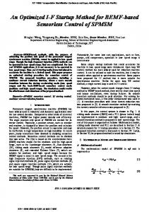
Fig. 1. Comparison of presumptive positive enumerations of animal feces. Error bars represent 95% confidence intervals. HBSA, Human Bifid Sorbitol agar; BM, Beerens Medium; modBM, modified Beerens Medium.
© 2005 NRC Canada
418
Can. J. Microbiol. Vol. 51, 2005
Fig. 2. Comparison of presumptive positive enumerations of domestic wastewater. Error bars represent 95% confidence intervals (95% confidence limits all less than ±9 × 10⁵ cfu/100 mL). HBSA, Human Bifid Sorbitol agar; modHBSA3, modified Human Bifid Sorbitol agar 3; BM, Beerens Medium; modBM, modified Beerens Medium; modBIM, modified BIM-25.
results from the animal fecal sample tests are presented in Fig. 1. Sorbitol-fermenting Bifidobacteria should be absent from the feces of adult animals. HBSA consistently yielded the lowest numbers of fermenting organisms (falsepositives) with the smallest variation. The coefficient of variation for results on HBSA ranged from 60%–70% for the 3 sample types, compared with 75%–149% for BM, and 88%–190% for modBM. No isolates from the buffalo feces or cow manure were confirmed as sorbitol-fermenting Bifidobacteria. No isolates from pig feces on HBSA were confirmed as sorbitol-fermenting Bifidobacteria, which may be an artifact of the relatively low number of isolates tested (n = 6). However, 4 out of 6 pig isolates from BM were confirmed as Bifidobacteria, of which 2 were sorbitolfermenters. One out of eight isolates from pig feces plated on modBM was confirmed as a sorbitol-fermenting Bifidobacteria. The limited number of fecal analyses conducted certainly are not statistically representative of human and animal populations. The presence and levels of sorbitol-fermenting Bifidobacteria detected in this study are generally consistent with results of other studies analyzing animal feces for sorbitol-fermenting Bifidobacteria, indicating that they are a good candidate indicator of human and pig fecal contamination (Mara and Oragui 1983; Jagals et al. 1995; Lynch et al. 2002). A careful sampling regime is suggested for watershed areas where human septage and swine agriculture are both present. The composite results for enumerations of 6 different wastewater influent samples are presented in Fig. 2. Generally, all media yielded presumptive colonies on the same order of magnitude (2 × 10⁷ cfu/100 mL) with the exception of modHSBA3, which was an order of magnitude lower. Up to 10 well separated presumptive colonies were selected for confirmation. For 1 wastewater sample on HBSA and modHBSA3, 6 and 4 colonies, respectively, were isolated, which represented all presumptive colonies because of the dilution levels plated. The confirmation rate for wastewater, expected to contain sorbitol-fermenting Bifidobacteria, averaged 66% for HBSA, 50% for the limited number of colonies that regrew from modHBSA3 (60% of transferred colonies did not
regrow), 20% for BM, 25% for modBM, 16% for BIM-25, and 33% for modBIM. The confirmation rates for all media tested indicate a relatively high rate of false-positive colonies (34%–84%) at the presumptive step. The presence of bromocresol purple in the HBSA medium allowed for easier discrimination between fermenting and non-fermenting organisms in the presumptive step. The percent confirmed colonies was consistently highest from HBSA plates. The specificity of HBSA medium was less than optimal, requiring a suite of confirmation tests to definitively report the presence of sorbitol-fermenting Bifidobacteria in an environmental sample. Therefore, the antibiotic neomycin was tested. Neomycin is an aminoglycoside antibiotic active primarily against Gram-negative bacteria, with activity against a wide variety of Gram-positive bacteria as well (Hill 2000). Cultures of B. breve 15700 and B. adolescentis 15074 were enumerated on modHBSA1 containing 5 mg/L and modHBSA2 containing 15 mg/L neomycin. Colonies from each medium were transferred onto RCA and HBSA for enrichment, and subjected to confirmation tests. All colony formation of the pure cultures was suppressed at the 15 mg/L concentration. Seventy-five percent of B. breve and 95% of B. adolescentis colony formation was suppressed at the 5 mg/L level when compared with colony forming units on HBSA controls. Upon transfer to either RCA or unmodified HBSA, none of the 8 colonies from the modHSBA1 plates with 5 mg/L neomycin grew, while 94% of the colonies transferred from the HBSA (unmodified) control did grow. The addition of 1 mg/L neomycin (modHBSA3) was compared with HBSA using wastewater. The modHBSA3 recovered fewer confirmed sorbitol-fermenting Bifidobacteria by a factor of 50 compared with unmodified HSBA, but performed better than modHBSA1 or 2. Overall, neomycin at the recommended concentrations was too harsh for use in media for enumeration of Bifidobacteria from environmental drinking water sources. Method optimization It can be hypothesized that longer incubation times allow for greater extent of colony development and for greater © 2005 NRC Canada
Long et al.
fermentation product accumulation. The GasPak system states that anaerobic conditions can be maintained for 48 h. Incubations were extended to 90 h, assuring that the oxygen indicator did not change color, for comparison. When B. adolescentis, the variable sorbitol-fermenter, was incubated on HBSA, there was no appreciable difference in the number of colonies with incubation time, but the colonies were larger after 90 h when compared with 48 h of incubation. The change in color of the bromocresol purple indicator in HSBA occurred only around groups of tightly spaced colonies after 48 h of incubation, while the color change on the whole plate was more extensive after 90 h. When incubated on modBM with bromocresol green added, the indicator did not change color after either 48 or 90 h of incubation. When B. breve 15700 was incubated on HBSA, most colonies exhibited a dark-yellow color after 48 h of incubation. These results suggest that the prevalence of false-negatives when using HBSA would be less than when using modBM with bromocresol green. For true sorbitol-fermenters, extended incubation times, and thus extended analysis time, do not seem warranted. Aeration caused by repeated inversion of the sample before subsampling for filtration could injure the indicator organisms because of their strict anaerobic character, and result in false-negatives. Maintaining reduced dissolved oxygen conditions in the samples could be hypothesized to prolong the survival of Bifidobacteria. To extend the amount of time samples may be held prior to laboratory processing, the dissolved oxygen in the water samples must be maintained at very low levels, below 2 mg/L and preferably around 0.5 mg/L. Sodium sulfite was identified as a possible sample preservative, and experiments to assess its suitability for Bifidobacteria enumeration were conducted. Phosphate buffered saline (PBS) solutions with final sodium sulfite concentrations of 0.02 to 0.04 mol/L were inoculated with B. breve 15700 and chilled for 4 h before enumeration. Inoculated PBS without sodium sulfite was enumerated immediately and after 4 h (also chilled). As shown in Table 5, B. breve 15700 concentrations in sodium sulfite preserved samples were not significantly different from the unpreserved sample that was enumerated immediately. However, the difference in enumerated sorbitol-fermenting Bifidobacteria was statistically significant at greater than the 95% confidence level for the unpreserved sample between time 0 h and 4 h. Split samples of Connecticut River water were inoculated with B. breve 15700, and 2 of the 3 subsamples were preserved using sodium sulfite (final concentration of 0.02 mol/L) and chilled for enumeration after 24 and 48 h. The unpreserved subsample was filtered and enumerated immediately. The results of this experiment, summarized in Table 6, indicated that the B. breve concentrations in the preserved subsample were not significantly different from the unpreserved subsample after 24 h (p = 0.144, Student’s t test). After 48 h, however, the concentrations were significantly different (p = 0.040) at a 95% confidence level. The final experiment examining the use of sodium sulfite as a sample preservative involved enumerating the presence of sorbitol-fermenting Bifidobacteria from environmental samples. A surface water sample downstream from septic leach pits was collected and split. One subsample was preserved using sodium
419 Table 5. Effect of sodium sulfite concentration on dissolved oxygen and B. breve. Time (h)
Sodium sulfite (M)
Dissolved oxygen (mg/L)
B. breve (cfu/100 mL)
p valuea
0 4 4 4 4
none none 0.02 0.03 0.04
8.32 8.48 0.43 0.30 0.44
3140 1690 2510 2910 2590
— 0.012 0.383 0.650 0.355
a
Compared to t = 0 h, using pooled standard deviation.
Table 6. Effect of sodium sulfite on dissolved oxygen and B. breve over time. Time (h)
Dissolved oxygen (mg/L)
B. breve (cfu/100 mL)
Percent reductiona
p valueb
0 24 48
10.85 0.54 0.51
873 000 708 000 553 000
— 19 37
— 0.144 0.04
a b
Compared to t = 0 h. Using pooled standard deviation.
sulfite (final concentration of 0.02 mol/L) and one was not. After a 48 hour incubation on HBSA, the number of presumptive colonies was identical for both samples (33 cfu/100 mL). However, the sodium sulfite preservative acted to inhibit approximately 30% of the background bacteria. This effect would aid in isolating the target indicator organism. The goal of testing the oxygen quenching agent sodium sulfite was to reduce continued oxygen inactivation of the target strict anaerobic organisms and specifically sorbitolfermenting strains. Other studies have been conducted aimed at recovery of injured Bifidobacteria from sewage using preexposure to L-cysteine, sodium pyruvate and sodium thioglycolate (Nebra et al. 2002). While this work demonstrated greater recoveries of Bifidobacteria spp. from sewage with use of the reducing agent treatments tested, enumeration of the strain of B. adolescentis tested resulted in no growth on Bifidobacterium selective medium (BFM). Overall, it is suggested that a 0.02 mol/L final concentration of sodium sulfite be used to reduce dissolved oxygen concentrations in water samples intended for sorbitol-fermenting Bifidobacteria enumeration. The relatively high percentage of non-confirming colonies from fecal and wastewater samples indicated a need for an even more differential medium. As neomycin was selective against the organism of interest, a different pH indicator was investigated. HBSA and modBM media containing bromocresol green, which has a color change at a more acidic pH (pH 3.8–5.4 vs. 5.2–6.8 for bromocresol purple), was tested in an attempt to develop a more sensitive medium formulation. Experiments were conducted with the variable and non-fermenting strains of Bifidobacteria. No color change was detected using bromocresol green as a pH indicator and it appeared to inhibit growth of the desired organisms over HBSA and modBM containing bromocresol purple. Studies of the sorbitol metabolic pathways in E. coli indicate that D-sorbitol is activated to D-glucitol-6-phosphate through an inducible pathway (Neidhardt et al. 1996). The genes for the series of enzymes involved in bacterial sorbitol © 2005 NRC Canada
420
metabolism comprise the gut operon. D-sorbitol itself functions as the inducer. The operon is also subject to catabolite repression by D-glucose. If metabolism of sorbitol in Bifidobacteria is similar to E. coli, this biochemical fact is significant in choosing the appropriate method for detection and confirmation of sorbitol-fermenting Bifidobacteria as a MST indicator of human sewage and fecal contamination. Initial work with the ATCC type strains (data not shown) and work presented below confirmed that the sorbitol biodegradation pathway in Bifidobacteria is inducible/repressible in some strains, as indicated in the literature. The transfer of sorbitol-fermenting organisms onto sorbitol-free/glucosecontaining medium resulted in the loss of sorbitolfermenting metabolism upon a second transfer back onto sorbitol containing medium. Therefore, pre-incubation in the presence of sorbitol was explored. Strains of Bifidobacteria with different sorbitol fermentation abilities were incubated on either sorbitol-containing or glucose-containing media. The cultures were spiked into stream water and analyzed. B. adolescentis, the variable sorbitol fermenter, was grown in RC medium broth with and without sorbitol. For B. adolescentis enumeration on HBSA showed no difference in total colony recovery at the 95% confidence level for cultures grown in RC medium with and without sorbitol. However, the colonies on HBSA from the sorbitol-grown culture were yellow while the colonies from the glucose-grown culture were not yellow. In both treatments, the B. adolescentis were confirmed as Bifidobacteria but not as sorbitol fermenters, indicating repression of sorbitol fermentation by glucose in the RC medium. The recovery of B. breve 16598, the non-sorbitol fermenter, was approximately 2 orders of magnitude higher with preincubation with sorbitol (865 vs. 20 cfu/mL); however, in both cases colonies on HBSA were not yellow. Growth and sorbitol fermentation (yellow color) by B. breve 15700 and an environmental isolate G89, a sorbitol fermenter, were not affected by preincubation with sorbitol. The results suggest that exposure to sorbitol, or the absence of that exposure, will affect the detection and enumeration of certain strains of Bifidobacteria. The results of these experiments indicate that when a variable sorbitol fermenter had been exposed to sorbitol immediately preceding testing, the organism demonstrated a strong fermentation ability (yellow colonies). The lack of exposure to sorbitol can reduce the sorbitol fermentation response within a 48 h incubation. Therefore, it was determined that RCA containing sorbitol should be used as the enrichment medium for growing isolates for confirmation tests. The presentation of yellow presumptive colonies on sorbitol containing media (HBSA) from an environmental sample is indicative of previous exposure to sorbitol. Therefore, such variable fermenters can be taken as an indicator of human-related wastes. For enumeration of environmental water samples, processing larger sample volumes results in lower detection limits. However, optimal growth of the desired organisms must not be sacrificed. It could be hypothesized that diffusion of nutrients and fermentation products through filter membranes could slow colony development and color change when compared with direct plating on the agar surface (spreadplating). Therefore, comparisons were conducted between spread-plating water samples, membrane-filtration with
Can. J. Microbiol. Vol. 51, 2005
transfer onto the medium (placing the membrane face down onto the medium for 1 min before removal), and membranefiltration with membrane incubation on the medium. Experiments were conducted using B. adolescentis 15704 to assess the methodology for variable fermenters that are difficult to detect. The results indicate that nutrient transfer and indicator color change was not inhibited by the presence of the membrane. Transfer efficiency using the transfer method was low compared with membrane incubation (3 colonies versus too numerous to count, respectively). Spread-plates were limited to 500 µL maximum volume per plate, resulting in detection limits on the order of 200 cfu/100 mL. Membrane filtration with incubation of the membrane on the medium surface promotes colony development and allows for analysis of larger water samples (100 to 200 mL depending on water quality), resulting in detection limits on the order of 1 cfu/100 mL. As a strict anaerobe with mild tolerance to oxygen, rapid development of anaerobic conditions within the incubation vessel (jar or paks) for Bifidobacteria manipulated in ambient air conditions is important. In initial work with the ATCC type strains, it was observed that inconsistent numbers of colony forming units were enumerated when membranes were incubated on tight-fit lid 50 mm petri dishes (Gelman Sciences, Ann Arbor, Mich.) typically used for coliform analyses. It was hypothesized that these plates were designed for limited gas flow to minimize dehydration of the medium, and therefore resulted in slow and variable rates of anaerobic condition development within the interior of the plate. Thus, 3 plate types were tested including 50 mm tight lid petri dishes typically used by utilities for coliform membrane filtration analyses, 50 mm tight lid petri dishes with holes pierced through the plate lid for ventilation, and 100 mm loose lid petri dishes. Experiments were conducted using B. adolescentis owing to its variable sorbitolfermentation ability and thus its difficult detection. Results indicate that rapid gas transfer using loose lid plates or plates with ventilation performed better, resulting in more consistent colony counts (smaller coefficient of variation). Owing to concerns about aseptic integrity, the use of loose lid plates is recommended. After extensive laboratory testing, it was determined that sodium sulfite preserved samples with enumeration via membrane-filtration with 48 h incubation on HBSA in loose lid plates followed by confirmation tests is most appropriate for enumerating sorbitol-fermenting Bifidobacteria in environmental drinking water sources. The factors considered included recovery rates of pure ATCC cultures, ease of colony identification, and percentage of confirmed positive enumerations. HBSA, BIM-25, and modBIM provided good recovery rates and differential colonies: yellow on HBSA and pink on BIM-25 and modBIM. However, confirmation rates of isolates from wastewater samples were consistently higher for HBSA, averaging 66%, compared with 16% and 33% for BIM-25 and modBIM, respectively. Field validation The membrane filtration method with enumeration on HBSA was used to analyze monthly water samples from 3 highly characterized sites located in a Massachusetts drinking water watershed to field validate the method. As described © 2005 NRC Canada
Long et al.
421
Table 7. Results of drinking water source monitoring for indicator organisms.
March April May June July August September October November December January February 2 March
Cosgrove Intake
French Brook
Gates Brook
Rain
FC
SFB
FC
SFB
FC
SFB
* * * *