Mj. Int. J. Sci. Tech., 2007, 01(02), 120-136
120
Maejo International Journal of Science and Technology ISSN 1905-7873 Available online at www.mijst.mju.ac.th Review Article
An overview of aerosol particle sensors for size distribution measurement Panich Intra 1,* and Nakorn Tippayawong 2 1*
Energy Research Center, Maejo University, Chiang Mai 50290, Thailand, E-mail:
[email protected] 2 Department of Mechanical Engineering, Faculty of Engineering, Chiang Mai University, Chiang Mai 50200, Thailand * Author to whom correspondence should be addressed. Received: 15 June 2007 / Accepted: 4 August 2007 / Published: 18 August 2007
Abstract: Fine aerosols are generally referred to airborne particles of diameter in submicron or nanometer size range. Measurement capabilities are required to gain understanding of these particle dynamics. One of the most important physical and chemical parameters is the particle size distribution. The aim of this article is to give an overview of recent development of already existing sensors for particle size distribution measurement based on electrical mobility determination. Available instruments for particle size measurement include a scanning mobility particle sizer (SMPS), an electrical aerosol spectrometer (EAS), an engine exhaust particle sizer (EEPS), a bipolar charge aerosol classifier (BCAC), a fast aerosol spectrometer (FAS) a differential mobility spectrometer (DMS), and a CMU electrical mobility spectrometer (EMS). The operating principles, as well as detailed physical characteristics of these instruments and their main components consisting of a particle charger, a mobility classifier, and a signal detector, are described. Typical measurements of aerosol from various sources by these instruments compared with an electrical low pressure impactor (ELPI) are also presented. Keywords: aerosol, electrical mobility, nanoparticles, size distribution, spectrometer
Mj. Int. J. Sci. Tech., 2007, 01(02), 120-136
121
Introduction A recent particulate air pollution episode has affected Chiang Mai adversely. Aerosol is one of the most important environmental topics and particulate air pollution has become a major national concern. Emissions to air arise from human activities and natural processes. Anthropogenic emissions occur during extraction, distribution and combustion of fossil fuels from various industrial processes, from waste treatment and disposal, from agriculture, and from a range of consumer products. High fine particle concentrations in Chiang Mai were identified to be associated with a number of open fires observed during the months from February to April. Additionally, automotive vehicles have long been recognized as a major source of particulate air pollution. Emissions from road transport are growing steadily in line with increasing traffic. Any solid or liquid material suspended in air with diameter in the range of 1 nm to 100 μm can be considered as particulate matter [1]. It is a complex mixture of liquid and solid particles that exist in dynamic equilibrium with gaseous components in surrounding air and can be classified into two categories, viz. primary (directly emitted to the atmosphere) and secondary (formed at a later stage with atmospheric reactions) particles. The aerosol undergoes a number of complex reactions, affecting both the physical and chemical characteristics of ambient particulate matter. These particles have significant effects on the human health, Earth’s climate, air quality and processes in various industries such as food, pharmaceutical and medical, electronic and semiconductor industries. Important physical properties of airborne particles are size, number, surface area, density and shape. Knowledge of these properties of aerosols is of great practical importance in aerosol science, air pollution, process control industry and epidemiological studies among others. In air pollution studies, photochemical reactions in the atmosphere often begin with nanometer-sized particles. In nucleation and condensation processes which are the basic of many technological applications, nanometer particles serve as the incipient nuclei for many processes. In the electronic and semiconductor industry, the contamination control of nanometer-sized particles is needed to prevent the formation and deposition of these particles on semiconductor devices during fabrication, because the size of the next generation 1-gigabit DRAM devices will have a minimum feature size smaller than 180 nm [2]. Using the common 1/3 or 1/5 rule for micro-contamination control, particles as small as 35 nm need to be measured and controlled. In industrial hygiene and epidemiological studies, the health consequences of these particulates depend on their ability to penetrate and deposit in the respiratory system. Particles with size larger than 10 µm are screened out quite easily at the upper inhalation system. For particles in the size range between 1 to 10 µm, they can penetrate the alveoli and bypass the upper respiratory tract. These particles are small enough to allow deposition at places where they can do the most damage. Particles with a diameter of less than 1 μm can penetrate deep into the respiratory system in the human lungs and are difficult to remove by lung clearance mechanisms, and for this reason they are considered to be the most dangerous. There is therefore a need to develop efficient respirators for protecting workers exposed to environments containing nanometer sized particles. Measurement and characterization of these particles is also needed in order to better understand and control them. A particle size instrument is one of the valuable tools for these applications. There are several common instruments using various methods of measuring particle sizes. The techniques include diffusion method, inertial impaction method, light scattering method, electron microscopy method and aerodynamic method. However, they can only be used to measure super-
Mj. Int. J. Sci. Tech., 2007, 01(02), 120-136
122
micrometer sized particles or are painstakingly difficult and slow to obtain results for sub-micrometer or nanometer-sized particles. The most efficient and widely used technique for measuring these submicron particles is essentially an electrical mobility determination method. Electrical mobility method was initially developed to measure ions in gases [3, 4] and in the atmosphere [5, 6]. Rohmann [7] later investigated and employed the method to measure atmospheric airborne particles. There have been numerous studies and developments on the electrical aerosol measurement in the past several decades. Among the first studies were carried out by Whitby and Clake [8], Tammet et al. [9], Knutson and Whitby [10] and Liu and Pui [11] over a few decades ago. An electrical aerosol analyzer and a differential mobility analyzer (DMA), capable of guaging the diameter to 10 nm - 1 μm were developed and later improved and refined by a number of researchers such as Kousaka et al. [12], Lehtimaki [13, 14], Stolzenburg [15] and Winklmayr et al. [16]. Chen et al. [17] employed the numerical method to improve DMA design. The results were in good agreement with experimental data [18 - 20]. Seto et al. [21] studied the performance of DMA under low pressure conditions to measure nanometer-sized particles in the size range between 4 - 10 nm. New developments were constantly tested and compared to assess their performance and to expand the measurement range [2226]. Kulon et al. [27 - 29] described similar development concerning a bipolar charge aerosol classifier (BCAC). It uses electrostatic technique and is capable of simultaneously measuring particle charge as well as size. With respect to recent development on a similar instrument, Tammet et al. [30 - 31] designed and developed an electrical aerosol spectrometer (EAS) which can classify particles in a similar fashion to, but faster than a typical DMA due to its multi-channel measurement capability. In his PhD work, Graskow [32] developed a fast aerosol spectrometer (FAS) to measure nanometer-sized particles. His FAS prototype has better time response than the EAS. Reavell et al. [33 -35] and Biskos [36 - 41] reported a further development of a differential mobility spectrometer (DMS), derived from Graskow’s concept. Based on similar principle to previously mentioned instruments, Intra and Tippayawong [42 - 46] designed, built and tested a multi-channel electrical mobility sensor for aerosol size distribution measurement. Although these instruments are all designed to measure airborne particle size distribution using the same principles, they are also different in terms of specific applications, construction, cost, measurement range, as well as time response and resolution. In this paper, an overview of the state-of-the-art electrical mobility sizers for nanoparticle size measurement is described. Some of these instruments are commercially available and others are still laboratory prototypes. A detailed description of the operating principle of these instruments as well as main components, including the particle charger, the mobility classifier, and the signal detector are presented. Typical measurements of aerosol from various sources are also shown.
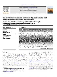
Electrical Mobility Operating Principle This section describes the operating principles of the particle sizer based on the electrical mobility technique. The basic principle of the electrical mobility technique is shown in Figure 1. This instrument consists of the corona charger, the mobility classifier, and the particle detector. Particle charging is accomplished by exposing aerosol sample to the cloud of unipolar corona ions inside the particle charger, and then charged via ion-particle collisions. The charged aerosol passes into the electrical mobility classifier, configured as coaxially cylindrical electrodes. A DC high voltage ranging
Mj. Int. J. Sci. Tech., 2007, 01(02), 120-136
123
from 1 to 5 kV is applied to the inner electrode. There are two separate streams: aerosol and sheath air flow. The charged aerosols enter the analyzer column. An electric field formed between the electrodes makes the particles deflect radially outward, and particles having specific mobility are collected on electrically-isolated electrometer rings, positioned at the inner surface of the outer electrode. Electrometers connected to these electrode rings measure currents corresponding to the number concentration of particles of a given mobility which is related to the particle size. Signal currents are Isolation Housing
Electrometer Rings
Isolation Rings
Sheath Air Flow (Qs) Charged Particles (Qa)
High Voltage Central Electrode
Outlet
uz
ur
Outer Housing
i Electrometers
+
Figure 1. Basic principle of the electrical mobility technique [43]. then recorded and processed by a data acquisition system. The particle mobility diameter deposited on each electrometer ring can be calculated by the following expression [45]:
d p ,i =
2VLi n p eCc
3μ Qt ln ( r2 r1 )
(1)
where V is the central rod voltage, Li is the axial position between the aerosol entry location and the midpoint of the electrometer ring, np is the net number of charges per particle, e is the elementary charge on an electron, Cc is the Cunningham slip correction factor, μ is the air viscosity, Qt is the total flow rate through the classifier column (sum of aerosol flow, Qa and sheath air flow, Qs), r1 is the central rod electrode radius, and r2 is the outer electrode radius. The electric signal current from deposited charged particles on each electrometer ring along the inner surface of the outer electrode of the classifier column corresponds to the total number concentration of particles. The aerosol number concentration, N p ,i , of particles in relation to the signal current, I i , at each electrometer ring is given by
N p,i =
Ii n p (d p )eQa
(2)
where n p (d p ) is the average number of elementary charges carried by particles with diameter dp and is
given by following equation [44]:
Mj. Int. J. Sci. Tech., 2007, 01(02), 120-136
124
⎛ π K E d p ci e 2 N i t ⎞ ln ⎜1 + n p (d p ) = ⎟⎟ 2 K E e 2 ⎜⎝ 2kT ⎠
(3)
d p kT
where ci is the mean thermal speed of the ions (240 m/s), k is the Boltzmann’s constant (1.380658 × 10⁻²³ J/K, for air), T is the temperature, KE is the constant of proportionality, Ni is the ion concentration, and t is the residence time of the charger. For the corona-wire charger [44], an approximate expression for the Nit product can be derived: Ni t (r ) =
(r22c − r12c ) I c 2rZ i eEc (r )Qc
(4)
where r1c and r2c are the radial position of the outer and inner charger cylinder, respectively, Ic is the charging current, Zi is the mobility of ion (equal to 0.00014 m2/V.s for the positive ion), Ec(r) is the charging electric field as a function of radial position, and Qc is the total flow rate through the charger. To obtain size distribution, the geometric midpoint diameter d pmid ,i is calculated as [44]: max d pmid d pmin ,i = ,i × d p ,i
(5)
max is the particle where d pmin ,i is the particle diameter with minimum mobility in channel i, and d p ,i
diameter with maximum mobility in channel i. The geometric midpoint particle number concentration in channel i can be approximated as [44]: N p ,i =
I e ,i
(6)
n p ( d pmid ,i ) eQa
The measured size channel i distribution results correspond to the channel concentration Np,i divided by the channel geometric width [44]:
N p ,i (d pmid ,i ) d log(d p ,i )
=
N p ,i
log ( d pmax d pmin ,i ,i )
(7)
Available Aerosol Particle Sizers
Scanning Mobility Particle Sizer The Scanning Mobility Particle Sizer (SMPS) is commercially available from Thermo-System, Incorporated (TSI) [47] and used to measure number-weighted particle size distributions in the range of 3 – 1000 nm. A schematic diagram of the SMPS is shown in Figure 2. The SMPS consists of three main parts: the particle charger, the differential mobility analyzer (DMA) and the detection system. As shown on the diagram, the SMPS uses a bipolar charger (neutralizer) before the DMA in order to bring particle charge levels to a Boltzmann equilibrium charge distribution. The polydisperse aerosols are then passed through the DMA near the inner surface of the outer electrode, which consists of a high voltage inner electrode concentrically surrounded by a grounded outer electrode. A laminar flow of particle-free sheath air (usually at a 10:1 ratio with respect to the aerosol flow rate) is passed through
Mj. Int. J. Sci. Tech., 2007, 01(02), 120-136
125
the DMA, near the inner electrode. Depending on their charge (positive and negative), particles are attracted or repelled by the inner electrode at different rates, depending on the electrical mobility of each particle. Particles of high electrical mobility precipitate close to the aerosol inlet while particles of lower mobilities precipitate further down the column. Particles within a narrow range of electrical mobility diameter exit the DMA through the monodisperse sample flow (a circumferential slit on the inner electrode located downstream of the aerosol inlet). These particles are referred to as the monodisperse aerosol. The monodisperse aerosol is then transferred to the condensation particle counter (CPC) where the number concentration of the particles is measured. All the remaining particles exit the DMA via the excess flow. The time response of the SMPS is typically 60 – 120 seconds. The particle number concentration that the SMPS can measure is approximately 104 – 109 particles/cm3.
Figure 2 Schematic diagram of the Scanning Mobility Particle Sizer [47]. Electrical Aerosol Spectrometer The Electrical Aerosol Spectrometer (EAS) developed at Tartu University, Estonia, is an instrument for measuring number-weighted particle size distributions in the range of 10 nm to 10 μm using electrical mobility methods [30 - 31]. The design of the EAS is shown in Figure 3. The EAS is designed on the full parallel measuring principle. It contains two mobility analyzers, one provided with a weak electric field or diffusion charger (D-analyzer), and the other with a strong electric field
Mj. Int. J. Sci. Tech., 2007, 01(02), 120-136
126
charger (E-analyzer). High sensitivity has been achieved by unipolar charging of particles. Positive polarity of charge is used. Unipolar diffusion charging is the dominant mechanism for particles less than 0.5 μm in diameter, whereas unipolar field charging is the dominant mechanism for particles larger than 0.3 μm in diameter. For the particles in the size range of 0.3-0.5 μm in diameter, the combined field and diffusion charging is used in this size range. The mobility analyzers are cylindrical capacitors consisting of particle repulsive inner electrode and particle collecting outer electrode. The collecting electrodes of the mobility analyzers are divided into isolated sections each of which is provided with an electrometric amplifier (electrometer). Each section together with its electrometer corresponds to a measuring channel of the spectrometer. The EAS has 32 measuring channels. The aerosol is sucked into the analyzers through preconditioning facilities (laminarizers, dischargers) and through charging zones of the chargers close to the inner (repulsive) electrodes. Charging conditions in the chargers are stabilized by feedback circuits. Charged particles moving in radial electric field of the mobility analyzers precipitate on the different sections of the mobility analyzers according to their electrical mobility. The electric currents carried over these sections by particles are measured by electrometers and form output signals vector of the apparatus (apparatus record). The time uncertainty of measurement is reduced by measuring the same aerosol sample, synchronously with its movement in the analyzer. A special controller has been designed to control the measurement and record data. Maximum resolution of the electrometric signal is 0.25 mV making about 2.5 × 10⁻¹⁶ A on the scale of the aerosol electric current. The main spectrometer parameters such as charging currents and air flow rates are also controlled. The time response of the EAS is approximately 1 second for fastest sampling rate. However, for statistically significant size distribution measurement a typical time of 5 seconds is required. Particle number concentration that the EAS can measure ranges between 102 - 105 particles/cm3 for particles as small as 10 nm, and between 2 × 10⁻² - 5 × 101 particles/cm3 for particle size up to 10 μm.
Figure 3 Schematic diagram of the Electrical Aerosol Spectrometer of Tartu University [30 - 31]. Engine Exhaust Particle Sizer
Mj. Int. J. Sci. Tech., 2007, 01(02), 120-136
127
The Engine Exhaust Particle Sizer (EEPS) is a very recent instrument for fast response aerosol measurement in the diameter range of 5.6 to 560 nm [48]. The EEPS commercially available from TSI is similar to the EAS described previously. Schematic diagram of the EEPS is shown in Figure 4. It employs a unipolar diffusion charger to charge the incoming aerosol sample, and an “inside-out” electrostatic classifier to separate particles according to their electrical mobility. It detects and measures particle concentration by a series of 22 electrometer rings along the column classifier. The particle size distribution is then estimated using the current measurements from individual channel and a data inversion algorithm. The time response of the EEPS is approximately 100 milliseconds.
Figure 4 Schematic diagram of the Engine Exhaust Particle Sizer [48]. Bipolar Charge Aerosol Classifier The Bipolar Charge Aerosol Classifier (BCAC) developed at Brunel University, UK, is an instrument for measuring aerosol bipolar charge distribution by electrical mobility technique [27 - 29]. A schematic diagram of the BCAC is shown in Figure 5. The BCAC system incorporates a front-end cylindrical arrangement consisting of 10 well-insulated sections. Each of these sections is composed of a cylindrical capacitor with a coaxial wire electrode maintained at a DC high potential. Aerosol is drawn through a separator using a suction pump, and subjected to an appropriate electric field. Depending on the electric field distribution and air flow rate, charged particles of the same polarity as that of the potential applied to the wire electrodes are deflected toward an outer collecting wall and give up their charge. Accumulated charge on each of the electrodes are measured with a Keithley 6517A electrometer incorporating low current, 10-channel 6522 scanner card. All devices are
Mj. Int. J. Sci. Tech., 2007, 01(02), 120-136
128
interfaced to a personal computer via an IEEE-488 interface and port input/output (PIO) card and controlled by TestPoint software. The time response of the BCAC is approximately 10 seconds.
Figure 5 Schematic diagram of the Bipolar Charge Aerosol Classifier [27 - 29]. Fast Aerosol Spectrometer The Fast Aerosol Spectrometer (FAS) developed at University of Cambridge, UK, is an instrument for fast measurement of number-weighted aerosol size distribution for particles in the size range of approximately 1 – 100 nm by electrical mobility method [32]. A schematic diagram of the FAS is shown in Figure 6. The overall design and operating principle of the FAS is similar to the EAS. However, there is a difference between the two instruments in that particle charging of the FAS is accomplished by photoelectric charger, resulting in bipolarly charged aerosol particles. The FAS is composed of an aerosol charger, a size classification column and a signal current detector. Particle charging is carried out by exposing the aerosol sample to intense monochromatic UV light which results in photoelectric ejection of electrons from particle surfaces. The UV lamp used is a KrCl excimer which produces photons at a wavelength of 222 nm (hv = 5.6 eV) [32]. It is powered by an AC source of a ±4 kV triangle wave pulse at a frequency of 40 kHz with charging residence time of approximately 100 ms. Downstream of the charger, the charged particles then enter the size classification section similar to the DMA. The FAS classifier consists of two coaxial electrodes with the central rod being maintained at a positive high voltage range varying between 1 and 10 kV and the outer chassis of the classification section being grounded. The central rod is made of a stainless steel rod with 20 mm diameter and the outer chassis is made of a stainless steel tube with 50 mm diameter and 150 mm in length. There are two streams which are the aerosol and the sheath air flow. The
Mj. Int. J. Sci. Tech., 2007, 01(02), 120-136
129
charged particles are introduced into the classification section near the central rod by a continuous flow of air, and surrounded by a sheath air flow. Since the central rod is kept at a positive voltage, the charged particles are deflected outward radially according to their electrical mobility and they are collected on a series of eleven isolated electrode rings at the inner surface of the outer chassis of the FAS classifier. Each electrode ring is 10 mm wide with a 0.6 mm gap between the electrometer rings for isolation. The electric currents due to the charged particles are measured by an electrometer and are then translated to particle number concentrations corresponding to the size range collected on each electrode ring. It was reported that the time response of the FAS is approximately 38 ms.
Figure 6 Schematic diagram of the Fast Aerosol Spectrometer [32]. Differential Mobility Spectrometer The Differential Mobility Spectrometer (DMS) manufactured by Cambustion, UK, is similar to the FAS [33 - 41]. However, there are two main differences between the two instruments. Firstly, particle charging is accomplished by a corona-wire diffusion charger resulting in unipolarly charged aerosol particles. Secondly, the DMS operates at pressure below ambient (0.25 atm). Figure 7 shows a schematic diagram of the DMS. It is capable of fast response aerosol measurements in the range of 5 to 1000 nm in diameter, with a time response of 200 ms. The DMS consists of three main parts: the particle charger, the classification column, and the detection system. Aerosol particles are passed through a corona-wire diffusion charger, a Hewitt-type single-wire corona charger, to charge the sample aerosol prior to entrance to the column. The charger is maintained at 0.25 bar in order to increase mobility resolution for particles with diameter greater than 100 nm [33]. Following the charger, the charged particles enter the classification column, which is 700 mm long with an internal diameter of 53 mm. The DMS column (operating at the same pressure as the charger) consists of two concentric electrodes with an axially increasing electric field established in between. This varying electric field is created by a linearly increasing (from the aerosol inlet to the end of the column) potential along the central rod. This varying electric field results in a better resolution of particle size distribution. The actual number concentration of the charged particles is determined by a series of 26 metallic rings connected to sensitive electrometers placed in the inner surface of the outer electrode of the column. The first eight rings from inlet are 14.5 mm wide, while the rest have a width of 29.5 mm. The start of the first electrometer ring is located 18.5 mm downstream of the aerosol inlet, and a 0.5 mm gap is allowed between the electrometer rings for isolation. The actual size distribution of the
Mj. Int. J. Sci. Tech., 2007, 01(02), 120-136
130
input aerosol can be determined by deconvoluting the electrometer current readings. The DMS can measure particle size with the number concentration in the range of approximately 1 × 10³ – 4 × 10⁷ particles/cm3.
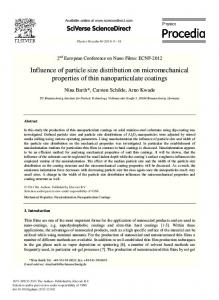
Figure 7 Schematic diagram of the Differential Mobility Spectrometer [36 - 41]. Electrical Mobility Spectrometer The Electrical Mobility Spectrometer (EMS) has been developed at Chiang Mai University, Thailand, under the support from the National Electronic and Computer Technology Center (NECTEC), National Science and Technology Development Agency [42 - 46]. The EMS is a multichannel size analyzer capable of measuring the aerosol size distribution near real time in the size range of approximately 10 – 1000 nm. Nonetheless, there are collective differences between the EMS and each of the existing instruments, which are as follows: (i) the concept of the present instrument is based on a compact, inexpensive and portable unit. Short column classifier and a small number of detection channels are used to reduce diffusion effect of the particle inside the classifier. Overall dimensions and weight are such that it is easy to handle and move around; (ii) the instrument adopts a tangential aerosol inlet upstream of the first electrode ring to ensure uniform particle distribution across the annular aerosol entrance to the classifier column; (iii) rather than diffusion charging, the instrument employs unipolar corona (diffusion and field) charging method; and (iv) the applied voltage is set to maintain at low level, well below the corona onset voltage, to avoid unintentional charging of the particles inside the classifier. The schematic diagram of the EMS is shown in Figure 8. The spectrometer has one short column which consists of coaxially cylindrical electrodes. The advantage of cylindrical geometry is that distortion of electric field between electrodes is minimal due to the absence of corners and edges. Operation and performance of the instrument depend upon aerosol transport under the influence of flow and electric fields. It is important to ensure that both flow and electric fields are laminar and uniformly distributed inside the classifying column. There are two streams which are the aerosol and sheath air flow. The two flows are regulated and controlled via mass flow controllers. The inside of the annular is constructed in such a way that smooth wall and turbulence free merging of the two gas flows are ensured. The flow field inside the apparatus is checked by solving numerically the continuity and Navier-Stokes equations using a commercial computational fluid dynamic software package. The charged particles enter the analyzer column near
Mj. Int. J. Sci. Tech., 2007, 01(02), 120-136
131
the central rod by a continuous flow of air. Since the central rod is kept at a positive high voltage, the charged particles are deflected radially outward. They are collected on electrically isolated electrometer rings positioned at the inner surface of the outer electrode of the column. A Keithley 6517A electrometer incorporating a Keithley 6522 low current scanner card connected to these electrodes measures currents corresponding to the number concentration of particles of a given mobility which is related to the particle size. The signal current from the electrometer is interfaced to an external personal computer via RS-232 serial port interface. Electrical current detection method is considered to be easier and faster than direct particle detection measurements. In addition, the applied high voltage is maintained at a lower value than the corona onset voltage to avoid unintentional charging of the particles within the classifier. DC positive high voltage power supply Positive High Voltage Supply
Impactor
Flowmeter Filter High voltage electrode
Electrometer rings
Sheath air
Silica gel dryer
Filter
Excess air Vacuum pump
E1 E2 E3 E4 E5 E6 E7 E8 E9 E10
Keithley 6522 scanner card
Corona charger
Silica gel dryer
Mass flow controller
Combustion aerosol generator
Keithley 6517A electrometer
External computer, Data logging, User interface
Mass flow controller Positive High Voltage Supply DC positive high voltage power supply
Figure 8 Schematic diagram of the Electrical Mobility Spectrometer developed at Chiang Mai University [42 - 46].
Comparison of Available Instruments’ Characteristics
Comparison of these instruments is made and shown in Table 1. Recent instrumentation developments allow the size distribution measurement of nanometer particles in the size range from 1 to 10,000 nm using the electrical mobility technique. Applications of these instruments are found in such diverse fields as materials synthesis, biotechnology, semiconductor manufacturing, pharmaceutical products, nano-composites and ceramics, electronics and computing, and exhaust gas
Mj. Int. J. Sci. Tech., 2007, 01(02), 120-136
132
Table 1 Comparison of available aerosol particle sensors. SMPS (TSI [47])
EAS (Tammet et al. [30 - 31])
EEPS (TSI [48])
DMS (Biskos et al. [33 - 41])
FAS (Graskow [32])
BCAC (Kulon et al. [27 - 29])
EMS (Intra and Tippayawong [42 - 46])
Electrical mobility
Electrical mobility
Electrical mobility
Electrical mobility
Electrical mobility
Electrical mobility
Electrical mobility
Size range
3 - 1,000 nm
5.6 - 560 nm
5 - 1,000 nm
1 - 100 nm
< 3,000 nm
10 - 1,000 nm
Concentration range
104 – 1011 particles/m3
10 - 10,000 nm 104 - 1011 particles/m3
109 - 1013 particles/m3
109 - 1013 particles/m3
n/a
n/a
1011 - 1013 particles/m3
Time response
60 – 120 s