G3: Genes|Genomes|Genetics Early Online, published on June 30, 2017 as doi:10.1534/g3.117.041848
1
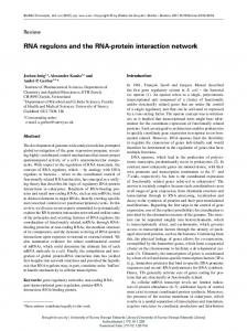
An RNA recognition motif-containing protein functions in meiotic silencing by unpaired DNA.
2 3
Dilini A. Samarajeewa*, Pennapa Manitchotpisit*, Miranda Henderson*, Hua Xiao†, David G.
4
Rehard†, Kevin A. Edwards*, Patrick K. T. Shiu†, and Thomas M. Hammond*,1
5 6
*School of Biological Sciences, Illinois State University, Normal, Illinois, 61790.
7
†
Division of Biological Sciences, University of Missouri, Columbia, Missouri, 65211.
1
Corresponding author: School of Biological Sciences, Illinois State University, Normal, IL
8 9 10
61790 E-mail:
[email protected].
11
1 © The Author(s) 2013. Published by the Genetics Society of America.
12
Running title
13
SAD-7 is an MSUD protein
14 15
KEYWORDS
16
meiosis, chromosome pairing, RNA silencing, homology search, RRM domain
17 18
1
19
61790. E-mail:
[email protected].
Corresponding author: School of Biological Sciences, Illinois State University, Normal, IL
20
2
21
ABSTRACT
22
Meiotic silencing by unpaired DNA (MSUD) is a biological process that searches pairs of
23
homologous chromosomes (homologs) for segments of DNA that are unpaired. Genes found
24
within unpaired segments are silenced for the duration of meiosis. In this report, we describe the
25
identification and characterization of Neurospora crassa sad-7, a gene that encodes a protein
26
with an RNA recognition motif. Orthologs of sad-7 are found in a wide range of ascomycete
27
fungi. In N. crassa, sad-7 is required for a fully-efficient MSUD response to unpaired genes.
28
Additionally, at least one parent must have a functional sad-7 allele for a cross to produce
29
ascospores. Although sad-7-null crosses are barren, sad-7Δ strains grow at a wild-type rate and
30
appear normal under vegetative growth conditions. With respect to expression, sad-7 is
31
transcribed at baseline levels in early vegetative cultures, at slightly higher levels in mating-
32
competent cultures, and at its highest level during mating. These findings suggest that SAD-7 is
33
specific to mating-competent and sexual cultures. Although the role of SAD-7 in MSUD remains
34
elusive, green fluorescent protein (GFP)-based tagging studies place SAD-7 within nuclei,
35
perinuclear regions, and cytoplasmic foci of meiotic cells. This localization pattern is unique
36
among known MSUD proteins and raises the possibility that SAD-7 coordinates nuclear,
37
perinuclear, and cytoplasmic aspects of MSUD.
3
38
INTRODUCTION
39
During meiosis in eukaryotic organisms, homologous chromosomes are grouped into pairs,
40
aligned, recombined, and segregated to produce genetically variable nuclei for reproduction.
41
Chromosome alignment during this process may provide meiotic cells with an opportunity to
42
identify potential problems within pairs of homologs. For example, consider a transposon that
43
exists between two genes on one chromosome but not between the same two genes on the
44
chromosome’s homolog. Alignment of the homologs would pair the genes flanking the
45
transposon, while the transposon itself would remain unpaired. A few fungi have been shown to
46
possess the ability to detect unpaired DNA, like the aforementioned transposon, and silence it for
47
the duration of meiosis. In these fungi, the process is called meiotic silencing by unpaired DNA
48
(MSUD) (Aramayo and Metzenberg 1996; Shiu et al. 2001; Son et al. 2011; Nagasowjanya et al.
49
2013; Wang et al. 2015).
50 51
MSUD was initially discovered in Neurospora crassa (Aramayo and Metzenberg 1996;
52
Shiu et al. 2001), a filamentous fungus made famous as a research model by Beadle and Tatum
53
(1941). Although N. crassa is haploid for most of its life cycle, it possesses a brief diploid phase
54
that occurs during sexual reproduction. MSUD begins during this diploid phase and continues
55
throughout meiosis; thus, a brief introduction to N. crassa’s sexual cycle is necessary (please see
56
Raju (1980) for a comprehensive review of the N. crassa sexual cycle).
57 58
In N. crassa, the sexual cycle begins with the formation of an immature fruiting body
59
called a protoperithecium. A hair-like cell structure called a trichogyne then extends from the
60
protoperithecium towards an asexual spore (conidium) or a hyphal segment of a strain of the
4
61
opposite mating type. Fertilization begins when fusion occurs and a “male” nucleus travels
62
through the trichogyne to the protoperithecium. The protoperithecium becomes a perithecium
63
after fertilization. Within the perithecium, the parental nuclei replicate and, through a series of
64
coordinated events, a nucleus from each parent is sequestered at the top of a cell structure called
65
a crozier. The two haploid parental nuclei fuse to form a single diploid nucleus while the tip of
66
the crozier elongates to form a tube-like meiotic cell. After nuclear fusion, the seven
67
chromosomes from each parent are paired, aligned, and recombined. Segregation during meiosis
68
I returns the haploid state and meiosis II produces four meiotic products. These meiotic products
69
then undergo a single round of mitosis to produce a total of eight nuclei in a single meiotic cell.
70
Cell walls and membranes develop around each of the eight nuclei during a process called
71
ascosporogenesis. At this stage, the meiotic cell is generally referred to as an ascus (“spore sac”).
72
A perithecium can have hundreds of asci, each formed from a unique meiotic event. At maturity,
73
ascospores are shot from the perithecium. This results in the accumulation of ascospores on the
74
underside of a crossing lid when mating is conducted in a standard petri dish.
75 76
The path to MSUD discovery began with deletion of a gene called ascospore maturation-
77
1 (asm-1) (Aramayo and Metzenberg 1996; Hammond 2017). The asm-1 gene is required for
78
proper ascospore maturation and its loss results in the production of white ascospores (Aramayo
79
et al. 1996). Interestingly, even in asm-1+ × asm-1Δ crosses, where four of eight ascospores in
80
each ascus inherit an asm-1+ allele, asci typically contain eight white ascospores instead of four
81
black and four white ascospores (Aramayo and Metzenberg 1996). This phenotype occurs
82
because MSUD detects asm-1+ as unpaired and silences it throughout meiosis (Shiu et al. 2001;
83
Shiu and Metzenberg 2002).
5
84 85
Genes like asm-1 are often targeted in MSUD experiments because they allow for MSUD
86
efficiency to be quantified with ascospore phenotype. For example, if a mutation that suppresses
87
MSUD is included in an asm-1+ × asm-1Δ cross, the strength of MSUD suppression can be
88
determined by the percentage of black ascospores produced by the cross (Lee et al. 2003, 2010;
89
Xiao et al. 2010; Hammond et al. 2011a; Hammond et al. 2013b; Samarajeewa et al. 2014). A
90
strong MSUD suppressor will produce a high percentage of black ascospores (because the
91
unpaired asm-1+ allele is expressed), while a weak MSUD suppressor will produce a low
92
percentage of black ascospores (because the unpaired asm-1+ allele is mostly silenced). In
93
addition to asm-1, a gene called Round spore (r) is often utilized in MSUD research because it
94
must be expressed during meiosis for a cross to produce spindle-shaped ascospores. Accordingly,
95
MSUD causes r+ × rΔ crosses to produce round ascospores (Shiu et al. 2001; Pratt et al. 2004).
96
The level of MSUD suppression can be quantified in r+ × rΔ crosses similar to the way it is
97
quantified in asm-1+ × asm-1Δ crosses (Xiao et al. 2010; Hammond et al. 2011a; Hammond et al.
98
2013b; Samarajeewa et al. 2014). In an r+ × rΔ crossing background, a strong MSUD suppressor
99
will produce a high percentage of spindle ascospores (because r+ is expressed) and a weak
100
MSUD suppressor will produces a low percentage of spindle ascospores (because r+ is mostly
101
silenced).
102 103
In asm-1+ × asm-1Δ and r+ × rΔ crosses, the corresponding unpaired regions are only 2.3
104
kb and 3.5 kb, respectively (Colot et al. 2006). Furthermore, there is evidence suggesting that
105
MSUD can identify unpaired DNA segments as short as 1.3 kb (Lee et al. 2004). Efforts to
106
determine how MSUD detects such relatively small segments of unpaired DNA have focused on
6
107
identifying silencing factors through genetic screens. These efforts have so far identified eleven
108
silencing proteins. A model for the MSUD mechanism has been developed from cytological
109
analyses of these fluorescently-tagged proteins and inferences based on the functions of their
110
homologs in other systems (Aramayo and Selker 2013; Hammond 2017).
111 112
MSUD begins with the identification of unpaired DNA through an undetermined
113
mechanism, possibly involving nuclear MSUD proteins SAD-5 and SAD-6 (Hammond et al.
114
2013b; Samarajeewa et al. 2014). While SAD-5 lacks characterized domains and homologs with
115
described functions, SAD-6 contains an SNF2 helicase domain and is related to proteins that
116
mediate DNA homology search processes. After an unpaired DNA is detected, hypothetical
117
molecules called aberrant RNAs (aRNAs) are thought to be transcribed from the unpaired region
118
and delivered to perinuclear MSUD proteins docked along the nuclear envelope (Bardiya et al.
119
2008; Decker et al. 2015). These molecules are called aRNAs because they are assumed to be
120
unique or marked in a manner that allows the cell to distinguish them from “normal” RNAs. The
121
perinuclear MSUD proteins include SAD-1, an RNA-directed RNA polymerase (Shiu et al.
122
2001, 2006); DCL-1, a Dicer homolog (Alexander et al. 2008); QIP, an exonuclease (Maiti et al.
123
2007; Lee et al. 2010; Xiao et al. 2010); and SMS-2, an Argonaute protein (Lee et al. 2003).
124
Presumably, SAD-1 synthesizes double-stranded (ds)RNAs from aRNAs, DCL-1 dices dsRNAs
125
into MSUD-associated small interfering RNAs (masiRNAs) (Hammond et al. 2013a), QIP
126
processes masiRNAs into single strands, and SMS-2 uses the single-stranded masiRNAs as
127
guides to identify complementary RNA molecules for silencing. Other perinuclear MSUD
128
proteins include SAD-2, which is required for recruiting most if not all known perinuclear
129
MSUD proteins to the perinuclear region of the meiotic cell (Shiu et al. 2006; Decker et al.
7
130
2015); SAD-3, which has a helicase-like domain and is a homolog of a protein involved in
131
RNAi-mediated heterochromatin formation in Schizosaccharomyces pombe (Hammond, Xiao,
132
Boone, et al. 2011); and SAD-4, a novel protein required for masiRNA production (Hammond et
133
al. 2013a). The most recent additions to the MSUD model are the nuclear cap-binding proteins
134
CBP20 and CBP80, both of which may help deliver capped RNA molecules to the perinuclear
135
silencing machinery (Decker et al. 2017).
136 137
Although the hypothetical aRNAs and dsRNAs of MSUD have not been detected
138
biochemically, masiRNAs have been identified by RNA sequencing (Hammond et al. 2013a;
139
Wang et al. 2015). These molecules are predominantly 25 nucleotides long with a bias for
140
uridine at their 5' ends. It seems likely that masiRNAs are used to silence any complementary
141
RNA molecules, not just those derived from the unpaired DNA. This would explain why
142
unpaired genes trigger silencing of paired copies of the same genes at other locations in the
143
genome (Shiu et al. 2001; Lee et al. 2004).
144 145
While the working model of MSUD is consistent with current observations, it leaves
146
many questions unanswered. For example, how are homologous chromosomes scanned and how
147
is unpaired DNA identified? If aRNAs exist, how are they transferred from unpaired DNA to the
148
perinuclear region? How do perinuclear MSUD proteins distinguish aRNAs from mRNAs?
149
Answering these and the many other outstanding questions on MSUD will likely require the
150
discovery of additional MSUD proteins through genetic screens for silencing suppressors.
151
8
152
Previously, Hammond et al. (2011a) described a high-throughput reverse-genetic screen
153
to identify suppressors of MSUD. The screen involves crossing strains from the N. crassa
154
knockout library (Colot et al. 2006) with strains that have been genetically-engineered to unpair
155
asm-1 or r during meiosis. A strain from the knockout library is marked as a candidate MSUD
156
suppressor if it increases the production of black ascospores (when asm-1 is unpaired) or spindle
157
ascospores (when r is unpaired). A suppressor candidate is then examined with a series of
158
experiments designed to purify the knockout strain from possible contaminants and confirm that
159
the gene deletion associated with the knockout strain is responsible for the MSUD suppression
160
phenotype. The gene is then characterized to help understand why its loss suppresses MSUD.
161
Here, we report using the screen to identify sad-7, a gene required for a fully-efficient MSUD
162
response to unpaired DNA.
163
9
164
MATERIALS AND METHODS
165
Strains, media, culture conditions, crosses, and general techniques
166
The key strains used in this study are listed along with genotype information in Table 1. The N.
167
crassa knockout collection (Colot et al. 2006) and various markers were obtained from the
168
Fungal Genetics Stock Center (FGSC) (McCluskey et al. 2010). Strains were cultured on
169
Vogel’s medium (Vogel 1956), except when performing a cross. Crosses were conducted on
170
synthetic crossing medium (pH 6.5; Westergaard and Mitchell 1947) with 1.5% sucrose.
171
Experiments and sexual crosses were performed on a laboratory benchtop at room temperature
172
with ambient lighting unless otherwise indicated. Genomic DNA was isolated from lyophilized
173
mycelia using IBI Scientific’s Mini Genomic DNA Kit (Plant/Fungi). When necessary, PCR
174
products were purified with IBI Scientific’s Gel/PCR DNA Fragment Extraction Kit. PCR was
175
generally performed with Thermo Scientific's Phusion High Fidelity DNA Polymerase.
176 177
Genetic modification of N. crassa
178
Transformation of conidia was performed by electroporation with the method of Margolin et al.
179
(1997). Conidia were filtered through a 100 μm nylon filter (EMD Millipore, SCNY00100)
180
before collection by centrifugation. Strain P8-43 was used as the transformation host to attach
181
the gene for green fluorescent protein (gfp) to sad-7 at its endogenous location on chromosome I
182
as previously described (Hammond et al. 2011b). Four different gfp-sad-7 fusions were
183
constructed. The gfp coding region was fused to either the first codon, 68th codon, 119th codon,
184
or 207th codon of sad-7. In each case, the GFP tag was placed on the N terminus of SAD-7 (or
185
truncated SAD-7). After transformation, the gfp-sad-7 coding regions were PCR-amplified from
186
the transgenes and determined to be free of mutations by Sanger sequencing (data not shown).
10
187
Transgene vectors were constructed with double-joint polymerase chain reactions (DJ-PCR) (Yu
188
et al. 2004; Hammond et al. 2011b) using primers described in supporting information (Table
189
S1).
190 191
Gene expression analysis
192
A total of 23 RNA sequencing datasets were downloaded from the Sequence Read Archive
193
(SRA) of the National Center for Biotechnology Information (NCBI) (Leinonen et al. 2011).
194
Ellison et al. (2011) generated datasets 1–3 from poly-A RNA; Wu et al. (2014) generated
195
datasets 4–13 from poly-A RNA; Wang et al. (2014) generated datasets 14–21 from poly-A
196
RNA; and Samarajeewa et al. (2014) generated datasets 22 and 23 from rRNA-reduced total
197
RNA. Although MSUD gene expression levels for some of the 23 datasets were previously
198
examined by Samarajeewa et al. (2014), Wang et al. (2014), and Decker et al. (2017), sad-7
199
expression levels were not examined in any of the three studies, so the datasets were reanalyzed
200
and presented here. The culture methods used to generate each dataset are briefly described in
201
the Results section. Complete culture methods can be obtained from the original reports. Reads
202
from each dataset were aligned to all primary transcripts located in version 12 of the N. crassa
203
genome annotation, which was provided by the Broad Institute of MIT and Harvard (Galagan et
204
al. 2003). Read alignments were performed with Bowtie 2 v.2.2.5 (Langmead and Salzberg
205
2012) using the local alignment setting. RPKM (Reads per kilobase exon model per million
206
mapped reads; Mortazavi et al. 2008) values were calculated from read alignments with custom
207
Perl scripts. Reads aligning to more than one location and/or containing more than one mismatch
208
were ignored. Accession numbers for the analyzed datasets are as follows: 1) SRR090363, 2)
209
SRR090364, 3) SRR090366, 4) SRR1055985, 5) SRR1055990, 6) SRR1055986, 7)
11
210
SRR1055991, 8) SRR1055987, 9) SRR1055992, 10) SRR1055988, 11) SRR1055993, 12)
211
SRR1055989, 13) SRR1055994, 14) SRR585661, 15) SRR585662, 16) SRR585663, 17)
212
SRR585664, 18) SRR585665, 19) SRR585666, 20) SRR585667, 21) SRR585668, 22)
213
SRR957218, and 23) SRR957223.
214 215
Confocal microscopy
216
Six-day-old perithecia were harvested from crossing plates and fixed in a solution of 4%
217
paraformaldehyde, 100 mM PIPES at pH 6.9, 10 mM EGTA, and 5 mM MgSO4 at room
218
temperature for 20 minutes before washing and storing in sodium phosphate buffer (80 mM
219
Na2HPO4, 20 mM NaH2PO4). Asci were dissected from perithecia in 25% glycerol and
220
transferred by pipette to a drop of mounting medium (25% glycerol, 10 mg/ml DABCO, 100
221
mM potassium phosphate buffer at pH 8.7) on a microscope slide. Cover slips were placed over
222
the samples and sealed with clear nail polish after excess mounting medium was wicked away
223
with tissue paper. Slides were stored at -20 °C before analysis with a Leica SP2 confocal
224
microscope.
225 226
Statement on data and reagent availability
227
All strains generated during this study are available upon request.
12
228
RESULTS
229
Identification of an MSUD suppressor
230
Strain FGSC 13880 from the N. crassa knockout library was marked as a putative MSUD
231
suppressor during a screen of mutants in the N. crassa knockout collection. FGSC 13880 is an
232
ncu01917 deletion mutant (ncu01917Δ), where ncu01917 refers to a gene encoding a
233
hypothetical protein on chromosome I. To confirm that loss of ncu01917 from one parent of a
234
cross suppresses MSUD, we performed quantitative MSUD suppression assays by crossing a
235
sad-2Δ strain, an ncu01917Δ strain (a descendant of FGSC 13880), and a wild-type control strain
236
(designated wt) with an rΔ tester. The sad-2Δ allele is among the strongest known suppressors of
237
MSUD (Shiu et al. 2006). We found that sad-2Δ, ncu01917Δ, and wt produced 96.1%, 53.2%,
238
and 1.7% spindle ascospores, respectively (Figure 1 and Table 2), thus silencing of r+ in a cross
239
was inefficient when sad-2Δ was a parent (most ascospores were spindles), more efficient when
240
ncu01917Δ was a parent (approximately half of the ascospores were spindles), and most efficient
241
when wt was a parent (few ascospores were spindles). These results suggest that ncu01917Δ
242
suppresses silencing of unpaired r+, albeit not as strongly as sad-2Δ does. Next, we repeated the
243
crosses with an asm-1Δ tester. We found that sad-2Δ, ncu01917Δ, and wt produced 60.2%, 67.3%,
244
and 5.9% black ascospores in these crosses, respectively (Table 2); thus, silencing of unpaired
245
asm-1+ was inefficient with sad-2Δ, similarly inefficient with ncu01917Δ, and highly efficient
246
with wt. These results suggest that ncu01917Δ suppresses silencing of asm-1+ as well as sad-2Δ
247
does because approximately equal percentages of black ascospores were produced in both
248
ncu01917Δ × asm-1Δ and sad-2Δ × asm-1Δ crosses. A hypothesis to explain why ncu01917Δ and
249
sad-2Δ suppress MSUD equally with respect to unpaired asm-1+ but not unpaired r+ is discussed
250
below. Overall, these results suggest that ncu01917Δ is a genuine suppressor of MSUD. To be 13
251
consistent with the historical naming system for suppressors of MSUD (Shiu et al. 2001), we
252
refer to ncu01917 as sad-7 (suppressor of ascus dominance-7) hereafter.
253 254
SAD-7 is required for sexual development
255
The above findings demonstrate that heterozygous sad-7Δ crosses are MSUD-deficient despite
256
the presence of a sad-7+ allele in one parent. These findings are consistent with at least two
257
hypotheses: (1) SAD-7 is critical for MSUD and decreased levels of the protein during meiosis
258
interfere with MSUD function, and (2) SAD-7 is an auxiliary MSUD protein that improves the
259
efficiency of MSUD but is not absolutely required for the process. In a sad-7Δ × sad-7Δ cross, the
260
first hypothesis is supported if MSUD is completely inactive, while the second is supported if
261
MSUD is partially active. We attempted to distinguish between these two hypotheses by first
262
examining the ability of sad-7Δ strains to complete the sexual cycle. In this experiment, sad-7Δ ×
263
sad-7Δ crosses were compared side-by-side with sad-7+ × sad-7+ crosses. We examined the
264
perithecia of both at 20 days post-fertilization and found that sad-7+ perithecia had normal beaks
265
while sad-7Δ perithecia were beakless (Figure 2, A and B). Upon dissection, we found that
266
perithecia of sad-7+ × sad-7+ crosses contained hundreds of asci, most with mature or maturing
267
ascospores (Figure 2E), while sad-7Δ × sad-7Δ perithecia were lacking asci and ascospores (data
268
not shown; we encountered a few asci with immature ascospores in one rosette of a sad-7Δ × sad-
269
7Δ cross, but we were unable to identify others during an examination of over 100 perithecia).
270
These results are consistent with our inability to detect ascospores in a quantitative assay of
271
sexual reproduction by sad-7Δ × sad-7Δ crosses (Table 3). Unfortunately, because at least one
272
sad-7+ allele is required for completion of the sexual cycle, we were unable to determine if SAD-
273
7 plays a critical or auxiliary role in MSUD.
14
274 275
SAD-7-null cultures are indistinguishable from wild-type cultures under standard growth
276
conditions
277
The discovery of SAD-7 brings the current number of known MSUD proteins to twelve. Of the
278
previous eleven, none have been found to be required for normal growth or conidiogenesis under
279
standard growth conditions. We thus examined if the loss of SAD-7 would affect growth or
280
conidia production. First, when sad-7+ and sad-7Δ strains were point-inoculated to the center of
281
petri dishes containing standard medium and cultured for several days on a laboratory bench top,
282
both grew at similar rates and produced qualitatively similar levels of conidia (Figure 3, A and
283
B). Second, when linear growth rate was examined by inoculating sad-7+ and sad-7Δ to the ends
284
of 30 cm glass tubes containing standard growth medium (race tubes), both strains grew with the
285
same maximum linear growth rate (Figure 3C). These data demonstrate that the deletion of sad-
286
7+ does not alter growth rate or conidiogenesis (at least with respect to macroconidia) under
287
standard growth conditions.
288 289
The gene expression pattern of SAD-7 is similar to that of SAD-4.
290
At least 28 morphologically-distinct cell types exist in N. crassa (Bistis et al. 2003), some of
291
which are restricted to specific stages of the N. crassa life cycle. The expression pattern of sad-7
292
may help infer the cell types in which it is most active. We thus obtained N. crassa RNA
293
sequencing datasets from four independent studies (Ellison et al. 2011; Wang et al. 2014; Wu et
294
al. 2014; Samarajeewa et al. 2014) to compare sad-7’s expression pattern with those of known
295
MSUD genes. We first analyzed three RNA sequencing datasets produced from a study of wild
296
isolates (Ellison et al. 2011). In this study, the wild isolates were transferred in hyphal plugs of
15
297
actively growing mycelium to the center of a sheet of cellophane over Bird’s medium and
298
cultured under constant light for 24 hours (Ellison et al. 2011). The sad-7+ expression levels
299
were close to 0 RPKM in all three strains under these conditions (Figure 4, columns 1–3). This is
300
similar for all other MSUD genes except dcl-1, qip, and sad-6 (Figure 4, columns 1–3), the
301
former two of which have been shown to have roles in vegetative processes (Catalanotto et al.
302
2004; Maiti et al. 2007). We next analyzed datasets from a study on the effect of light on liquid
303
shaking cultures of N. crassa (Wu et al. 2014). In this study, a standard laboratory strain was
304
cultured in the dark at 25 ºC and 150 RPM in liquid Bird’s medium for 24 hours and then
305
exposed to cool white fluorescent light for durations of up to four hours. Interestingly, expression
306
levels were near baseline for every time point in these datasets for all MSUD genes except dcl-1
307
and sad-6 (Figure 4, columns 4–13). We next examined datasets from a study on gene expression
308
changes during sexual development (Wang et al. 2014). For these datasets, a standard laboratory
309
strain was allowed to develop protoperithecia on cellophane over carrot agar medium at 26 ºC
310
under constant light. On day seven, protoperithecia were fertilized with a standard laboratory
311
strain of the opposite mating type and perithecia were allowed to develop. RNA was sequenced
312
from protoperithecia at “time point 0” and from perithecia at seven different time points after
313
fertilization. The relative expression levels of qip, sad-4, sad-6, and sad-7 were elevated in
314
protoperithecia with respect to the other MSUD genes in the analysis (Figure 4, column 14), and
315
expression levels of all MSUD genes increased as sexual development progressed (Figure 4,
316
columns 14–21). The last study included in our analysis examined RNA transcripts from crosses
317
between rid+ or rid- (Freitag et al. 2002) laboratory strains at 144 hours post-fertilization
318
(Samarajeewa et al. 2014). Unlike the Wang et al. (2014) study, which also examined the 144
319
hour time point, Samarajeewa et al. (2014) performed crosses over miracloth on synthetic
16
320
crossing medium at room temperature and ambient light conditions. Despite these changes,
321
expression levels of MSUD genes were less than 2-fold different across all 144 hour datasets
322
from both studies (Figure 4, columns 21–23).
323 324
Overall, the above analysis of MSUD gene expression patterns indicates that sad-7’s
325
expression pattern is most similar to that of sad-4. For example, both sad-7 and sad-4 are
326
expressed poorly under early vegetative conditions (Figure 4, columns 1–13), upregulated in
327
protoperithecial cultures (Figure 4, column 14), and reach maximum expression levels after
328
fertilization (Figure 4, columns 15–23).
329 330
SAD-7 homologs are present in a wide range of ascomycete fungi
331
A search of NCBI’s non-redundant protein database with the predicted sequence of N. crassa
332
SAD-7 found SAD-7 homologs in many classes of ascomycete fungi. A synteny analysis
333
suggests that many of these homologs are orthologous (related by speciation). For example,
334
homologs of genes flanking N. crassa sad-7 were found flanking genes of putative SAD-7
335
homologs in Sordariomycetes (14 of 14 species analyzed), Leotiomycetes (1 of 1 species
336
analyzed), Dothidiomycetes (1 of 1 species analyzed), and Eurotiomycetes (1 of 1 species
337
analyzed) (Figure S1).
338 339
To gain knowledge on relationships between SAD-7 homologs in ascomycete fungi, we
340
performed a phylogenetic analysis. A single clade representing 13 SAD-7 homologs in the
341
Sordariales order of fungi is shown in Figure 5A. The clade contains three subclades that are
342
consistent with current designations of the taxa into three families: the Sordariaceae, the
17
343
Chaetomiaceae, and the Lasiosphaeriaceae (Federhen 2003). It should be noted that two of the
344
four SAD-7 homologs in the Chaetomiaceae are unusually short (< 419 amino acids) (Figure
345
5B). We are unclear on whether this is a biologically meaningful finding or a result of errors in
346
the available genome sequences and/or annotation for these two fungi. A search of NCBI’s
347
conserved domain database (Marchler-Bauer et al. 2010) identified an RNA recognition motif
348
(RRM) in all 13 SAD-7 homologs. The RRM motif is found in the C-terminal half of each
349
protein (when ignoring the two unusually short Chaetomiaceae proteins). The SAD-7 homologs
350
in the Sordariaceae are longer than those from the other two families. For example, the shortest
351
SAD-7 homolog in the Sordariaceae has 827 amino acids, while the longest one in the
352
Chaetomiaceae and the Lasiosphaeriaceae has 763 amino acids. Pairwise alignments were made
353
to identify a reason for this family-specific length difference. Alignments between Podospora
354
anserina and Madurella mycetomatis SAD-7 revealed a high level of identity along the C-
355
terminal half of the proteins and a comparatively low level of identity along the N-terminal half,
356
despite both proteins being approximately the same length (Figure 5C, top pair). In contrast, the
357
SAD-7 homologs in Neurospora discreta and N. crassa have a high level of identity along their
358
entire lengths (Figure 5C, bottom pair). Interestingly, alignments between N. crassa and P.
359
anserina SAD-7 homologs, and N. crassa and M. mycetomatis SAD-7 homologs, reveal a series
360
of gaps along their N-terminal halves. These data suggest that the N-terminal half of SAD-7
361
lengthened in the lineage leading to the Sordariacea and/or experienced deletions in the lineage
362
leading to Chaetomiacea and Lasiosphaeriaceae fungi.
363 364 365
We also examined the sequence of the SAD-7 RRM domain. RRM domains are approximately 100 amino acids long and contain a conserved β1α1β2β3α2β4 fold (Maris et al.
18
366
2005). A consensus sequence called RNP1 ([RK]-G-[FY]-[GA]-[FY]-[ILV]-X-[FY]) is typically
367
found within β3, while another called RNP2 ([ILV]-[FY]-[ILV]-X-N-L) is typically found within
368
β1. The aromatic residues at the 2nd position in RNP2 and the 3rd and 5th positions in RNP1
369
play critical roles in RNA binding for many RRM-containing proteins. Interestingly, the RRM-
370
domain of SAD-7 lacks an aromatic residue at position 3 in RNP1 and at position 2 in RNP2
371
(Figure 5D). This is not a general feature of RRM domains in N. crassa because other N. crassa
372
RRM-containing proteins (NCU04182, NCU04799, and NCU09193) have aromatic residues at
373
these positions. NCU04182 is a homolog of HSH49, a spliceosomal protein (Igel et al. 1998);
374
NCU04799 is a homolog of PAB1, a poly(A)-binding protein (Brune et al. 2005); and
375
NCU09193 is a homolog of NOP12, a protein involved in ribosome assembly (Konikkat and
376
Woolford 2017). However, similar to SAD-7, NCU08046 has an RRM domain that lacks an
377
aromatic residue at position 2 in RNP2. NCU08046 is a homolog of eIF3g, a protein involved in
378
translation (Hinnebusch 2014). The biological relevance of the amino acid differences in SAD-
379
7’s RRM domain relative to canonical RRM domains is unclear, but the residue exchanges at
380
these positions are likely important for SAD-7 function because they are conserved among the
381
SAD-7 orthologs in Sordariaceae, Chaetomiaceae, and Lasiosphaeriaceae fungi (Figure S2).
382 383
GFP-SAD-7 fusion proteins are found in the nucleus, the perinuclear region, and
384
cytoplasmic foci of meiotic cells
385
The detection of unpaired DNA must occur in the nucleus. However, of the eleven previously-
386
characterized MSUD proteins, only four have been detected within this region of the meiotic cell
387
(SAD-5, SAD-6, CBP20, and CBP80, see Introduction). To examine SAD-7’s localization
388
pattern during meiosis, we tagged its N-terminus with GFP, performed crosses, and examined the
19
389
meiotic cells with confocal microscopy. For these crosses, we included an mCherry-tagged
390
version of SPO76 (mCherry-SPO76), which localizes to meiotic chromosomes (van Heemst et
391
al. 1999; Samarajeewa et al. 2014). In the gfp-sad-7 × mCherry-spo76 crosses, GFP-SAD-7 was
392
detected at three locations: 1) within the nucleus, 2) in a ring around the nucleus, and 3) within
393
randomly distributed cytoplasmic foci (Figure 6, A–C). To eliminate the possibility that our
394
detection of GFP-SAD-7 within nuclei was due to an experimental artifact, we also examined the
395
localization patterns of GFP-SAD-3 and GFP-SMS-2 (Hammond et al. 2011a, b). As reported in
396
previous studies, GFP-SAD-3 and GFP-SMS-2 both formed perinuclear rings and were not
397
detected within nuclei (Figure 6, D–I). We also noticed that GFP-SAD-7 and GFP-SAD-3 were
398
both associated with randomly distributed cytoplasmic foci throughout the cytoplasm (Figure 6,
399
A–F), while GFP-SMS-2 was rarely observed with such foci (Figure 6, G–I). The biological
400
significance of these cytoplasmic foci is currently unclear.
401 402
The identification of an MSUD protein that travels between the nucleus and the
403
cytoplasm could shed light on how nuclear events are linked to silencing processes outside of the
404
nucleus. For example, CBP20 and CBP80 appear to be nuclear-cytoplasmic shuttling proteins
405
involved in MSUD (Decker et al. 2017). If GFP-SAD-7 were also a shuttling protein, it could be
406
possible to isolate sad-7 mutants that are unable to travel between the nucleus and the cytoplasm.
407
To test if N-terminal truncations of SAD-7 could disrupt the normal localization pattern, we
408
fused GFP to positions 68 (GFP-SAD-7Δ1-67), 119 (GFP-SAD-7Δ1-118), and 207 (GFP-SAD-7Δ1-
409
206
410
point were deleted). We then examined the ability of the GFP-tagged full-length protein (i.e.,
411
GFP-SAD-7) and each GFP-tagged truncated protein to complete the sexual cycle when neither
) of the 875-amino acid SAD-7 protein (for each GFP tag, the amino acids prior to the fusion
20
412
parent of the cross carried an untagged sad-7+ allele. While sad-7Δ × gfp-sad-7 crosses produced
413
phenotypically normal perithecia and asci with ascospores (Figure 2, C and F), sad-7Δ × gfp-sad-
414
7Δ1-67 crosses produced beakless and barren perithecia (Figure 2D and data not shown). Beakless
415
and barren perithecia were also produced by sad-7Δ × gfp-sad-7Δ1-118 and the sad-7Δ × gfp-sad-
416
7Δ1-206 crosses (data not shown). These findings suggest that at least some of the amino acids
417
prior to position 68 are necessary for SAD-7’s function in sexual reproduction (although we
418
cannot discount the possibility that the GFP tag is inhibitory to the truncated SAD-7 protein but
419
not the full length protein). Surprisingly, despite the inability of the truncated proteins to
420
complement the barren phenotype, all three truncated proteins displayed a meiotic localization
421
pattern that was indistinguishable from that of full length SAD-7 (Figures 7 and S3), suggesting
422
that some of the amino acids prior to position 68 are required for SAD-7’s function in sexual
423
development but none are required for its proper localization.
424 425
21
426
DISCUSSION
427
In this report, we present evidence demonstrating that N. crassa SAD-7 (NCU01917) is an
428
MSUD protein. The strongest support for this hypothesis is seen in heterozygous crosses
429
between sad-7+ and sad-7Δ, which are deficient in MSUD. This deficiency phenotype could be
430
due to haploinsufficiency, where one copy of sad-7+ does not supply enough SAD-7 protein to
431
the meiotic cell, and/or it could be due to a process called “silencing the silencer”, where the
432
unpairing of sad-7+ turns the MSUD machinery against itself (i.e., sad-7+) (Shiu et al. 2001). In
433
either case, decreased levels of SAD-7 likely cause MSUD deficiency because it is a silencing
434
protein.
435 436
Like many MSUD proteins, SAD-7 is required for sexual reproduction. DCL-1, QIP,
437
SAD-1, SAD-2, SAD-3, and SMS-2 are other examples (Shiu et al. 2001; Lee et al. 2003; Shiu
438
et al. 2006; Alexander et al. 2008; Lee et al. 2010; Xiao et al. 2010; Hammond et al. 2011a).
439
When an MSUD protein is required for sexual reproduction, it is not possible to determine if the
440
protein is required (critical) for MSUD or if it simply improves its efficiency (nonessential). To
441
understand this distinction, it is useful to consider MSUD proteins that are not required for
442
sexual reproduction, such as SAD-5 and SAD-6. MSUD is partially suppressed in sad-5+ × sad-
443
5Δ crosses but completely absent in sad-5Δ × sad-5Δ crosses (Hammond et al. 2013b); therefore,
444
SAD-5 is a critical MSUD protein. In contrast, MSUD is only partially suppressed in both sad-6+
445
× sad-6Δ and sad-6Δ × sad-6Δ crosses (Samarajeewa et al. 2014); therefore, SAD-6 improves the
446
efficiency of MSUD but is not strictly required for the process. The reason why SAD-6 is
447
nonessential for MSUD is unknown, but it could be that its role in MSUD is shared with another
448
protein. With respect to SAD-7, it is not possible to determine if it is more like SAD-5 (critical)
22
449
or more like SAD-6 (nonessenital) because sexual development stalls before meiosis when a
450
cross is completely devoid of SAD-7.
451 452
The sad-7Δ allele suppresses silencing of unpaired asm-1+ as strongly as the sad-2Δ allele
453
does; however, it suppresses silencing of unpaired r+ less well than the sad-2Δ allele (Table 2).
454
This finding is consistent with previous research on alleles that partially suppress silencing of
455
unpaired asm-1+ and/or r+ in heterozygous crosses. For example, sad-4Δ, sad-5Δ, and sad-6Δ
456
alleles are all stronger suppressors of unpaired asm-1+ silencing than they are of unpaired r+
457
silencing (Hammond et al. 2013b; Samarajeewa et al. 2014). The Neurospora Spore killers Sk-2
458
and Sk-3, which are MSUD suppressors, also suppress unpaired asm-1+ silencing better than
459
unpaired r+ silencing (Raju et al. 2007). These differences could be related to the level of effort
460
MSUD must exert to silence asm-1+ and r+ alleles. For example, perhaps asm-1+ is expressed
461
higher than r+ in meiotic cells. In such a scenario, MSUD may need to work harder to silence
462
unpaired asm-1+.
463 464
SAD-7 appears to have at least one role in addition to its function in MSUD. This role
465
should occur at or before the initiation of meiosis because sexual reproduction stalls before the
466
appearance of elongated meiotic cells in sad-7Δ × sad-7Δ crosses. The fact that SAD-7 has been
467
conserved across a diverse range of ascomycete fungi (Figure S1) and MSUD has only been
468
described in three species, two from Neurospora and one from Fusarium (Shiu et al. 2001;
469
Ramakrishnan et al. 2011b; Son et al. 2011), suggests that SAD-7’s non-MSUD role in sexual
470
reproduction is more broadly conserved than its role in MSUD. However, MSUD may be more
471
common in ascomycete fungi than it is currently suggested to be by the available literature. For
23
472
example, research on wild N. crassa isolates has shown that MSUD can be difficult to detect
473
even when it is known to exist within a species (Ramakrishnan et al. 2011a). Therefore, it is
474
possible that SAD-7 performs similar functions in MSUD and sexual reproduction in a diverse
475
range of ascomycete fungi.
476 477
By comparing SAD-7 orthologs from three families in the Sordariales class of
478
ascomycete fungi, we found that while the N-terminal halves of SAD-7 proteins have undergone
479
the most diversification, the C terminal halves have changed comparatively little. The simplest
480
explanation for this is that the N-terminal halves mediate interactions with lineage-specific
481
proteins, while the C-terminal halves perform a similar function among the various lineages.
482
Accordingly, the RRM domain of each SAD-7 is found in the C-terminal half of each protein
483
(ignoring the two unusually short Chaetomiaceae SAD-7s). A sequence level analysis of the
484
RRM domain in 12 SAD-7 orthologs revealed all to be missing two aromatic residues typical of
485
canonical RRM domains. The current reason for this is unclear, but similar variations have been
486
identified in other RRM sequences. These variations appear to be due to the diverse functions of
487
RRM domains, which include RNA binding, protein binding, and/or directing cellular
488
localization (Cléry et al. 2008; Cassola et al. 2010; Muto and Yokoyama 2012). Future
489
investigation on the binding affinities of SAD-7’s RRM domain could help us understand SAD-
490
7’s specific role in MSUD and sexual reproduction.
491 492
Perhaps the most intriguing finding concerning SAD-7 thus far is its peculiar localization
493
pattern relative to other MSUD proteins. Typically, MSUD proteins are localized by N-terminal
494
or C-terminal tagging of the protein with GFP (Lee et al. 2010; Hammond et al. 2011a, b;
24
495
Hammond et al. 2013b; Samarajeewa et al. 2014). This has generally produced localization
496
patterns that are nuclear or extranuclear, but not both. We propose that GFP-SAD-7’s nuclear
497
and extranuclear localizations are related to its role in MSUD and/or sexual reproduction.
498
Interestingly, SAD-7 function appears to require the 67 amino acids at its N-terminal end
499
because fusing GFP to the 68th amino acid (while eliminating the previous 67) abolishes spore
500
production. This finding was made during an attempt to alter the localization pattern of
501
GFP-SAD-7. For example, if the loss of the first 67 amino acids eliminated our ability to detect
502
SAD-7 in either the nucleus or the cytoplasm, it would have added credence to the hypothesis
503
that SAD-7 is a shuttling protein. Surprisingly, all three of the truncated SAD-7 proteins
504
examined in this study displayed a localization pattern similar to the full-length protein despite
505
none being sufficient for sexual reproduction. One possibility is that SAD-7 localization is
506
determined by residues at and after position 207, the site of the GFP fusion in GFP-SAD-7∆1-206.
507
However, because our analysis of the GFP-SAD-7 truncation proteins was performed in the
508
presence of the full-length SAD-7 (e.g., sad-7+ × gfp-sad-7∆1-67), one possibility is that SAD-7
509
forms a homodimer through interactions between residues after position 206. In this scenario,
510
localization signals from the full length SAD-7 would direct SAD-7/GFP-SAD-7∆1-67, SAD-
511
7/GFP-SAD-7∆1-118, and SAD-7/GFP-SAD-7∆1-206 dimers to the proper location in the cell.
512 513
The presence of SAD-7 in nuclear and extranuclear regions of meiotic cells provides a
514
clue towards understanding how the nuclear aspects of MSUD are linked to perinuclear and
515
cytoplasmic aspects of the process. The MSUD model suggests that aRNAs are transcribed from
516
unpaired DNA and delivered to silencing proteins present in a perinuclear ring around the
517
nucleus. Given SAD-7’s nuclear and extranuclear localization pattern, it is conceivable that
25
518
SAD-7 participates in the latter process. Future studies on the binding affinities of SAD-7’s
519
RRM domain and the identification of proteins that interact with SAD-7 could help determine if
520
and how SAD-7 links nuclear and extranuclear aspects of MSUD.
521 522
ACKNOWLEDGEMENTS
523
We thank members of the Hammond and Shiu laboratories for assistance with various technical
524
aspects of this work. We are pleased to acknowledge use of materials generated by P01
525
GM068087 “Functional Analysis of a Model Filamentous Fungus”. This project was supported
526
by start-up funding from Illinois State University (T.M.H) and a grant from the National
527
Institutes of Health (1R15HD076309-01, to T.M.H.). P.K.T.S was supported by the National
528
Science Foundation (MCB1157942/1715534) and the University of Missouri Research Board
529
and Research Council.
530 531
Literature Cited
532
Alexander, W. G., N. B. Raju, H. Xiao, T. M. Hammond, T. D. Perdue et al., 2008 DCL-1
533
colocalizes with other components of the MSUD machinery and is required for silencing.
534
Fungal Genet. Biol. 45: 719–727.
535
Aramayo, R., and R. L. Metzenberg, 1996 Meiotic transvection in fungi. Cell 86: 103–113.
536
Aramayo, R., Y. Peleg, R. Addison, and R. Metzenberg, 1996 Asm-1+, a Neurospora crassa
537 538 539
gene related to transcriptional regulators of fungal development. Genetics 144: 991–1003. Aramayo, R., and E. U. Selker, 2013 Neurospora crassa, a model system for epigenetics research. Cold Spring Harb. Perspect. Biol. 5: a017921.
26
540
Bardiya, N., W. G. Alexander, T. D. Perdue, E. G. Barry, R. L. Metzenberg et al., 2008
541
Characterization of interactions between and among components of the meiotic silencing
542
by unpaired DNA machinery in Neurospora crassa using bimolecular fluorescence
543
complementation. Genetics 178: 593–596.
544 545 546 547 548
Beadle, G. W., and E. L. Tatum, 1941 Genetic control of biochemical reactions in Neurospora. Proc. Natl. Acad. Sci. U. S. A. 27: 499–506. Bistis, G. N., D. D. Perkins, and N. D. Read, 2003 Different cell types in Neurospora crassa. Fungal Genet. Newsl. 50: 17–19. Brune, C., S. E. Munchel, N. Fischer, A. V. Podtelejnikov, and K. Weis, 2005 Yeast poly(A)-
549
binding protein Pab1 shuttles between the nucleus and the cytoplasm and functions in
550
mRNA export. RNA 11: 517–531.
551 552 553
Cassola, A., G. Noé, and A. C. Frasch, 2010 RNA recognition motifs involved in nuclear import of RNA-binding proteins. RNA Biol. 7: 339–344. Catalanotto, C., M. Pallotta, P. ReFalo, M. S. Sachs, L. Vayssie et al., 2004 Redundancy of the
554
two Dicer genes in transgene-induced posttranscriptional gene silencing in Neurospora
555
crassa. Mol. Cell. Biol. 24: 2536–2545.
556 557
Cléry, A., M. Blatter, and F. H.-T. Allain, 2008 RNA recognition motifs: boring? Not quite. Curr. Opin. Struct. Biol. 18: 290–298.
27
558
Colot, H. V., G. Park, G. E. Turner, C. Ringelberg, C. M. Crew et al., 2006 A high-throughput
559
gene knockout procedure for Neurospora reveals functions for multiple transcription
560
factors. Proc. Natl. Acad. Sci. USA 103: 10352–10357.
561
Decker, L. M., E. C. Boone, H. Xiao, B. S. Shanker, S. F. Boone et al., 2015 Complex
562
formation of RNA silencing proteins in the perinuclear region of Neurospora crassa.
563
Genetics 199: 1017–1021.
564
Decker, L. M., H. Xiao, E. C. Boone, M. M. Vierling, B. S. Shanker et al., 2017 The nuclear
565
cap-binding complex mediates meiotic silencing by unpaired DNA. G3 7: 1149–1155.
566
Ellison, C. E., C. Hall, D. Kowbel, J. Welch, R. B. Brem et al., 2011 Population genomics and
567
local adaptation in wild isolates of a model microbial eukaryote. Proc. Natl. Acad. Sci.
568
USA 108: 2831–2836.
569 570 571
Federhen, S., 2003 The taxonomy project, in The NCBI Handbook [Internet], National Center for Biotechnology Information (US). Freitag, M., R. L. Williams, G. O. Kothe, and E. U. Selker, 2002 A cytosine methyltransferase
572
homologue is essential for repeat-induced point mutation in Neurospora crassa. Proc.
573
Natl. Acad. Sci. USA 99: 8802–8807.
574 575 576 577
Galagan, J. E., S. E. Calvo, K. A. Borkovich, E. U. Selker, N. D. Read et al., 2003 The genome sequence of the filamentous fungus Neurospora crassa. Nature 422: 859–868. Hammond, T. M., 2017 Sixteen years of meiotic silencing by unpaired DNA. Adv. Genet. 97: in press (DOI: 10.1016/bs.adgen.2016.11.001).
28
578
Hammond, T. M., H. Xiao, E. C. Boone, T. D. Perdue, P. J. Pukkila et al., 2011a SAD-3, a
579
putative helicase required for meiotic silencing by unpaired DNA, interacts with other
580
components of the silencing machinery. G3 1: 369–376.
581
Hammond, T. M., H. Xiao, D. G. Rehard, E. C. Boone, T. D. Perdue et al., 2011b Fluorescent
582
and bimolecular-fluorescent protein tagging of genes at their native loci in Neurospora
583
crassa using specialized double-joint PCR plasmids. Fungal Genet. Biol. 48: 866–873.
584
Hammond, T. M., W. G. Spollen, L. M. Decker, S. M. Blake, G. K. Springer et al., 2013a
585
Identification of small RNAs associated with meiotic silencing by unpaired DNA.
586
Genetics 194: 279–284.
587
Hammond, T. M., H. Xiao, E. C. Boone, L. M. Decker, S. A. Lee et al., 2013b Novel proteins
588
required for meiotic silencing by unpaired DNA and siRNA generation in Neurospora
589
crassa. Genetics 194: 91–100.
590 591 592 593 594 595 596 597
Hinnebusch, A. G., 2014 The scanning mechanism of eukaryotic translation initiation. Annu. Rev. Biochem. 83: 779–812. Igel, H., S. Wells, R. Perriman, and M. Ares, 1998 Conservation of structure and subunit interactions in yeast homologues of splicing factor 3b (SF3b) subunits. RNA 4: 1–10. Konikkat, S., and J. L. Woolford, 2017 Principles of 60S ribosomal subunit assembly emerging from recent studies in yeast. Biochem. J. 474: 195–214. Langmead, B., and S. L. Salzberg, 2012 Fast gapped-read alignment with Bowtie 2. Nat. Methods 9: 357–359.
29
598
Lee, D. W., R. Millimaki, and R. Aramayo, 2010 QIP, a component of the vegetative RNA
599
silencing pathway, is essential for meiosis and suppresses meiotic silencing in
600
Neurospora crassa. Genetics 186: 127–133.
601 602 603 604 605 606 607
Lee, D. W., R. J. Pratt, M. McLaughlin, and R. Aramayo, 2003 An Argonaute-like protein is required for meiotic silencing. Genetics 164: 821–828. Lee, D. W., K.-Y. Seong, R. J. Pratt, K. Baker, and R. Aramayo, 2004 Properties of unpaired DNA required for efficient silencing in Neurospora crassa. Genetics 167: 131–150. Leinonen, R., H. Sugawara, and M. Shumway, 2011 The sequence read archive. Nucleic Acids Res. 39: D19-21. Maiti, M., H.-C. Lee, and Y. Liu, 2007 QIP, a putative exonuclease, interacts with the
608
Neurospora Argonaute protein and facilitates conversion of duplex siRNA into single
609
strands. Genes Dev. 21: 590–600.
610
Marchler-Bauer, A., S. Lu, J. B. Anderson, F. Chitsaz, M. K. Derbyshire et al., 2010 CDD: a
611
conserved domain database for the functional annotation of proteins. Nucleic Acids Res.
612
39: D225–D229.
613 614 615
Margolin, B. S., M. Freitag, and E. U. Selker, 1997 Improved plasmids for gene targeting at the his-3 locus of Neurospora crassa by electroporation. Fungal Genet. Newsl. 44: 34–36. Maris, C., C. Dominguez, and F. H.-T. Allain, 2005 The RNA recognition motif, a plastic
616
RNA-binding platform to regulate post-transcriptional gene expression. FEBS J. 272:
617
2118–2131.
30
618 619 620 621 622
McCluskey, K., A. Wiest, and M. Plamann, 2010 The Fungal Genetics Stock Center: a repository for 50 years of fungal genetics research. J. Biosci. 35: 119–126. Mortazavi, A., B. A. Williams, K. McCue, L. Schaeffer, and B. Wold, 2008 Mapping and quantifying mammalian transcriptomes by RNA-Seq. Nat. Methods 5: 621–628. Muto, Y., and S. Yokoyama, 2012 Structural insight into RNA recognition motifs: versatile
623
molecular Lego building blocks for biological systems. Wiley Interdiscip. Rev. RNA 3:
624
229–246.
625
Nagasowjanya, T., K. B. Raj, K. Sreethi Reddy, and D. P. Kasbekar, 2013 An apparent increase
626
in meiotic silencing strength in crosses involving inbred Neurospora crassa strains.
627
Fungal Genet. Biol. 56: 158–162.
628 629 630 631 632 633 634 635 636
Perkins, D. D., 2004 Wild type Neurospora crassa strains preferred for use as standards. Fungal Genet. Newsl. 51: 7–8. Perkins, D. D., A. Radford, and M. S. Sachs, 2000 The Neurospora Compendium: Chromosomal Loci. Academic Press. Perkins, D., and V. Pollard, 1986 Linear growth rates of strains representing 10 Neurospora species. Fungal Genet. Newsl. 33: 41–43. Pratt, R. J., D. W. Lee, and R. Aramayo, 2004 DNA methylation affects meiotic trans-sensing, not meiotic silencing, in Neurospora. Genetics 168: 1925–1935. Raju, N. B., 1980 Meiosis and ascospore genesis in Neurospora. Eur. J. Cell Biol. 23: 208–223.
31
637 638 639
Raju, N. B., R. L. Metzenberg, and P. K. T. Shiu, 2007 Neurospora Spore killers Sk-2 and Sk-3 suppress meiotic silencing by unpaired DNA. Genetics 176: 43–52. Ramakrishnan, M., T. N. Sowjanya, K. B. Raj, and D. P. Kasbekar, 2011a A factor in a wild
640
isolated Neurospora crassa strain enables a chromosome segment duplication to suppress
641
repeat-induced point mutation. J. Biosci. 36: 817–821.
642
Ramakrishnan, M., T. N. Sowjanya, K. B. Raj, and D. P. Kasbekar, 2011b Meiotic silencing by
643
unpaired DNA is expressed more strongly in the early than the late perithecia of crosses
644
involving most wild-isolated Neurospora crassa strains and in self-crosses of N.
645
tetrasperma. Fungal Genet. Biol. 48: 1146–1152.
646
Samarajeewa, D. A., P. A. Sauls, K. J. Sharp, Z. J. Smith, H. Xiao et al., 2014 Efficient
647
detection of unpaired DNA requires a member of the Rad54-like family of homologous
648
recombination proteins. Genetics 198: 895–904.
649 650 651 652 653
Shiu, P. K. T., and R. L. Metzenberg, 2002 Meiotic silencing by unpaired DNA: properties, regulation and suppression. Genetics 161: 1483–1495. Shiu, P. K., N. B. Raju, D. Zickler, and R. L. Metzenberg, 2001 Meiotic silencing by unpaired DNA. Cell 107: 905–916. Shiu, P. K. T., D. Zickler, N. B. Raju, G. Ruprich-Robert, and R. L. Metzenberg, 2006 SAD-2
654
is required for meiotic silencing by unpaired DNA and perinuclear localization of SAD-1
655
RNA-directed RNA polymerase. Proc. Natl. Acad. Sci. USA. 103: 2243–2248.
32
656 657 658
Son, H., K. Min, J. Lee, N. B. Raju, and Y.-W. Lee, 2011 Meiotic silencing in the homothallic fungus Gibberella zeae. Fungal Biol. 115: 1290–1302. Thompson, J. D., D. G. Higgins, and T. J. Gibson, 1994 CLUSTAL W: improving the
659
sensitivity of progressive multiple sequence alignment through sequence weighting,
660
position-specific gap penalties and weight matrix choice. Nucleic Acids Res. 22: 4673–
661
4680.
662
van Heemst, D., F. James, S. Pöggeler, V. Berteaux-Lecellier, and D. Zickler, 1999 Spo76p is a
663
conserved chromosome morphogenesis protein that links the mitotic and meiotic
664
programs. Cell 98: 261–271.
665 666 667
Vogel, H. J., 1956 A convenient growth medium for Neurospora (Medium N). Microb. Genet Bull 13: 42–43. Wang, Z., F. Lopez-Giraldez, N. Lehr, M. Farré, R. Common et al., 2014 Global gene
668
expression and focused knockout analysis reveals genes associated with fungal fruiting
669
body development in Neurospora crassa. Eukaryot. Cell 13: 154–169.
670
Wang, Y., K. M. Smith, J. W. Taylor, M. Freitag, and J. E. Stajich, 2015 Endogenous small
671
RNA mediates meiotic silencing of a novel DNA transposon. G3 Bethesda Md 5: 1949–
672
1960.
673 674
Westergaard, M., and H. K. Mitchell, 1947 Neurospora V. A synthetic medium favoring sexual reproduction. Am. J. Bot. 34: 573–577.
33
675
Wu, C., F. Yang, K. M. Smith, M. Peterson, R. Dekhang et al., 2014 Genome-wide
676
characterization of light-regulated genes in Neurospora crassa. G3 Bethesda Md 4:
677
1731–1745.
678
Xiao, H., W. G. Alexander, T. M. Hammond, E. C. Boone, T. D. Perdue et al., 2010 QIP, a
679
protein that converts duplex siRNA into single strands, is required for meiotic silencing
680
by unpaired DNA. Genetics 186: 119–126.
681
Yu, J.-H., Z. Hamari, K.-H. Han, J.-A. Seo, Y. Reyes-Domínguez et al., 2004 Double-joint
682
PCR: a PCR-based molecular tool for gene manipulations in filamentous fungi. Fungal
683
Genet. Biol. 41: 973–981.
684
34
Table 1 Strains used in this study Strain name
Genotype
F2-26 (RTH1005.2)
rid; fl a
F2-27 (RTH1027.3)
rid r∆::hph; fl a
F3-24 (RTH1083.17)
rid his-3+::asm-1; fl; asm-1Δ::hph a
FGSC 13880
sad-7∆::hph a
ISU-3329 (RDS19.3)
rid; fl; mus-52∆::bar mCherryNC-spo76::hph; sad-2∆::hph A
ISU-3334 (RDS19.9)
rid; fl; mus-52∆::bar mCherryNC-spo76::hph; sad-2∆::hph a
ISU-3817 (HDS30.1.1)
rid gfp-sad-7::hph his-3; mus-52∆::bar A
ISU-4078 (HDS34.1.2)
rid gfp-sad-7∆1-67::hph his-3; mus-52∆::bar A
ISU-4079 (HDS35.1.1)
rid gfp-sad-7∆1-118::hph his-3; mus-52∆::bar A
ISU-4134 (RAB1.8)
rid A
ISU-4217 (HDS36.1.1)
rid gfp-sad-7∆1-206::hph his-3; mus-52∆::bar A
ISU-4261 (RTH0061.2.1.6)
rid his-3 gfp-sad3::hph; mus-52∆::bar?; mus-51∆::bar? a
ISU-4262 (RTH1035.7)
rid sad-7∆::hph A
ISU-4263 (P16-17)
a
ISU-4264 (F5-23)
fl A
ISU-4265 (RTH1080.19)
sad-7∆::hph; fl a
P6-07
rid A
P6-08
rid a
P8-01
sad-2Δ::hph A
P8-42
rid his-3; mus-52∆::bar a
P8-43
rid his-3; mus-52∆::bar A
P15-22
rid his-3; mus-52∆::bar; gfp-sms2::hph A
All strains in this study are descendants of lines 74-OR23-1VA (FGSC 2489) and 74-ORS-6a (FGSC 4200) (Perkins 2004). The mCherryNC-spo76::hph allele was obtained from ISU-3123 (Samarajeewa et al. 2014). ISU-4261 carries a gfp-sad3::hph allele identical to the one contained in strain F4-31 (Hammond, Xiao et al. 2011a). The gfp-sms2::hph allele in P15-22 was described in Hammond et al. (2011b). The r∆, asm-1∆, sad-7∆, mus-51∆, and mus-52∆ alleles are as described by Colot et al. (2006). Mutant rid alleles suppress repeat-induced point mutation (Freitag et al. 2002). The fl allele eliminates macroconidia production (Perkins et al. 2000).
685 686 687 688
35
Table 2 MSUD is suppressed by sad-7∆
wt ♂
sad-7∆ ♂ sad-2
∆
♂
wt ♀ Total (×106)
rΔ ♀ Spindle (%)
asm-1Δ ♀ Black (%)
8.3 ± 1.0
1.7 ± 1.1
5.9 ± 0.7
8.9 ± 0.2
53.2 ± 6.5
67.3 ± 4.3
8.7 ± 0.3
96.1 ± 1.1
60.2 ± 21.6
Unidirectional crosses were performed between MSUD-testers (females) and wt, sad-7∆, or sad-2∆ (males) as previously described (Samarajeewa et al. 2014). In short, crosses were performed in triplicate and ascospores were collected from the lids of crossing plates at 21 days post-fertilization. Ascospores were suspended in water for analysis under magnification. The following phenotypes were analyzed: total ascospores (column 2), percent spindle ascospores (column 3), and percent black ascospores (column 4). Only the pertinent genotype is provided for each crossing parent. Note that “wt” is not a true wild type but carries wild-type alleles for all genes related to MSUD, r, and asm-1. Strains: wt ♀ F2-26, r∆ ♀ F2-27, asm-1∆ ♀ F3-24, wt ♂ P6-07, sad-7∆ ♂ ISU-4262, and sad-2∆ ♂ P8-01.
689 690
36
691 Table 3 Homozygous sad-7Δ crosses fail to produce ascospores Total ascospores (×106)
Cross sad-7Δ ♀ × wt ♂
3.6 ± 1.6
sad-7Δ ♀ × sad-7Δ ♂ sad-7
Δ
♀ × sad-2 ♂ Δ
0 3.7 ± 0.1
Unidirectional crosses were performed between sad-7Δ (female) and wt, sad-7Δ, or sad-1Δ strains (males) as described in Table 2 to determine how sad-7Δ affects ascospore production. Only the pertinent genotype is provided for each crossing parent. Please see Table 1 for complete genotype information. Strains: sad-7Δ ♂ ISU-4265, wt ♂ P607, sad-7Δ ♂ ISU-4262, and sad-2Δ ♂ P8-01.
692 693 694
37
695
Figure Legends
696
Figure 1 MSUD is suppressed in sad-7Δ heterozygous crosses. (A) Asci from an rΔ × wt
697
perithecium. Mature (black-pigmented) ascospores are round because of MSUD. (B) Asci from
698
an rΔ × sad-7Δ perithecium. Some mature ascospores are spindle-shaped because MSUD is
699
partially suppressed by sad-7Δ. (C) Asci from an rΔ × sad-2Δ perithecium. Mature ascospores are
700
predominantly spindle-shaped because sad-2Δ is a strong suppressor of MSUD. The designated
701
female strain in all crosses (rΔ ♀) is F2-27. The designated male strains are wt ♂ P6-07, sad-7Δ
702
♂ ISU-4262, and sad-2Δ ♂ P8-01. Bars are 50 µm. Quantitative analysis of ascospore
703
phenotypes from each cross is provided in Table 2.
704 705
Figure 2 SAD-7 is required for ascus and ascospore development. (A–D) Perithecia were
706
isolated from crosses 20 days post-fertilization and examined in water under magnification. (A)
707
Perithecia from a sad-7+ × sad-7+ cross (ISU-4264 × ISU-4263). Perithecial beaks (bk) are
708
present, and two are highlighted in the image. (B) Perithecia from a sad-7Δ × sad-7Δ cross (ISU-
709
4265 × ISU-4262). No beaks are observed. (C) Perithecia from a sad-7Δ × gfp-sad-7 cross (ISU-
710
4265 × ISU-3817). Perithecial beak development appears normal. (D) Perithecia from a sad-7Δ ×
711
gfp-sad-7Δ1-67 cross (ISU-4265 × ISU-4078). No beaks are observed. (E) Asci from a sad-7+ ×
712
sad-7+ cross (ISU-4264 × ISU-4263). Phenotypically normal asci and ascospores are detected.
713
(F) Asci from a sad-7Δ × gfp-sad-7 cross (ISU-4265 × ISU-3817). Phenotypically normal asci
714
and ascospores are detected. In summary, these results demonstrate that (1) at least one parent of
715
a cross must have a functional SAD-7 protein for a cross to complete the sexual cycle and (2)
38
716
tagging the full length SAD-7 with GFP at its N-terminal end does not prevent the protein from
717
performing its function in sexual reproduction.
718 719
Figure 3 A sad-7+allele is not required for conidiogenesis or linear growth. (A and B) Cultures
720
of sad-7+ (P6-08) and sad-7Δ (ISU-4262) are indistinguishable when incubated on standard
721
growth medium at room temperature on a laboratory benchtop. (C) sad-7+ (ISU-4134, red
722
circles) and sad-7Δ [ISU-4262, blue triangles] have similar growth rates on standard growth
723
medium at room temperature on a laboratory bench top. Linear growth rate was measured with a
724
race tube assay (Perkins and Pollard 1986). Strains were allowed approximately two days to
725
colonize the race tubes before data collection. Error bars are standard deviation values.
726 727
Figure 4 Expression pattern of sad-7 is most similar to that of sad-4. The transcript levels of
728
MSUD genes under different culture conditions (according to RNA sequencing analysis) are
729
shown. The Y-axis marks the expression level of each gene in “reads per kilobase exon model
730
per million mapped reads” (RPKM). The 23 datasets included in the analysis (Ellison et al. 2011;
731
Wang et al. 2014; Wu et al. 2014; Samarajeewa et al. 2014) are plotted along the X axis. See
732
Materials and Methods and Results for a full description of each dataset. In short, datasets 1–3
733
are of vegetative cultures on solid medium, datasets 4–13 are of vegetative cultures in liquid
734
medium, and datasets 14–23 are of sexual cultures on solid medium. For datasets 1–3, time refers
735
to the age of the vegetative tissue; datasets 4–13, time refers to hours after exposure to light; and
736
datasets 14–23, time refers to hours post-fertilization. The “A” and “B” designations refer to
737
replicate datasets that were generated with slightly different methods after RNA isolation (Wu et
738
al. 2014). Five of the datasets (1–3, 22, and 23) used in this study were examined by
39
739
Samarajeewa et al. (2014) and eight (14–21) were examined by Wang et al. (2014) with respect
740
to MSUD gene expression, but sad-7 expression was not examined in either study. Overall, sad-
741
7’s expression pattern is most similar to that of sad-4, with barely detectable expression during
742
early vegetative culture conditions (datasets 1–13), elevated expression in protoperithecial
743
cultures (dataset 14), and maximum expression in sexual cultures (datasets 15–23). Gene
744
numbers dcl-1 (ncu08270); qip (ncu00076); sms-2 (ncu09434); sad-1 (ncu02178); sad-2
745
(ncu04294); sad-3 (ncu09211); sad-4 (ncu01591); sad-5 (ncu06147); sad-6 (ncu06190); sad-7
746
(ncu01917); actin (ncu04173).
747 748
Figure 5 SAD-7 is a widely conserved RRM protein in ascomycete fungi. (A) A tree diagram
749
depicting relationships between SAD-7 homologs in a clade of Sordariales. The diagram
750
contains a subset of homologs from a more complete phylogenetic analysis presented in Figure
751
S1. The families of 12 of the 13 taxa in Sordariaceae, Chatomiaceae, and Lasiosphaeriaceae
752
families are indicated (Federhen 2003). (B) RRM (NCBI CDD: cl17169) locations in SAD-7
753
homologs. RRM domains are indicated with gray boxes. The predicted number of amino acids in
754
each protein is listed along the right side of the panel. (C) Graphical depictions of Clustal W
755
(Thompson et al. 1994) alignments between pairs of SAD-7 homologs. Identical amino acids are
756
indicated with red shading. Different amino acids are indicated with black shading. Gap
757
positions are indicated with gaps. The locations of the RRM domains are indicated with a gray
758
box. The blue scale bar is equivalent to 50 amino acids. In summary, these results show that
759
SAD-7 is an RRM domain-containing protein conserved across a wide range of ascomycete
760
fungi. They also suggest that the N-terminal halves of the protein have changed more than the C-
761
terminal halves during evolution of Sordariaceae, Chatomiaceae, and Lasiosphaeriaceae fungi.
40
762
(D) A manual alignment of RRM domains from five N. crassa proteins is shown. The residues
763
are shaded according to the PAM120 similarity matrix. The positions of RNP1 and RNP2 are
764
indicated. Sequences can be obtained from GenBank or FungiDB with the following accession
765
numbers: Podospora anserina (Pa) CAP60824.1; Madurella mycetomatis (Mm) KXX77199.1;
766
Chaetomium thermophilum EGS22685.1; Thielavia terrestris AEO64981.1; Myceliophthora
767
thermophila AEO61061.1; Neurospora crassa (Nc) EAA36312.1; Neurospora tetrasperma
768
EGO51840.1; Neurospora discreta (Nd) NEUDI 136685; Neurospora africana GCA
769
000604205.2; Neurospora africana GCA 000604205.2; Neurospora sublineolata GCA
770
000604185.2; Neurospora terricola GCA 000604245.2; Neurospora pannonica GCA
771
000604225.2; Sordaria macrospora XP 003349025.1.
772 773
Figure 6 GFP-SAD-7 is detected at three different locations in the ascus. (A-C) Asci from a gfp-
774
sad-7 × mCherry-spo76 sad-2Δ cross are shown. When SAD-7 is tagged with GFP (GFP-SAD-
775
7), a GFP signal is detected throughout the nucleus (n) except for a spherical-subnuclear domain
776
representing the nucleolus. The GFP-SAD-7 signal is most intense around the nucleus and within
777
cytoplasmic foci (f). mCherry-SPO76 is used to depict the position of the chromosomes (van
778
Heemst et al. 1999; Samarajeewa et al. 2014), while sad-2Δ is used to allow the expression of
779
tagged and unpaired alleles during meiosis. ISU-3334 × ISU-3817. (D-F) Asci from a gfp-sad-3
780
× mCherry-Spo76 sad-2Δ cross are shown. When SAD-3 is tagged with GFP (GFP-SAD-3), the
781
GFP signal is similar to that of GFP-SAD-7 except that there is no signal within the nucleus.
782
ISU-3329 × ISU-4261. (G-I) Asci from a gfp-sms-2 × mCherry-Spo76 sad-2Δ cross are shown.
41
783
When SMS-2 is tagged with GFP, the GFP signal is strong around the nucleus but absent from
784
within the nucleus. Cytoplasmic foci are uncommon for GFP-SMS-2. ISU-3334 × P15-22.
785 786
Figure 7 The meiotic localization pattern of GFP-SAD-7 is independent of the first 206 amino
787
acids of the protein. A series of truncated SAD-7 proteins was created by fusing GFP to different
788
positions from the N-terminal end of SAD-7. Amino acids prior to the fusion point were deleted
789
in the process. Representative images of GFP signal within asci in meiotic prophase I from
790
crosses between ISU-3334 (an mCherry-spo76, sad-2Δ strain) and various GFP-SAD-7
791
truncation strains are shown. In these crosses, GFP signal was detected within the nucleus, the
792
perinuclear region, and cytoplasmic foci despite the loss of up to 206 amino acids from the N-
793
terminal end of SAD-7. (A) ISU-3334 × ISU-3817, (B) ISU-3334 × ISU-4078, (C) ISU-3334 ×
794
ISU-4079, and (D) ISU-3334 × ISU-4217.
795
42
Figure 1
A
C
rΔ
rΔ
× wt
× sad-2Δ
B
rΔ
× sad-7Δ
Figure 2
A
B
sad-7+ × sad-7+
sad-7Δ × sad-7Δ
C
sad-7Δ × gfp-sad-7
D
sad-7Δ × gfp-sad-7Δ1-67
bk
bk
E
sad-7+ × sad-7+
F
sad-7Δ × gfp-sad-7
1 mm
100 µm
Figure 3
A
C
sad-7+
B
sad-7Δ
) 25 m c( 20 h t 15 w o r g10 r a e 5 in L
0
0
20
440 60 Tim me (hours)
80
100
Figure 4
45 dcl-1 30 15 0
1
2
3
4
5
6
7
8
9
10
1 12 11
13
14
15
16
17
18
19
20
21 22
23
150 qip 100 50 0 690 sms-2 460 230 0 30 sad-1 20 10 0
sad-2
42 28 14 0
sad-3
30 20 10 0
sad-4
18 12 6 0
sad-5
18 12 6 0
sad-6
27 18 9 0
13.5 9 sad-7 4.5 0 2700 1800 act 900 0
g ev h 4 2 . 1
g ev h 4 2 . 2
g ev h 4 2 . 3
A t h gil h 0 . 4
B t h gil h 0 . 5
A t h gil m 5 1 . 6
B t h gil m 5 1 . 7
A t h gil h 1 . 8
B t h gil h 1 . 9
A t h gil h 2 . 0 1
B t h gil h 2 . 1 1
A t h gil h 4 . 2 1
B t h gil h 4 . 3 1
x es h 0 . 4 1
x es h 2 . 5 1
x es h 4 2 . 6 1
x es h 8 4 . 7 1
x es h 2 7 . 8 1
x es h 6 9 . 9 1
x es h 0 2 1 . 0 2
x es h 4 4 1 . 1 2
x es h 4 4 1 . 2 2
x es h 4 4 1 . 3 2
Figure 5
B
A
P. anserina
704
M. mycetomatis
693
C. thermophilum
763
T. terrestris
418
M. thermophila N. crassa
355 875
N. tetrasperma
875
N. discreta
863
N. africanca
835
N. sublineolata
827
N. terricola
831
N. pannonica
884
S. macrospora
886
C
50 aa
D NCU04182 NCU04799 NCU09193 NCU08046 NCU01917
NCU04182 NCU04799 NCU09193 NCU08046 NCU01917
SAP49 PAB1 NOP12 eIF3g SAD-7
SAP49 PAB1 NOP12 eIF3g SAD-7
93 142 311 298 499
RNP2 123456 KTVDIGAELFINNLDPQVDEKI----------------------------LRKTGAGNIFIKNLDAAIDNK-----------------------------VLQVKVDEDGKEVTEKKKRTKQPMDVEEGL APVDHKRCVFVGNLGFVDDETV GERDDLATLRVTNVSEMAEEQE----------------------------ASASDAGVVKITNLPYTTTHQEIK---------------------------
115 163 362 320 523
RNP1 12345678 NNISKGYGFVSFGSFEASD LYDTFSQFGQILRQPNIVRDDN ALHDTFAAFGNILSCKVAQDEHGNSKGYGFVHYETDEAAS WRVFGKEGGKVESVRVVRDPVTRVGKGFAYVQFCDENAVE LRDMFERFGRVTRVFLAKDRDTGLAKGFAFISFADRSDAV ALLGRNAKLLTEESVHVIMERINGKTQDAYIEFCSQDDAI
154 202 401 359 562
114 162 361 319 522
Figure 6
A. GFP-SAD-7
B. mCherry-SPO PO76
C. merge
D. GFP-SAD-3
E. mCherry-SPO PO76
F. merge
G. GFP-SMS-2
H. mCherry-SPO PO76
I. merge
n
f
Figure 7
A. GFP-SAD-7
B. GFP-SAD-7 7Δ1-67
C. GFP-SAD-7Δ1-118
D. GFP-SAD-7 7Δ1-206