noticed dyspnea on exertion, orthopnea, and paroxysmal nocturnal dyspnea. At the time of admission, chest exaniination revealed bilateralinspiratory rales; and ...
AN UNUSUAL ECHOCARDIOGRAPHIC PATTERN OF A LEFT ATRIAL MYXOMA Jean-Robert Mathurin, M.D., Ajit V. Adyanthaya, M.D., Lawrence J. Petrovich, M.D., Mauricio Franco, M.D., Kenneth L. Mattox, M.D., and James K. Alexander, M.D.
ABSTRACT Unusual echocardiographic findings in a 58-year-old woman with a history of rheumatic fever and an angiographically demonstrated prolapsing left atrial myxoma are presented. With variations of gain and damping controls, it was possible to isolate a more distinct anterior mitral leaflet echo, or a more posterior linear echo, thought to represent the prolapsing tumor. The tumor, instead of presenting as a cloud of echoes behind the anterior mitral valve leaflet, demonstrated an alternate pattern of a single linear dense echo at this location. Echocardiography, though very useful in the diagnosis of left atrial tumors, can be fallible at times.
INTRODUCTION Echocardiagraphy is a well established technique that can be used in the diagnosis of left atrial myxomas.1 2 These tumors are found in approximately one of every 300 operations for mitral valve stenosis3 and may go undetected for a significant period of time. The echocardiographic appearance of left atrial myxoma is well known,4 6 and little has been added to its initial description. This is a report of a case with unusual and misleading echocardiographic findings.
CASE REPORT A 58-year-old white female was first seen at the Ben Taub General Hospital in January, 1976, at which time she was noted to have mild pulmonary edema. The patient had a history of rheumatic fever at 15 years of age, but was apparently in relatively good health until 1974, when she noticed dyspnea on exertion, orthopnea, and paroxysmal nocturnal dyspnea. At the time of admission, chest exaniination revealed bilateral inspiratory rales; and cardiac examination revealed sinus rhythm S4 and S3 gallops, and a grade 3/6 pansystolic murmur of mitral regurgitation. Left atrial From the Cardiology Division, Department of Medicine, Baylor College of Medicine and the Ben Taub General Hospital. Address for reprints: Ajit V. Adyanthaya, M.D., Cardiology Division, Department of Medicine, Baylor College of Medicine, 1200 Moursund Avenue, Houston, Texas 77030.
Cardiovascular Diseases, Bulletin of the Texas Heart Institute, Vol. 4, Number 4
409
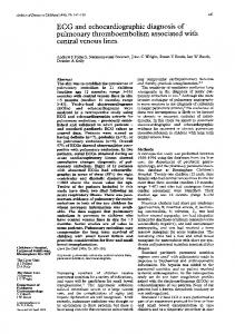
and left ventricular enlargement, as well as vascular congestion, were noted on her chest X ray. The congestive heart failure responded well to treatment, and she was discharged receiving digitalis and diuretics. One week later she returned to the emergency center with severe dyspnea, dry cough, fever and chills. On physical examination, the temperature was 40.3°C, a maculopapular rash extend.ed over the extremities with petechiae on the face and eyelids. She had a few inspiratory rales, and the heart examination was unchanged. There was no hepatospl.enomegaly. The hematocrit was 40.3%o, the sedimentation rate was 46 mm/hr, the platelet count was 226,000 per cubic mm, and the white blood cell count was 17,000 per cubic mm, with a shift to the left. Over the following 48 hours, the temperature returned to normal. Six blood cultures yielded no growth. A skin biopsy showed changes consistent with subacute dermatitis. After seven days she was discharged afebrile and improved without a specific diagnosis for the f.ebrile illness. During a routine follow-up visit six months later, engorged neck veins, congested lungs, moderate hepatomegaly, and atrial fibrillation were noted. She was readmitted for treatment of congestive heart failure and cardiac catheterization. Laboratory studies at that time were unremarkable. An echocardiogram was obtained with the use of a Smith-Kline Ekoline 20 Ultrasonoscope, utilizing a 2.25 MHz focused transducer 0.5 inch in diameter, interfaced with an Electronics for Medicine DR8 multichannel recorder.7 A dense, well-defined linear echo was seen posterior to the anterior leaflet of the mitral valve during ventricular diastole. The E to F slope was normal (Fig. 1). The left atrium appeared unremarkable. Scant i
AWOW^U10"I
$
--A:
I
I,
Fig. 1. Echogram of a dense, well-defined linear echo posterior to the anterior leaflet echo. 410
t'
ning the heart in different positions and planes did not significantly alter the findings.
Cardiac catheterization data are reported in the Table. Prominent pulmonary wedge "V" waves were noted, but notching on the upstroke and downstroke of the ventricular pressure tracing, reported by others to be present with atrial myxomas,9"10 was not seen. The left ventricular angiogram demonstrated gross mitral regurgitation, and a filling defect in the left atrium, that was seen to prolapse into the left ventricle in diastole, was noted (Fig. 2). At open heart surgery, a 5.5 x 2 x 1.3 cm lobulated myxoma was found attached to the interatrial septum above the fossa ovalis (Fig. 3). It was resected and the scarred rheumatic mitral valve was replaced with a Beall mitral prosthesis.
TABLE
Cardiac Catheterization Data
Pressure (mm Hg) Right atrial mean
(12)
Right ventricle
55/10
Pulmonary artery
55/24 (38)
Pulmonary Capillary Wedge
"V" wave
38
Mean
25
Left Ventricle
120/19
Aorta
120/75 (97)
Cardiac Output (L/Min)
2.62
Cardiac Index (L/Min/M2)
1.51
A-V 02 Difference (Vol%)
7.6
Pulmonary Vascular Resistance
(dynes/sec-cm-5)
397
Systemic Vascular Resistance
(dynes/sec-cm-5)
2595
Fig. 2. A prolapsing filling defect from a frame of the left ventricular angiogram in diastole.
2I
SPECIMEN d-
2-
Fig. 3. Post-surgical specimen of the excised left alrial myxoma. 412
45
DATE_
6!~
~~
The patient's postoperative course was uneventful, and she was discharged on the seventh postoperative day in stable condition. DISCUSSION
Previous reports of atrial myxoma have stressed the presence of a cloud of echoes behind the anterior leaflet of the mitral valve, which appear shortly after the mitral valve opens. This delay in the appearance of myxoma echoes is a result of the time interval between the opening of the mitral valve and the prolapsing of the atrial tumor. Previous reports have also emphasized a psudomitral stenosis pattern and systolic echoes in the left atrium.' 2'6 It is clear that echoes posterior to the mitral valve are dependent on movement of the atrial tumor into and through the mitral valve annulus. In order to visualize this movement, the heart should be scanned in at least three sections: both leaflets of the mitral valve, the mitral valve and left atrium, and the aorta and the left atrium.13 The incidence of false negative echograms is presumed to be high in nonprolapsing left atrial myxoma and thrombi14; however, their incidence in prolapsing myxoma is unknown. This case is unusual in that the most striking echoca,rdiographic finding was a single dense echo posterior to the anterior mitral valve echo (Tig. 1). All repeated attempts to demonstrate the usual cloud of echoes behind the anterior leaflet of the mitral valve were unsuccessful, even after angiographic demonstration of the presence of a tumor. It was possible, with variations of gain and damping, to isolate the mor.e posterior echo, in which case mitral stenosis was simulated (Fig. 4); or to visualize only
t~~~~~~~~~~~~~~~~~
s~~~-j
t
r ts
.
IT^l
~
,/4s
e. .J.
a
_
Is
_
I
I
*~~~~~~~~~~~~~~~~~~~~~~~~~~~~~4
Fig. 4. Echogram of the posterior isolated linear echo simulating anterior leaflet in mitral stenosis.
:_.*~ INw'/|.
the more anterior true mitral valve echo, in which case an essentially normal pattern was seen (Fig. 5). The parallel echoes seen in Figure 1 were initially interpreted as being a combination of chordal and mitral valve echoes, which is not an uncommon pattern. However, in retrospect, the density of the posterior echo and its anterior movement slightly after the anterior movement of the mitral valve should permit proper diagnosis in the future. In conclusion, a case of left atrial myxoma, instead of presenting a cloud of echoes representing the prolapsing atrial tumor, demonstrated an alternate pattern of a single dense echo. Echocardiography, though very useful in the diagnosis of left atrial tumors, may at times be fallible. Further studies on the incidence of false negative findings are needed.
1. 2.
3. 4.
REFERENCES Efferts DE: The diagnosis of intra-atrial tumors and thrombi by the ultrasonic echo method. Med Monatsschr 4:1-3, 1959 Schattenberg TT: Echocardiographic diagnosis of the left atrial myxoma. Mayo Clin Proc 43:620-627, 1968 Bass NM, Sharatt GP: Left atrial myxoma diagnosed by echocardiography, with observations on tumor movement. Br Heart J 35: 1332-1335, 1973 Selzer A, Sakai J, Popper R: Protean clinical manifestations of primary tumors of the hleart. Am J Med 52:9-18, 1972
.go!
7
e~ih Xi ,
4
AK
II KL 9l,..-
-
zi
.At.-
Fig. 5. Echogram of the isolated true anterior mitral leaflet echo. 414
j
\
5. Nassar W, Davis R, Dillon J, Tavel M, Hellman C, Feigenbaum H, Fisch G: Atrial myxoma I: Clinical and pathological features in 9 cases. Am Heart J 83:694-704, 1972 6. Cohen A, McIntosh HD, Orgain E: The mimetic nature of left atrial myxomas. Am J Cardiol 11:802-807, 1963 7. Wolfe S, Popp R, Feigenbaum H: Diagnosis of atrial tumor by ultrasound. Circulation 34:615622, 1969 8. Ghahramani AR, Arnold JR, Hildner FJ, Sommer LS, Samet P: Left atrial myxomahemodyanmic and phonocardiographic features. Am J Med 52: 525-532, 1972 9. Penny JL, Gregory JJ, Ayres SM, Giannelli S, Rossi P: Calcified left atrial myxoma simulating mitral insufficiency. Circulation 36:417-491, 1967 10. Goodwin JF: Diagnosis of left atrial myxoma. Lancet 1:464467, 1963 11. Adams WC, Collins HA, Dummit ES, Allen JH: Intracardiac myxomas and thrombi-clinical manifestations, pathology and treatment. Am J Cardiol 7:176-187, 1961 12. Martinez CM, Giles TD, Burch GE: Echocardiographic diagnosis of left atrial myxoma. Am J Cardiol 33:281-285, 1974 13. Tallury VK, DePasquale NP: Ultrasound cardiography in the diagnosis of left atrial thrombus. Chest 59:501-503, 1971
415