Commentary
2337
An update on nuclear calcium signalling Martin D. Bootman1, Claire Fearnley1, Ioannis Smyrnias1, Fraser MacDonald2 and H. Llewelyn Roderick1,3,* 1
Laboratory of Molecular Signalling, The Babraham Institute, Babraham, Cambridge CB22 3AT, UK KCL Dental Institute, Department of Orthodontics, St Thomas Street, London SE1 9RT, UK 3 Department of Pharmacology, University of Cambridge, Tennis Court Road, Cambridge CB2 1PD, UK 2
*Authors for correspondence (e-mails:
[email protected];
[email protected])
Journal of Cell Science 122, 2337-2350 Published by The Company of Biologists 2009 doi:10.1242/jcs.028100
Journal of Cell Science
Summary Over the past 15 years or so, numerous studies have sought to characterise how nuclear calcium (Ca2+) signals are generated and reversed, and to understand how events that occur in the nucleoplasm influence cellular Ca2+ activity, and vice versa. In this Commentary, we describe mechanisms of nuclear Ca2+ signalling and discuss what is known about the origin and physiological significance of nuclear Ca2+ transients. In particular, we focus on the idea that the nucleus has an
Introduction Calcium (Ca2+) is a key player in signal transduction. It modulates diverse cellular activities ranging from fertilisation to cell death (Berridge et al., 2000). It is well known that different cell types use various elements from a broad Ca2+ signalling ‘toolkit’ (Box 1); consequently, the characteristics of global Ca2+ signals, and their physiological effects, can vary considerably (Berridge et al., 2000). This might also be the case for nuclear Ca2+ signalling, which is essentially a complex form of local Ca2+ signalling. As described below, the mechanisms of nuclear Ca2+ signalling are extensive, and it is likely that there is considerable flexibility in how Ca2+ signals are triggered in the nucleus and in the downstream targets that are affected. In this Commentary, we discuss what is known about the origin and physiological significance of nuclear Ca2+ transients. In particular, we focus on the idea that the nucleus has an autonomous Ca2+ signalling system that can generate its own Ca2+ transients, which modulate processes such as gene transcription. We also discuss the role of nuclear pores and the nuclear envelope in controlling ion flux into the nucleoplasm. It should be noted that nuclear Ca2+ signalling has been the subject of previous reviews, some of which have presented different points of view (Bootman et al., 2000; Gerasimenko and Gerasimenko, 2004; Gomes et al., 2006; Lee et al., 1998; Santella and Carafoli, 1997). The physiological significance of nuclear Ca2+ signalling It has been well established that Ca2+ signals occurring inside cells affect activities within the nucleus. For example, the amplitude and frequency of global cellular Ca2+ signals can regulate the transcription of various genes (Dolmetsch et al., 1998). But what is the significance of the nuclear component of these Ca2+ transients? Do patterns of nuclear Ca2+ signalling simply track cytosolic Ca2+ signalling, or are there independent nuclear Ca2+ signalling mechanisms that are necessary for proper cell function? A precise method for analysing the function of nuclear Ca2+ is to specifically localise a buffering molecule within the nucleoplasm, which prevents changes in nuclear Ca2+ levels but does not affect cytosolic Ca2+ signals. Nathanson and colleagues employed this
autonomous Ca2+ signalling system that can generate its own Ca2+ transients that modulate processes such as gene transcription. We also discuss the role of nuclear pores and the nuclear envelope in controlling ion flux into the nucleoplasm.
Key words: Calcium, Gene transcription, InsP3, Nucleus, Paclitaxel, Ryanodine, Signalling, Spindle checkpoint, Taxol
approach to examine the involvement of nuclear Ca2+ in regulating the proliferation of hepatocyte cell lines (Rodrigues et al., 2007). They observed that targeting the Ca2+-binding protein parvalbumin to the nucleus significantly reduced cell proliferation and altered the proportions of cells that were in the S and G2-M phases of the cell cycle. By contrast, the expression of parvalbumin in the cytosol did not alter cell proliferation or cell cycle distribution. Most interestingly, the expression of the parvalbumin in the nucleus, but not in the cytosol, reduced the development of tumours when HepG2 cells were subcutaneously implanted into nude mice. Using a similar approach, the nuclear expression of parvalbumin was found to inhibit activation of the Elk1 transcription factor following stimulation of hepatoma cells with epidermial growth factor (EGF), whereas buffering cytosolic Ca2+ did not have the same effect (Pusl et al., 2002). Collectively, these data suggest that nuclear Ca2+signals are important for regulating cell proliferation in some contexts. Ca2+ is a central player in many cellular signal-transduction cascades that modulate gene transcription. Signalling cascades mediate changes in gene expression by stimulating the translocation of transcription factors from the cytosol to the nucleus, or by causing the translocation and/or activation of enzymes that regulate the activity of nuclear transcription factors or the structure of chromatin. Although an increase in the concentration of nuclear Ca2+ might not be the initiating signal for such events, it can be necessary for gene transcription to occur or to be sustained. For example, nuclear Ca2+ can activate gene transcription via the nuclear factor of activated T cells (NFAT) family of proteins. In quiescent cells, NFAT is mainly found in the cytosol, but in response to an increase in cytosolic Ca2+, it rapidly (~10 minutes) translocates to the nucleus (Tomida et al., 2003). The translocation of NFAT occurs following its dephosphorylation by the Ca2+-calmodulin-sensitive phosphatase calcineurin (Mellstrom et al., 2008), which exposes an intrinsic nuclear localisation sequence (NLS) in NFAT. NFAT and calcineurin associate in a Ca2+-dependent manner, and it has been reported that they translocate into the nucleus as a complex (Shibasaki et al., 1996). When Ca2+ is elevated in the nucleus, calcineurin remains active and associated with NFAT, and thereby sustains its transcriptional activity. Calcineurin mediates this effect by outcompeting kinases that promote NFAT re-phosphorylation
2338
Journal of Cell Science 122 (14)
Box 1. Cellular Ca2+-signalling machinery Buffers Ca2+ channels: receptor operated store operated voltage operated second-messenger operated Hormone Neurotransmitter Growth factor Messenger
Sensors (e.g. calmodulin, troponin C) [Ca2+]high
Ca2+ pump Na+/Ca2+ exchanger
[Ca2+]low
Ca2+ pump Ca2+ release channel
Ca2+ store
Journal of Cell Science
Buffers
Cytosolic Ca2+ signals can be evoked by chemical stimuli (such as hormones, growth factors and toxins), environmental changes (such as changes in pH or temperature shifts), mechanical deformation or depolarisation. Depending on the cell type, the nature of the stimulus and the extent of stimulation, Ca2+ signals can be transient, oscillatory or sustained (Thomas et al., 1997), and can occur globally or as subcellular events (Bootman et al., 2001). To generate such a diverse spectrum of signals, cells employ a range of messengers and mechanisms to evoke Ca2+ fluxes between various cellular compartments. For example, cells can access Ca2+ from a variety of intracellular stores (including the sarcoplasmic or endoplasmic reticulum, the Golgi, acidic vesicles and the NE) and via influx from the extracellular space. The figure depicts the basic Ca2+ cycle present in all mammalian cell types. The pathways that increase cytosolic Ca2+ levels are shown with red arrows and the mechanisms that reduce Ca2+ levels are depicted with blue arrows. Ca2+ influx can occur via several different types of channels that have diverse activation mechanisms. The characteristics of the Ca2+ signals evoked by these channels depends on their biophysical properties, their expression levels and their location within the plasma membrane of the cells. Release of Ca2+ from intracellular stores is also mediated by several different types of Ca2+ channel, of which InsP3Rs (see Box 2) and RyRs are the best characterised. The magnitude of Ca2+ signals can be limited by various buffers inside cells, such as Ca2+-binding proteins and mitochondria. These buffers shape the spatial domain, timecourse and amplitude of Ca2+ rises. Elevation of cytosolic Ca2+ concentration is transduced into functional changes in cellular activity by numerous sensors that bind Ca2+. Cellular Ca2+ sensors have varying affinities and locations, allowing them to specifically respond to different Ca2+ patterns.
(which would cause its nuclear export) and through a physical interaction that occludes a nuclear-export sequence (NES) in the NFAT protein. An example of a nuclear-localised transcription factor that is regulated by both nuclear and cytosolic Ca2+ signals is cyclic AMP response element-binding protein (CREB). The signalling mechanisms that underlie CREB activation have been worked out particularly well in neurons, in which it has been established that depolarisation leads to the rapid phosphorylation of CREB on Ser133 by Ca2+-calmodulin-dependent kinase IV, which provides a necessary, but not sufficient, signal for CREB-mediated gene transcription (Dolmetsch et al., 2001). The triggering event is an
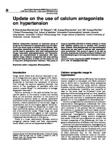
increase in cytosolic Ca2+ levels in the immediate vicinity of L-type Ca2+ channels or N-methyl-D-aspartate receptors that are found in the synapse, at a site that is distal to the nucleus (Shaywitz and Greenberg, 1999). As CREB is only found in the nucleus, the phosphorylation of Ser133 is mediated by one of several Ca2+sensitive signal transduction cascades that convey information from the remote synapses into the nucleus. In the nucleus, phosphorylated CREB binds additional proteins to form a transcriptionally active complex (Shaywitz and Greenberg, 1999). Although the synaptic Ca2+ signal does not need to propagate to the nucleus to induce phosphorylation of Ser133 on CREB, an increase in the level of nuclear Ca2+ is required for CREB-mediated gene transcription to occur. This is because additional Ca2+-dependent phosphorylation of CREB and its co-activators is required (Chawla et al., 1998; Kornhauser et al., 2002). Indeed, nuclear injection of an artificial Ca2+ buffer prevents CREB-mediated gene transcription, but not transcription induced by the serum-response element, which is sensitive to cytosolic Ca2+ buffering (Hardingham et al., 1997). Another well-known target of nuclear Ca2+ signalling is the transcriptional regulator downstream regulatory element antagonist modulator (DREAM). It is well established that DREAM represses the expression of prodynorphin, an opiate-receptor precursor, and that mice that lack DREAM have a constitutive analgesic condition (Cheng et al., 2002). DREAM contains four Ca2+-binding EF hands (a widely expressed structural motif in proteins that bind Ca2+), and is directly regulated by an increase in nuclear Ca2+ levels (Mellstrom et al., 2008). When the concentration of Ca2+ in the nucleus is low, DREAM represses gene transcription by binding to DNA and displacing other transcriptional activators such as CREB (Ledo et al., 2002). An increase in the concentration of nuclear Ca2+ causes DREAM to dissociate from its DNA binding sites and thereby allows the transcription of its target genes. Space limitations prevent a comprehensive discussion of the downstream targets of Ca2+ signalling pathways in the nucleus. However, it is evident from the in vitro and in vivo experiments described above that nucleoplasmic and cytosolic Ca2+ signals can synergistically control important cellular events. Furthermore, the nuclear component of global Ca2+ signals – or an independent increase in nuclear Ca2+ alone – can regulate specific nuclear processes. Mechanisms that regulate the level or function of Ca2+ in the nucleus Changes in nuclear Ca2+ concentration clearly impact on many cellular functions, but what are the mechanisms involved in regulating nuclear Ca2+ levels, and how are they related to those that operate in the cytosol? In the following section, we describe the mechanisms by which Ca2+ moves into and out of the nucleus and how intranuclear Ca2+ stores are triggered. Together, these mechanisms contribute to the regulation of Ca2+-dependent nuclear signalling pathways (Fig. 1). InsP3Rs, RyRs and NAADPRs
To date, the most widely implicated mechanism for nuclear Ca2+ release is via the activation of inositol(1,4,5)-trisphosphate receptors (InsP3Rs) (see Box 2). Evidence in support of a role for nuclear InsP3Rs in regulating nuclear Ca2+ release has been obtained by using InsP3-binding assays (Humbert et al., 1996), InsP3R-specific antibodies (Leite et al., 2003; Malviya, 1994; Stehno-Bittel et al., 1995), electrophysiological recordings from nuclear envelope (NE) patches or from whole nuclei (Bustamante et al., 2000; Stehno-Bittel
Nuclear calcium signalling
2339
Hormone or growth factor Plama membrane
Receptor
PtdIns(4,5)P2
Cytosol
Endoplasmic reticulum InsP3 NAADP cADPR
PLC
DAG
PKC
InsP3 PLC Ca2+- or phosphorylationinduced translocation
Nuclear envelope
? NAADP cADPR
InsP3 InsPx
Nucleoplasm
PLC Transcription factors and chromatin-remodelling enzymes
PKC
PtdIns(4,5)P2 Nuclear speckle
+ or – Nucleoplasmic reticulum
Transcription
Key
Journal of Cell Science
+
DAG
Ca2+ release channel
Nuclear pore complex
Ca2+ ions
Fig. 1. Summary of the pathways that regulate nuclear Ca2+ levels. Key aspects of this schematic are the separate Ca2+-releasing mechanisms in the cytosol and nucleus. In particular, the independent cytosolic and nuclear phosphoinositide cycles are depicted. Although Ca2+ release and the production of Ca2+-releasing messengers can be localised in the individual compartments, nuclear and cytosolic Ca2+ levels can also be coordinated via the diffusion of messengers through NPCs. Some of the putative pathways described in this figure are controversial. In particular, the translocation of growth-factor receptors into the nucleus has not been observed in all studies (marked by ‘?’). + and – indicate a positive or negative effect on a downstream process, respectively.
et al., 1995), nuclear microinjection of InsP3 (Hennager et al., 1995; Huh et al., 2007; Santella et al., 2003), direct visualisation of InsP3evoked nuclear Ca2+ signals (Higazi et al., 2009), reconstitution of nuclear InsP3Rs into lipid bilayers (Leite et al., 2003) and prevention of nuclear Ca2+ signals by using InsP3R antagonists (Kumar et al., 2008). Furthermore, the addition of InsP3 to nuclei that were isolated from Xenopus oocytes, Aplysia neurons, mammalian epithelial cells or pancreatic acinar cells (Bezin et al., 2008a; Gerasimenko et al., 1995; Quesada and Verdugo, 2005; Stehno-Bittel et al., 1995) has been shown to induce nucleoplasmic Ca2+ transients. Not all of these studies differentiated between expression of InsP3Rs on the inner or outer NE. However, it is clear that InsP3Rs are present on both the inner and outer NE, and that those channels expressed on the inner NE can trigger the release of Ca2+ from the NE lumen directly into the nucleoplasm. The actual proportion of the total cellular InsP3Rs that are found on the inner NE (or other putative nuclear stores; see below) is probably very small (