At the molecular level, NMDA receptors are linked to a voltage-dependent cation channel (Engberg et al., 1978;. MacDonald et al., 1982; Flatman et al., 1983).
The Journal
Anatomic Brain William
Correlation
F. Maragos,
Department
of NMDA and 3H-TCP-Labeled
John B. Penney,
of Neurology
of Neuroscience,
February
1988,
Receptors
8(2):
493-501
in Rat
and Anne B. Young
and the Neuroscience
Program, University
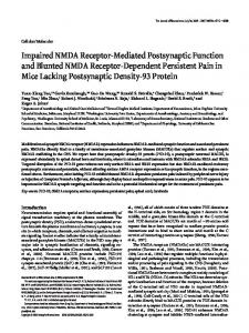
Using quantitative autoradiography, we have compared the regional distribution of N-methyl-D-aspartate (NMDA) receptors labeled with 3H-glutamate and dissociative anesthetic binding sites labeled with 3H-K( l -[2-thienyl]cyclohexyl)3,4piperidine (‘H-TCP). Binding of both ligands was highest in the hippocampal formation, with high concentrations in a number of cortical and olfactory regions. Intermediate amounts of binding for both ligands were measured in several thalamic and basal telencephalic structures. Very little binding was observed in the hypothalamus, some deep forebrain regions, and most brain-stem structures. Linearregression analysis comparing the binding at both sites revealed a marked concordance (R = 0.95; p -K 0.001; Pearson product-moment). The granule cell layer of the cerebellum was the only region in which this concordance was not observed. Scatchard analysis of 3H-glutamate binding to NMDA receptors in stratum radiatum of hippocampal formation revealed an apparent single binding site with a B,,, of 9.78 f 0.84 pmol/mg protein and K, of 158 f 37 nM. 3H-TCP also bound to an apparent single site with a B,,, of 2.07 f 0.18 pmol/mg protein and K, of 127 f 30 nM. Our results are consistent with the hypothesis that the dissociative anesthetic binding site is linked to the NMDA receptor, and the data suggest that there are approximately4-5 NMDA binding sites for each dissociative anesthetic binding site.
The dissociative anesthetics-e.g., phencyclidine (PCP), ketamine, and related drugs-comprise a novel group of drugs that were originally usedas surgicalanesthetics(Greifenstein et al., 1958;Johnstoneet al., 1959;Corssenand Domino, 1966).More recently, thesedrugs have become major substancesof abuse (Burns and Lemer, 1976). Drugs of this classproduce psychotomimetic effects in humans, including thought disorders, distortions of body image, depersonalization, mania, and sometimes catatonia (Luby et al., 1959; Domino, 1964). These behaviors are similar to those seen in chronic schizophrenia, and this observation had led someto postulate a common underlying mechanism. Recently, electrophysiologicalstudieshave indicated that the dissociative anestheticsantagonize the effects of N-methyl-Daspartate(NMDA)-sensitive glutamate receptorsin the cerebral Received Jan. 29, 1987; revised June 15, 1987; accepted July 29, 1987. Thanks to Gene11 Fries for preparation of the manuscript and to Zane Hollingsworth and Darrell DeBowey for technical assistance. Supported by USPHS Grants NS15655, NS19613, AG06155, and the ADRDA. Correspondence should be addressed to Dr. John B. Penney, Neuroscience Laboratory Building, University of Michigan, 1103 East Huron, Ann Arbor, MI 48101. Copyright 0 1988 Society for Neuroscience 0270-6474/88/020493-09$02.00/O
of Michigan, Ann Arbor, Michigan 48104
cortex (Harrison and Simmonds, 1985; Thomson et al., 1985), hippocampal formation (Raja and Guyenet, 1982), and spinal cord (Anis et al., 1983). The NMDA receptor is one of at least 3 receptor subtypes at which the excitatory amino acid, glutamate, is believed to exert its action (Watkins and Evans, 1981). NMDA receptors occur throughout the mammalian CNS (Greenamyre et al., 1985; Monaghan and Cotman, 1985). Although the exact behavioral role of thesereceptorsis unknown, they appear to be involved in learning (Morris et al., 1986), memory (Collingridge, 1985),and possiblyepilepsy (Meldrum, 1985). At the molecular level, NMDA receptors are linked to a voltage-dependent cation channel (Engberg et al., 1978; MacDonald et al., 1982; Flatman et al., 1983). This channel is gated by Mg2+ (Mayer et al., 1984; Nowak et al., 1984), which has been shown to inhibit the actions of NMDA noncompetitively (Harrison and Simmonds, 1985). The dissociative anesthetics exert inhibitory actions on the channel similar to Mg2+ (Duchen et al., 1985; Honey et al., 1985), perhapsthrough an allosteric site on the NMDA receptor-channel complex (Harrison and Simmonds, 1985; Martin and Lodge, 1985). If NMDA receptors and dissociative anesthetic (PCP) recognition sitesare linked, one prediction is that they shouldhave a similar anatomical distribution. Recently, autoradiographic assayshave been developed in several laboratories to visualize both NMDA (Greenamyreet al., 1985;Monaghan and Cotman, 1985)and dissociative anestheticreceptor distribution (Quirion et al., 1981; Sircar and Zukin, 1985; Gundlach et al., 1986; Vignon et al., 1986). Using 3H-glutamateand 3H-N-(l-[2-thienyl]cyclohexyl)3,4-piperidine (3H-TCP), we have applied the technique ofquantitative autoradiography to investigatethe distribution of NMDA and dissociativeanestheticrecognition sites, respectively, in the rat CNS (Maragos et al., 1986). 3H-TCP was selectedasthe ligand sinceit binds with higher affinity (Vignon et al., 1983)and 50-100 times greater specificity (Largent et al., 1986)to the dissociative anestheticsitethan PCP or other PCPlike drugs. Materials and Methods Male Sprague-Dawley ratsweighingapproximately200gm wereused in thisstudy. On the dayof the experiment,animalsweredecapitated, and the brains were rapidly removed and immediately frozen on dry ice. The frozen brains were mounted on aluminum chucks and equilibrated to the cryostat temperature (- 17°C) for a half-hour prior to sectioning. Assays of each receptor class were carried out on alternate 20 pm sections thaw-mounted onto gelatin-coated slides. ‘H- TCP binding assay. jH-TCP binding using autoradiographic techniques has been examined by several groups (Sircar and Zukin, 1985; Gundlach et al., 1986; Largent et al., 1986). In these studies, the pharmacology, distribution, and physiochemical properties of binding have been examined, and they fit closely with the known physiology and
494
Maragos
et al.
l
NMDA
and TCP
Receptors
behavior of dissociative anesthetics. In initial experiments, we found that the association rate of )H-TCP with the receptor at 4°C was 6.1 2 2.0 x lo3 M-I set’. The dissociation rate was determined as 8.8 + 3.0 x 1O-4 secl. The ratio of k-,/k, corresponded closely to the equilibrium-saturation constant, KD = 127 ? 30 nM. This finding suggested that the labeled ligand was binding via a bimolecular reaction and was unlikely to be sequestered by transport processes, as has been suggested for the binding of some labeled ligands (Kessler et al., 1987). The offrate of ‘H-TCP was slower than that of jH-glutamate, and rinse times of 3 x 1 min optimized the specific to nonspecific ratios. We also confirmed the pharmacolonical specilicitv of )H-TCP bindina as described by others (Largentet al.,*1986; W. F. Maragos, unpiblished observations). For routine assays, sections were washed for 30 min in cold 50 mM Tris-acetate, pH 7.4, and dried. For regional distribution studies, sections were incubated for 45 min in the same buffer at 4°C containing 1 mM magnesium acetate and 20 nM ‘H-TCP (52.9 Ci/mmol, New England Nuclear Corporation). Magnesium acetate was added to the incubation medium, as this has beenshown in preliminary experiments to enhance binding (D. C. M. Chu. unpublished observations). Nonspecific binding was ‘determined by’ incubating slides with IH-TCP in the presence of 20 PM unlabeled PCP. Saturation studies were carried out in 2 parts. For the lower concentrations, sections were incubated for 45 min in cold buffer containing concentrations of 3H-TCP ranging from 1 to 20 nM. Nonspecific binding was determined for each point in the presence of 20 MM PCP. For higher concentrations, sections were incubated for 45 min in 20 nM )H-TCP diluted with varying amounts of nonradioactive TCP ranging from 30 to 980 nM. For this group of sections, nonspecific binding was determined in 20 nM 3H-TCP in the presence of 20 FM PCP. Following incubation, sections were washed 3 times for 1 min each in cold buffer containing magnesium and rapidly dried. NMDA receptor assay. NMDA receptors were labeled with L-~Hglutamate under conditions that have been shown to select for binding to NMDA receptors (Greenamyre et al., 1985). Under these conditions, the kinetics of glutamate binding were identical to those determined for ‘H-glutamate binding in the presence of calcium and chloride (Greenamyre et al., 1983). For regional distribution studies, sections were washed for 30 min in cold 50 mM Tris-acetate, pH 7.4, and dried. Sections were then incubated in the same buffer at 4°C containing 200 nM L-3H-glutamate (specific activity, 5.24 Ci/mmol; Amersham Corporation) and 1 WM quisqualic acid. This concentration of quisqualate has been determined to inhibit >90% of the non-NMDA glutamate receptors. For saturation studies, sections were incubated at 4°C in buffer containing 26 nM r?H-glutamate (39 Ci/mmol) diluted with varying concentrations of nonradioactive glutamate ranging from 25 nM to 10 PM. Following a 45 min incubation all sections were rapidly rinsed 3 times with 3 ml of cold buffer followed by 1 rinse with a solution of cold glutaraldehyde and acetone (1: 19, vol/vol) and rapidly dried. The total rinse time of this assay did not exceed 10 sec. Nonspecific binding was determined in the presence of 100 PM NMDA or 1 mM glutamate, and values were similar using either displacer. Depending on the region, nonspecific binding varied from 5 to 50% of total glutamate binding. Autoradiographyanddataanalysis.Sections were placed in an x-ray cassette and apposed to a piece of Ultrofilm )H (LKB). A complete set of radioactive standards calibrated against brain paste with known amounts of tritium was co-exposed with each film. Following a 3 week exposure at 4°C films were developed in Kodak D19, fixed, and airdried. Autoradiographic analysis was carried out using previously described methods (Pan et al., 1983). At least 15 readings were made for each region studied. Anatomical areas were determined using the atlases of Paxinos and Watson (1982) and Zilles (1985). Scatchard analyses of saturation data were constructed using the computer program LIGAND (Munson and Rodbard, 1980). Binding of the 2 ligands to the various areas of the brain was compared with the Pearson product-moment coefficient and linear-regression analysis.
Results The binding pattern of L-3H-glutamate-labeledNMDA receptors and ‘H-TCP recognition sites was nearly identical in all forebrain regions(Fig. 1). When the bound values for NMDA and TCP receptorswere normalized to the stratum radiatum of the CA1 region of the hippocampal formation, the ratio of NMDA and TCP binding sitesin the forebrain and brain stem
never exceeded1.2, while in the cerebellargranulecell layer this ratio wasgreater than 6. On plotting the number of NMDA and TCP receptorsagainst each other and using linear regression,a highly significant correlation wasobserved(R = 0.95,~ < 0.001) (Fig. 2). The stratum radiatum ofthe CA1 division ofthe hippocampal formation contained the highest number of both NMDA and PCP receptorsin the CNS. Scatchardanalysisof L-3H-glutamate binding revealed a single binding site with a maximum bound value (B,,,) of 9.78 f 0.84 pmol/mg protein and an association constant (K,) of 158 f 37 nM (Fig. 3A). 3H-TCP binding displayed an apparent single binding site with a B,,, of 2.07 -+ 0.16 pmol/mg protein and KD of 127 + 30 nM (Fig. 3B). When the B,,, of NMDA receptors was compared with that of 3HTCP labeledreceptors, a stoichiometry of 4-5 NMDA to 1 TCP site was determined. Similar values were observed when saturation analysesof binding to layersI-II of cortex wereexamined. Comparative distribution of 3H-glutamate-sensitiveNMDA and ‘H- TCP-labeled PCP binding sites A distinctly “NMDA-like” laminar distribution wasrecognized for both ligands in the hippocampal formation (Fig. l&G). Binding was highest in stratum radiatum, followed by stratum oriens of the CAlKA2 region (Table 1). Binding in these 2 regionswashighly delineatedsinceboth of thesestructuresabut the stratum pyramidale, a region of relatively low binding. There was a relatively small number of receptors for both ligands within stratum oriens and stratum radiatum of CA3, which made the boundary between CAl/CA2 and CA3 readily apparent. Both bladesof the molecular layer of the dentate gyrus possessed very high densitiesof binding, while the stratum moleculare/lacunosumof CA1 exhibited intermediate levels. Densitiesof binding rangedwidely in the cerebralcortex (Table 1). The visual cortex clearly possessed the greatestnumber of NMDA and TCP receptors, being approximately 3/ias high asthe binding in the stratum radiatum (Fig. 1H). High densities werealsoobservedin layers I and II of somatosensoryand motor cortices, while layers V and VI of these regions had lessthan half of the number of binding sitesobservedin superficiallayers (Fig. 1D). In the somatosensorycortex, but not the motor region, an intermediate level of binding wasobservedasa narrow band in layer IV (Fig. 1D). The anterior cingulate (Fig. 1C) and entorhinal regions(Fig. 1H) possessed equally high levels of binding, which were more than 2 times higher than those in either the retrosplenial cortex (Fig. 1G) or the posterior cingulategyrus (Fig. 1E). The basal ganglia and precommissuralbasal forebrain also revealed heterogeneouspatterns of binding. In these regions, however, binding never exceeded 50% of that observed in the densestregion of the hippocampus.In the striatum binding was of intermediate density, while the adjacent globuspallidus possessed virtually no binding sites(Fig. 1D). Similarly, the nucleus accumbensand lateral septum possessednearly equal, intermediate levels of binding, while the closely situated medial septum and horizontal limb of the diagonal band were almost devoid of receptors (Fig. 1c). Numerous connectionshave been said to exist betweenvarious olfactory regions and the amygdala (Fig. 1, A-G). In this study, with only several exceptions, theseregions appearedto possess intermediate to high levels of NMDA and TCP binding. Relatively low levels of binding were measuredin the internal granule layer of the olfactory bulb and medial amygdaloid nu-
The Journal
m
of Neuroscience,
February
1988.
C?(2) 495
496
Maragos
et al. - NMDA
and TCP
Receptors
Table 1. Binding of 3H-TCP to PCP sites and of 3H-glutamate to NMDA
Area Olfactory region External plexiform layer (EPL) Internal granule layer (IGL) Anterior olfactory nucleus (AON) Amygdala Basolateral, anterior (BLa) Medial (M) Posterior cortical (PCoA) Basal ganglia Caudate-putamen (CPU) Globus-pallidus (GP) Septal area Lateral septum (LS) Medial septum (MS) Ventral pallidum Nucleus accumbens (NAc) Olfactory tubercle (TU) Cortex Somatosensory, layers I&II (5 1 8~ 2) Somatosensory, layer III (53) Somatosensory, layers V&VI (55 & 6) Motor, layers I&II (M 1 & 2) Motor, layers V&VI (M5 & 6) Anterior cingulate (AC& Posterior cingulate (PCg) Entorhinal (EC) Primary olfactory (POC) Retrosplenial (Rspl) Striate (Str) Hippocampal formation Dentate gyrus (DG) CAl, stratum radiatum (SR) CAl, stratum oriens (SO) CA3, stratum radiatum CA3, stratum oriens CAlKA3, stratum pyramidale (SP) Stratum lacunosum/moleculare (SML) Subiculum, dorsalis (SUB) Thalamus Habenula (Hb) Lateral dorsal (LD) Medial dorsal (MD) Medial geniculate (MG) Dorsolateral geniculate (DLG) Ventral posterior lateral/medial (VPL) Reticular (Rt) Other Bed nucleus stria terminalis (BST) Medial preoptic area (MPOA) Lateral hypothalamus (LH) Ventromedial hypothalamus (VH) Diagonal band, horizontal limb (HDb) Zona incerta (ZI)
sites in rat brain Binding relative to stratum radiatum of CA1 (%) NMDA TCP
Ligand bound (fmol/mg protein) NMDA TCP 1069 f 159 488 k 115 1368 I+ 346
189 + 46 57 + 6 211 * 43
33 15 42
52 16 58
1056 + 203 506 + 165 1346 + 330
141 iz 24 70 + 14 176 k 48
32 15 41
39 19 49
695 k 161 75 + 29
92k 18 18 + 6
21 2
25 5
1060 + 184 130 f 35
136 + 25 35 f 11
32 4
38 10
849 t 241 1135 k 290
106 + 18 198 k 35
26 35
29 54
2160 1184 1038 1932 713 1830 752 1553 1548 884 2416
-t 381 dr 216 + 257 f 315 + 162 k 472 + 119 k 299 +- 311 k 197 + 256
251 -t 128 + 97 + 251 f 97-t 198 k 70+ 242 + 216 f 79* 268 +
34 19 18 36 18 34 15 28 33 15 27
66 36 32 59 22 56 23 48 47 27 74
68 35 26 69 27 54 19 64 59 22 73
2473 3238 2886 1060 946 792 1839 1074
k + k k e k k +
262 367 327 169 305 200 268 141
251 365 312 176 101 97 216 97
f k + + + + + +
36 34 38 26 19 16 33 19
76 100 89 32 29 24 56 33
68 100 85 48 27 27 59 26
75 902 977 968 858 647 308
+ t + k k + +
44 167 168 239 153 178 119
13 f 88k 84 f 92+ 106 k 88k 31+3
6 17 17 15 21 11
2 27 30 29 26 19 9
3 24 23 25 28 24 8
497 286 48 84 190 31
+ + + t f +
127 119 27 31 67 13
53+ 40+ 18 f 31 2 35 * 13 zt
11 14 7 10 9 8
15 8 1 2 6 1
14 11 4 8 9 4
The Journal
of Neuroscience,
February
1988,
8(2)
497
Table 1. Continued
Bindingrelative to stratum radiatumof CA1 (%) NMDA TCP
Ligandbound (fmol/mgprotein) NMDA TCP
Area Brainstem Superiorcolliculus(SC) Periaqueductal gray (PAG) Interpeduncularnucleus(IPn) Pontinenuclei(Pn) Substantianigra(SN) Cerebellum Granulelayer(Gnl)
Values represent the mean f SEM of 7 animals. ‘H-glutamate 20 nw Autoradiography and quantification of autoradiograms
616 +- 132 242 + 65 198+ 144 110+ 101 163+ 81
53 k 10 40 + 6 26 f 8 18 +4 13 + I
18 8 6 3 5
15 10 I 5 4
633 I!I 97
13 + 5
19
3
concentration was 200 nM. ‘H-TCP was carried out as described in text.
cleus. The external plexiform layer of the olfactory bulb, the lateral anterior olfactory nucleus, the olfactory tubercle, pyramidal layer of cortex, and the basolateraland primary olfactory/ posterior amygdaloid nuclei contained 30-60% of the maximum number of NMDA and TCP recognition sites. In the thalamus (Fig. l&G), homogeneousbinding was observed in the laterodorsal and mediodorsalnuclei, as well as in the medial and dorsolateral geniculate and the ventroposterolateral nuclei. This density of binding is in sharpcontrast to that in the medial habenula, reticular nuclei, and ventrolateral geniculate nuclei (data not shown), which were nearly devoid of binding. Very low levels of binding were alsoobserved in other diencephalicstructures(Fig. 1, E, 0, including the zona incerta, and the lateral and ventromedial hypothalamus. The telencephalic medial preoptic area (Fig. lD), which is the anterior continuation of the hypothalamus, contained low but slightly higher levels than the hypothalamus (Fig. 1E). The bed nucleus of the stria terminalis contained slightly higher levels than the medial preoptic area (Fig. 1D).
concentration
was
7-
A 86432l0
I
0
2
,
4
’
I
.
6
I
8
.
I 10
BOUND(pmol/mgprotein) 2.6-1
400 n l
300 m
1
mm
n n
200 -
100 -
&’
im’
0
I 1000
I 2000
I 3000
[3~]-~~~~~~~~~ BOUNDTO NMDA RECEPTOR (fmol/mgprotein)
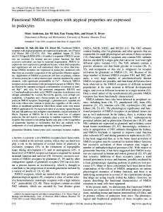
BOUND(pmol/mgprotein) Figure 3. Representative Scatchardplotsof ‘H-glutamatebinding(A)
and ‘H-TCP binding(B) in stratumradiatumof the CA1 subfieldof the hippocampalformation.Autoradiographyfor eachligandwasperScatter-plotof boundvalues(in fmol/mgprotein)of )H-TCP formedasdescribedin Materialsand Methods.Eachpoint represents (20 nM) versusbound valuesfor NMDA receptors(3H-glutamate, specificbinding (averageof 15 readingsminusreadingsfrom nearly 200nMasabove).Linear-regression analysisyieldeda correlationcoefadjacentsections)incubatedin eitherthe presence of 100PM NMDA ficientof 0.95(p < 0.001,Pearsonproduct-moment). for 3H-glutamate or 20 PMPCPfor ‘H-TCP binding.
499
Maragos
et al. * NMDA
and TCP Receptors
Brain stem and cerebellum In the brain stem, significant densities were observed only in the superior colliculus and periaqueductal gray region (Fig. 1H). Interestingly, as has been previously noted, the granule layer of the cerebellum had about 20% of the highest number of NMDA binding sites but had very few TCP binding sites (Fig. II).
Discussion The described distribution of NMDA receptors in the rat brain is in agreement with previous reports (Greenamyre et al., 1985; Monaghan and Cotman, 1985). Each region evaluated receives putative excitatory amino acid inputs (for a review, see Fagg and Foster, 1983). In hippocampal formation, for instance, NMDA receptors are densest in the stratum radiatum of the CA 1 division, which has dense neuropil consisting of the apical dendrites of CA1 pyramidal neurons (Lorente de No, 1934) upon which terminate fibers originating from the ipsi- and contralateral CA3 regions (Laurberg, 1979; Taxt and Storm-Mathisen, 1984). Both neurochemical (Storm-Mathisen and Iversen, 1979; Skrede and Malthe-Sorenssen, 198 1) and physiological (Collingridge et al., 1983a) studies indicate that these pathways use either glutamate or aspartate as their transmitter. In physiological studies, NMDA, quisqualate- and kainate-sensitive glutamate receptors exist on the apical dendrites of CA1 pyramidal neurons (Collingridge et al., 1983b). NMDA elicits longlasting, delayed responses suited for involvement in synaptic plasticity (Collingridge, 1985). Excitatory stimulation of CA 1 in vitro induces long-term potentiation (Alger and Tyler, 1976; Dunwiddie and Lynch, 1978) a model of information storage (Swanson et al., 1982). Dendritic responses elicited by excitation of the CA1 input pathways and long-term potentiation are inhibited by specific NMDA antagonists (Collingridge et al., 1983a; Harris et al., 1984; Wigstrom and Gustafsson, 1984). Various cortical regions are also densely populated with NMDA receptors. Layers I and II of somatosensory cortex, for instance, have more than twice as many NMDA receptors as intermediate and deep layers. Inter- and intrahemispheric corticocortical association fibers, which are thought to be primarily glutamatergic (Stone, 1979; Fonnum et al., 198 1; Streit, 1984; Manzoni et al., 1986; Peinado and Mora, 1986), synapse heavily in the superficial and, to a lesser degree, the deep layers (Lorente de No, 1933). The excitation of apical dendrites elicited by stimulation of these pathways is inhibited by NMDA antagonists (Thomson et al., 1985), suggesting a role for NMDA receptors in cortical synaptic transmission. The striatum receives a rich glutamatergic innervation from the cortex (Spencer, 1976; McGeer et al., 1977; Young et al., 198 1; Young and Bradford, 1986) which synapses in part on cholinergic interneurons where NMDA and its antagonists modulate the release ofACh (Scatton and Lehmann, 1982; Snell and Johnson, 1986). The hypothalamic nuclei and most brain-stem structures have very few NMDA receptors. L-‘H-glutamate binds sites in these areas (Greenamyre et al., 1984; Halpain et al., 1984); thus, they likely mediate actions of other classes of glutamate receptors, such as the quisqualate or kainate receptors. Dissociative anesthetics affect a number of chemically and physiologically defined systems in the CNS. For instance, they compete at both muscarinic and opiate receptors (Vincent et al., 1978) as well as inhibit high-affinity reuptake of dopamine (Snell and Johnson, 1986) 5-HT (Smith et al., 1977) and nor-
epinephrine (Gear and Heath, 1976). Moreover, PCP interacts with ion channels such as that associated with the nicotinic receptor (Eldefrawi et al., 1982) and certain potassium channels (Albuquerque et al., 198 1). The effects ofdissociative anesthetics on so many different chemical systems is not surprising in light of the myriad behavior effects they produce. Until recently, the dissociative anesthetics and the so-called sigma opiates-the compound SKF 10,047 being the prototype) (Martin et al., 1976)-were thought to act at the same site, the PCP/sigma opiate receptor. This assertion was based on binding (Zukin et al., 1983; Mendelsohn et al., 1985) and drug discrimination studies (Herling et al., 198 1; Brady et al., 1982). However, recent data have shown that SKF 10,047 binds to 2 separate sites (Largent et al., 1986; Sircar et al., 1986). The highaffinity site, or “sigma receptor,” has a distinct pharmacology, and drugs such as haloperidol and 3-[3-hydroxyphenyll-N-( lpropyl)piperidine display marked inhibitory actions at this site. The low-affinity site has an anatomical distribution distinct from the sigma site. The dissociative anesthetics, including TCP, are very potent inhibitors at this so-called dissociative anesthetic (PCP) site. Several groups have found that 3H-TCP binding sites are dense in the dendritic zones of CA1 of the hippocampal formation, with binding highest in the stratum radiatum (Gundlach et al., 1986; Sircar et al., 1986). Minimal binding was noted in the pyramidal cell layer of the hippocampal formation, which possesses a high concentration of non-PCP sigma opiate binding sites (Largent et al., 1986). Very high 3H-TCP binding was also found in the dentate gyrus, where dissociative anesthetics inhibit the actions of NMDA (Raja and Guyenet, 1982). ‘H-TCP binding in these and other regions paralleled the distribution of NMDA receptors in the cerebral cortex, striatum, and thalamus. The hypothalamus and brain-stem structures, with the exception of the superior colliculus and periaqueductal gray zone, revealed very little binding of either ligand. The greater density of binding in telencephalic versus the more primitive hypothalamic and brain-stem structures is compatible with the cognitive and behavioral abnormalities produced by PCP. Physiologically, PCP and PCP-like drugs inhibit noncompetitively the excitatory properties of NMDA in the cerebral cortex (Harrison and Simmonds, 1985) and spinal cord (Martin and Lodge, 1985). Like NMDA antagonists, the dissociative anesthetics inhibit long-term potentiation in vitro (Stringer et al., 1983) and in vivo (Stringer et al., 1983; Morris et al., 1986). The dissociative anesthetics also inhibit NMDA-mediated neural destruction (Olney et al., 1986; Weiss et al., 1986) and attenuate NMDA-enhanced release of ACh in the cortex (Lodge and Johnston, 1985). Behavioral studies indicate a unique relationship between NMDA receptors and PCP recognition sites. For instance, animals given the NMDA antagonist 2-amino-5phosphonovalerate behave as if treated with PCP (Koek et al., 1986). Moreover, animals generalize to NMDA antagonists as PCP-like in drug discrimination tests (Willets et al., 1986). These data support the concept that NMDA and PCP bind to the same receptor complex. The cerebellum was the only region in the rat brain where NMDA and 3H-TCP receptors were mismatched. NMDA receptors are concentrated in the granule cell layer and to a lesser extent in the molecular layer. Quinolinic acid, a tryptophan metabolite, preferentially stimulates NMDA receptors in the cerebral cortex, compared with those in the cerebellum or spinal cord (Perkins and Stone, 1983). Based on this observation, the
The Journal
existence of 2 NMDA receptor subtypes has been postulated. The loss of concordance between 3H-TCP and NMDA binding sites in the cerebellum adds further support to this hypothesis and suggests that the quinolinate-insensitive site may not be linked to a TCP-regulated site. The few 3H-TCP sites in the granular layer may represent labeling to the lower-affinity TCP site described by Vignon et al. (1986). The precise mechanism whereby the dissociative anesthetics inhibit the action of the NMDA receptor is unclear. NMDA causes the opening of a voltage-dependent cation channel that is gated by Mg2+ (Mayer et al., 1984; Nowak et al., 1984). Like PCP, Mg2+ inhibits noncompetitively the action ofNMDA (Nowak et al., 1984; Harrison and Simmonds, 1985). Pharmacological evidence suggests that Mg2+ and the dissociative anesthetics bind at allosteric sites on the receptor-channel molecule and that both of these sites are distinct from the NMDA recognition site (Harrison and Simmonds, 1985; Martin and Lodge, 1985). The 4-5:l stoichiometry of NMDA to 3H-TCP-labeled receptors suggests that several molecules of glutamate are needed to fully activate the receptor and that the dissociative anesthetics exert their inhibitory influences somewhere near the channel. Like Mg2+, ketamine and PCP produce a voltage-dependent blockade of the channel (Honey et al., 1985). Furthermore, it has been shown that 3H-TCP binding is enhanced in the presence of glutamate (Loo et al., 1986). Thus, the dissociative anesthetics, like Mg2+, may bind preferentially near the open channel site. However, pharmacological evidence indicates that Mg*+ and the dissociative anesthetic binding sites are not identical (Harrison and Simmonds, 1985; Martin and Lodge, 1985).
References Albuquerque, E. X., L. G. Aguayo, J. E. Wamick, H. Weinstein, S. D. Click, S. Maayani, R. K. Ickowicz, and M. P. Blaustein (198 1) The behavioral effects of phencyclidines may be due to their blockade of potassium channels. Proc. Natl. Acad. Sci. USA 12: 7792-7796. Alger, B. E., and T. J. Tyler (1976) Long-term and short-term plasticity in the CAI, CA3 and dentate regions of the rat hippocampal slice. Brain Res. 110: 463-480. Anis, N. A., S. C. Berry, N. R. Burton, and D. Lodge (1983) The dissociative anaesthetics, ketamine and phencyclidine, selectively reduce excitation of central mammalian neurones by N-methyl-aspartate. Br. J. Pharmacol. 79: 565-575. Brady, K. T., R. L. Balster, and E. L. May (1982) Stereoisomers of N-allylnormetazocine: Phencyclidine-like behavioral effects in squirrel monkeys and rats. Science 215: 178-l 80. Burns, R. S., and S. E. Lemer (1976) Phencyclidine: An emerging drug problem. Clin. Toxicol. 9: 473-475. Collingridge, G. L. (1985) Long term potentiation in the hippocampus: Mechanisms of initiation and modulation by neurotransmitters. Trends Pharmacol. Sci. 6: 407-411. Collingridge, G. L., S. J. Kehl, and H. McLennan (1983a) Excitatory amino acids in synaptic transmission in the Schaffer collateral-commissural pathway of the rat hippocampus. J. Physiol. (Lond.) 334: 33-46. Collingridge, G. L., S. J. Kehl, and H. McLennan (1983b) The antagonism of amino acid-induced excitations of rat hippocampal CA1 neurones in vitro. J. Physiol. (Lond.) 334: 19-3 1. Corssen, G., and E. F. Domino (1966) Dissociative anesthesia: Further pharmacologic studies and first clinical experience with the phencyclidine derivative CI-58 1. Anesth. Analg. 45: 29-40. Domino, E. F. (1964) Neurobiology of phencyclidine (Semyl), a drug with an unusual spectrum of pharmacological activity. Int. Rev. Neurobiol. 6: 303-347. Duchen, M. R., N. R. Burton, and T. J. Biscoe (1985) An intracellular study of the interactions of N-methyl-DL-aspartate with ketamine in the mouse hiDoocamDa1 slice. Brain Res. 342: 149-I 53. Dunwiddie, T.; and G: Lynch (1978) Long-term potentiation and
of Neuroscience,
February
1988,
8(2)
499
depression of synaptic responses in the rat hippocampus: Localization and frequency dependency. J. Physiol. (Lond.) 276: 353-367. Eldefrawi, A. T., E. R. Miller, D. L. Murphy, and M. E. Eldefrawi (1982) [3H]Phencyclidine interactions with the nicotinic acetylcholine receptor channel and its inhibition by psychotropic, antipsychotic, opiate, antidepressant, antibiotic, antiviral, and antiarrhythmic drugs. Mol. Pharmacol. 22: 72-8 1. Engberg, I., J. A. Flatman, and J. D. C. Lambert (1978) The action of N-methyl-D-aspartic and kainic acids on motoneurones with emphasis on conductance changes. Proc. Br. Physiol. Sot. 384P-385P. Fagg, G. E., and A. C. Foster (1983) Amino acid neurotransmitters and their pathways in the mammalian central nervous system. Neuroscience 9: 701-719. Flatman, J. A., P. C. Schwindt, W. E. Crill, and C. E. Stafstrom (1983) Multiple actions of N-methyl-D-aspartate on cat neocortical neurons in vitro. Brain Res. 266: 169-173. Fonnum, F., A. Soreide, I. Kvaile, J. Walker, and I. Walaas (198 1) Glutamate in cortical fibers. In Glutamate as a Neurotransmitter, G. DiChiara and G. L. Gessa, eds., pp. 29-41, Raven, New York. Gear, R. E., and R. G. Heath (1976) The effects of phencyclidine on the uptake of ‘H-catecholamines by rat striatal and hypothalamic synaptosomes. Life Sci. 18: 1105-l 110. Greenamyre, J. T., J. B. Penney, and A. B. Young (1983) Quantitative autoradiography of L-[)H]glutamate binding to rat brain. Neurosci. Lett. 37: 155-160. Greenamyre, J. T., A. B. Young, and J. B. Penney (1984) Quantitative autoradiographic distribution of L-[‘HIglutamate-binding sites in rat central nervous system. J. Neurosci. 4: 2133-2144. Greenamyre, J. T., J. M. M. Olson, J. B. Penney, Jr., and A. B. Young (1985) Autoradiographic characterization of N-methyl-D-aspartate-, quisqualate- and kainate-sensitive glutamate binding sites. J. Phannacol. Exp. Ther. 233: 254-263. Greifenstein, F. E., J. Yoshitake, M. DeVault, and J. E. Gajewski (1958) A study of a I-arylcyclohexylamine for anesthesia. Anaesth. Analg. 37: 283-294. Gundlach, A. L., B. L. Largent, and S. H. Snyder (1986) Phencyclidine (PCP) receptors: Autoradiographic location in brain with the selective ligand, [“H]TCP. Brain Res. 386: 266-279. Halpain, S., C. M. Wieczorek, and T. C. Rainbow (1984) Localization of L-glutamate receptors in rat brain by quantitative autoradiography. J. Neurosci. 4: 2247-2258. Harris, E. W., A. H. Ganong, and C. W. Cotman (1984) Long-term potentiation in the hippocampus involves activation of N-methyl-Daspartate receptors. Brain Res. 323: 132-137. Harrison, N. L., and M. A. Simmonds (1985) Quantitative studies on some antagonists of N-methyl-D-aspartate in slices of rat cerebral cortex. Br. J. Pharmacol. 84: 38 l-39 1. Herling, S., E. H. Coale, Jr., D. W. Hein, G. Winger, and J. H. Woods (198 1) Similarity of the discriminative stimulus effects of ketamine, cyclazocine, and dextrorphan in the pigeon. Psychopharmacology 73; 286-291. Honey, C. R., Z. Miljkovic, and J. F. MacDonald (1985) Ketamine and phencyclidine cause a voltage-dependent block of responses to L-aspartic acid. Neurosci. Lett. 61: 135-139. Johnstone, M., V. Evans, and S. Baigel (1959) Semyl (CI-395) in clinical anesthesia. Br. J. Anaesth. 31: 433-439. Kessler, M., G. Petersen, H. M. Vu, M. Baudry, and G. Lynch (1987) L-Phenyl-alanyl-L-glutamate-stimulated, chloride-dependent glutamate binding represents glutamate sequestration mediated by an exchange system. J. Neurochem. 48: 119 l-2000. Koek, W., E. Kleer, P. J. Mudar, and J. H. Woods (1986) Phencyclidine-like catalepsy induced by the excitatory amino acid antagonist DL-2-amino-5-phosphonovalerate. Behav. Brain Res. 19: 257-259. Largent, B. L., A. L. Gundlach, and S. H. Snyder (1986) Pharmacological and autoradiographic discrimination of sigma and phencyclidine receptor binding sites in brain with (+)-[)H]SKF 10,047, ( +)-[3H]-3-[3-hydroxyphenyl]-N-( 1-propyl)piperidine and [‘HI- I-[ l(2-thienyl)cyclohexyl]piperidine. J. Pharmacol. Exp. Ther. 238: 739748. Laurberg, S. (1979) Commissural and intrinsic connections of the rat hippocampus. J. Comp. Neurol. 184: 685-708. Lodge, D., and G. A. R. Johnston (1985) Effect of ketamine on amino acid-evoked release of acetylcholine from rat cerebral cortex in vitro. Neurosci. Lett. 56: 371-375. Loo, P., A. Braunwalder, J. Lehmann, and M. Williams (1986) Ra-
500
Maragos
et al. - NMDA
and TCP Receptors
dioligand binding to central phencyclidine sites is dependent on excitatory amino acid receptor agonists. Eur. J. Pharmacol. 123: 467468. Lorente de N6, R. (1933) Studies on the structure ofthe cerebral cortex. The Area Entorhinais. J. Psychol. Neurol. 45: 381-438. Lorente de No, R. (1934) Studies on the structure ofthe cerebral cortex. Continuation of the study of the ammonic system. J. Psychol. Neurol. 46: 113-117. Luby, E. D., B. D. Cohen, G. Rosenbaum, J. S. Gottleb, and R. Kelley (1959) Study of a new schizophrenomimetic drug-Semyl. Arch. Neurol. Psychiatr. 81: 363-369: MacDonald. J. F.. A. V. Porietis. and J. M. Woitowicz (1982) L-Aspartic acidinduces a region of negative slope conductance in the current-voltage relationship of cultured spinal cord neurons. Brain Res. 237: 248-253. Manzoni, T., P. Barbaresi, and M. Fabri (1986) D-[‘H]Aspartate retrograde labelling of association neurones in area SI of the cat. Neurosci. L&t. 67: 175-180. Maragos, W. F., D. C. M. Chu, J. T. Greenamyre, J. B. Penney, and A. B. Young (1986) High correlation between the localization of [‘H]TCP bindingand NMDA-receptors. Eur. J. Pharmacol. 123: 173-174. Martin. D.. and D. Lodae (1985) Ketamine acts as a non-competitive N-methyl-D-aspartate antagonist on frog spinal cord in vitro. Neuropharmacology 24: 999-1003. Martin, W. R., C. G. Fades, J. A. Thompson, R. E. Huppler, and P. E. Gilbert (1976) The effects of morphine- and nalorphine-like drugs in the nondependent and morphine-dependent chronic spinal dog. J. Pharmacol. Exp. Ther. 197: 5 17-532. Mayer, M. L., G. L. Westbrook, and P. B. Guthrie (1984) Voltagedependent block by Mg*+ of NMDA responses in spinal cord neurones. Nature 309: 26 l-263. McGeer, P. L., E. G. McGeer, U. Scherer, and K. Singh (1977) A glutamatergic corticostriatal path? Brain Res. 128: 369-373. Meldrum. B. (1985) Possible therapeutic applications of antagonists of excitatory‘amino acid neurotransmitters.Clin. Sci. 68: 113-122. Mendelsohn, L. G., V. Kalra, B. G. Johnson,andG. A. Kerchner (1985) Sigma opioid receptor: Characterization and co-identity with the phencyclidine receptor. J. Pharmacol. Exp. Ther. 233: 597-602. Monaghan, D. T., and C. W. Cotman (1985) Distribution of N-methylD-aspartate-sensitive L-[3H]glutamate-binding sites in rat brain. J. Neurosci. 5: 2909-29 19. Morris, R. G. M., E. Anderson, G. S. Lynch, and M. Baudry (1986) Selective impairment of learning and blockade of long-term potentiation by an N-methyl-D-aspartate receptor antagonist, AP5. Nature 319: 774-776. Munson, P. J., and D. Rodbard (1980) Ligand: A versatile computerized approach for characterization of ligand-binding systems. Anal. Biochem. 107: 220-239. Nowak, L., P. Bregestovski, P. Ascher, A. Herbert, and A. Prochiantz (1984) Magnesium gates glutamate-activated channels in mouse central neurones. Nature 307: 462-465. Olney, J. W., M. T. Price, T. A. Fuller, J. Labruyere, L. Samson, M. Carpenter, and K. Mahan (1986) The anti-excitotoxic effects of certain anesthetics, analgesics and sedative-hypnotics. Neurosci. Lett. 68: 29-34. Pan, H. S., K. A. Frey, A. B. Young, and J. B. Penney (1983) Changes in [3H]muscimol binding in substantia nigra, entopeduncular nucleus, globus pallidus, and thalamus after striatal lesions as demonstrated by quantitative receptor autoradiography. J. Neurosci. 3: 1189-l 198. Paxinos. G.. and C. Watson (1982) The Rat Brain in Stereotaxic Coorknaies, Academic, New‘York: Peinado, J. M., and F. Mora (1986) Glutamic acid as a putative transmitter of the interhemispheric corticocortical connections in the rat. J. Neurochem. 47: 1598-1603. Perkins, M. N., and T. W. Stone (1983) Quinolinic acid: Regional variations in neuronal sensitivity. Brain Res. 259: 172-l 76. Quirion, R., R. P. Hammer, Jr., M. Herkenham, and C. B. Pert (198 1) Phencyclidine (angel dust)/u“opiate” receptor: Visualization by tritium-sensitive film. Proc. Natl. Acad. Sci. USA 78: 5881-5885. Raja, S. N., and P. G. Guyenet (1982) Action of phencyclidine on synaptic transmission in the hippocampus. Brain Res. 236: 289-304. Scatton, B., and J. Lehmann (1982) N-methyl-D-aspartate-type receptors mediate striatal 3H-acetylcholine release evoked by excitatory amino acids. Nature 297: 422-424.
Sircar, R., and S. R. Zukin (1985) Quantitative localization of pH]TCP binding in rat brain by light microscopy autoradiography. Brain Res. 344: 142-145. Sircar, R., R. Nichtenhauser, J. R. Ieni, and S. R. Zukin (1986) Characterization and autoradiographic visualization of (+)-[3H]SKF10,047 binding in rat and mouse brain: Further evidence for phencyclidinej “sigma opiate” receptor commonality. J. Pharmacol. Exp. Ther. 237: 68 l-688. Skrede, K. K., and D. Malthe-Sorenssen (198 1) Differential release of D-[)H]aspartate and [14C]-y-aminobutyric acid following activation of commissural fibers in a longitudinal slice preparation of guineapig hippocampus. Neurosci. Lett. 21: 71-76. Smith, R. C., H. Y. Meltzer, R. C. Arora, and J. M. Davis (1977) Effects of phencyclidine on [3H]catecholamine and [‘Hlserotonin uptake in synaptosomal preparations from rat brain. Biochem. Pharmacol. 26: 1435-1439. Snell, L. D., and K. M. Johnson (1986) Characterization of the inhibition of excitatory amino acid-induced neurotransmitter release in the rat striatum by phencyclidine-like drugs. J. Pharmacol. Exp. Ther. 238: 938-946. Spencer, H. J. (1976) Antagonism of cortical excitation of striatal neurons by glutamic acid diethyl ester: Evidence for glutamic acid as a neurotransmitter in the rat striatum. Brain Res. 102: 9 l-10 1. Stone. T. W. (1979) Amino acids as neurotransmitters of corticofugal net&ones in the rat: A comparison of glutamate and aspartate. Brr J. Pharmacol. 67: 545-551. Storm-Mathisen, J., and L. L. Iversen (1979) Uptake of [3H]glutamic acid in excitatory nerve endings: Light and electron microscopic observations in the hippocampal formation of the rat. Neuroscience 4: 1237-1253. Streit, P. (1984) Glutamate and aspartate as transmitter candidates for systems of the cerebral cortex. In Cerebral Cortex, Vol. 2, E. G. Jones and A. Peters, eds., pp. 119-143, Plenum, New York. Stringer, J. L., L. J. Greenfield, J. T. Hackett, and P. G. Guyenet (1983) Blockade of long-term potentiation by phencyclidine and IJ opiates in the hiooocamous in vivo and in vitro. Brain Res. 280: 127-138. Swanson, L.-W., T.-J. Teyler, and R. F. Thompson (1982) Hippocampal long-term potentiation: Mechanisms and implications for memory. Neurosci. Res. Prog. Bull. 20: 613-769. Taxt, T., and J. Storm-Mathisen (1984) Uptake of D-aspartate and L-glutamate in excitatory axon terminals in hippocampus: Autoradiographic and biochemical comparison with y-aminobutyrate and other amino acids in normal rats and in rats with lesions. Neuroscience I I: 79-100. Thomson, A. M., D. C. West, and D. Lodge (1985) An N-methylaspartate receptor-mediated synapse in rat cerebral cortex: A site of action of ketamine? Nature 313: 479-48 1. Vignon, J., R. Chicheportiche, M. Chicheportiche, J. M. Kamenka, P. Geneste, and M. Lazdunski (1983) [‘H]TCP: A new tool with high affinity for the PCP receptor in rat brain. Brain Res. 280: 194-l 97. Vignon, J., A. Privat, I. Chaudieu, A. Thierry, J. M. Kamenka, and R. Chicheportiche (1986) [3H]Thienyl-phencyclidine ([3H]TCP) binds to two different sites in rat brain. Localization by autoradiographic and biochemical techniques. Brain Res. 378: 133-141. Vincent. J. P.. D. Cavev. J. M. Kamenka. P. Geneste. and M. Lazdunski (1978) Interaction of phencyclidines with the muscarinic and opiate receptors in the central nervous system. Brain Res. 152: 176-l 82. Watkins, J. C., and R. H. Evans (198 1) Excitatory amino acid transmitters. Annu. Rev. Pharmacol. Toxicol. 21: 165-204. Weiss, J., M. P. Goldberg, and D. W. Choi (1986) Ketamine protects cultured neocortical neurons from hypoxic injury. Brain Res. 380: 186-190. Wigstrom H., and B. Gustafsson (1984) A possible correlate of the postsynaptic condition for long-lasting potentiation in the guinea pig hippocampus in vitro. Neurosci. Lett. 44: 327-332. Willets, J., M. Y. Chapman, and R. L. Balster (1986) Discriminative stimulus effects of 2-amino-7-phosphonoheptanoic acid in phencyclidine-trained rats. Sot. Neurosci. Abstr. 12: 909. Young, A. B., M. B. Bromberg, and J. B. Penney, Jr. (198 1) Decreased glutamate uptake in subcortical areas deafferented by sensorimotor cortical ablation in the cat. J. Neurosci. 3: 241-249. Young, A. M. J., and H. F. Bradford (1986) Excitatory amino acid neurotransmitters in the corticostriate pathway: Studies using intracerebral microdialysis in vivo. J. Neurochem. 47: 1399-1404.
The Journal of Neuroscience,
Zilles, K. (1985) TheCortexof theRat: A StereotaxicAtlas, SpringerVerlag, New York. Zukin, S. R., M. L. Fitz-Syage, R. Nichtenhauser, and R. S. Zukin
February
1998, 8(2) 501
(1983) Specific binding of [‘Hlphencyclidine in rat central nervous tissue: Further characterization and technical considerations. Brain Res. 258: 277-284.