1 Ci = 3.7 X 101' Bq), podophyllotoxin was from Aldrich, DEAE- cellulose (DE-52) was from Whatman and was prewashed ac- cording to standard procedures, ...
Proc. Natt Acad. Sci. USA Vol. 80, pp. 3894-3898., July 1983 Biochemistry
Regulation of microtubule cold stability by calmodulin-dependent 'and -independent phosphorylation (tubulin/microtubule-associated proteins/protein kinase)
DIDIER JOB*, CHARLES T. RAUCHt, EDMOND H. FISCHERO, AND ROBERT L. MARGOLISt *lAboratoire de Biochimie Endocrinienne U244, Institut National de la Sante et de la Recherche Medicale, Universit6 Scientifique et Medicale de Grenoble BP 53X,
38041 Grenoble Cedex, France; tThe Fred Hutchinson Cancer Research Center, 1124 Columbia Street, Seattle, Washington 98104; and tDepartment of Biochemistry, SJ-70, University of Washington, Seattle, Washington 98195
Contributed by Edmond H. Fischer, March 14, 1983
stable microtubule protein had been passed. Because the factor was partially lost upon dialysis, they proposed that low molecular weight material might be involved. Similar data were obtained from DEAE-cellulose fractionation of sheep brain coldstable microtubules (11). A number of polypeptides specifically associated with cold-stable microtubules (referred to as STOPs for stable tubule only polypeptides) were retained on the column. Margolis and Rauch (12) have reported that cold-stable microtubules in rat brain crude extracts could be rendered coldlabile by the addition of MgATP. It was reasonable to expect that one or more polypeptides of the active fraction would become phosphorylated during this ATP-induced labilization. This report describes the calmodulin-dependent and -independent phosphorylation of cold-stable microtubules and the role these reactions may play in regulating microtubule assem-
ABSTRACT Cold-labile microtubule protein can be rendered cold-stable by addition of a fraction containing a small number of polypeptides that are derived from cold-stable microtubules. These polypeptides can be obtained from purified cold-stable microtubules by passage through a DEAE-cellulose (DE-52) ion.exchange column from which they emerge in the first eluate fraction. The stabilizing. activity of these proteins is abolished by phosphoryla.tion catalyzed by two types of protein kinases, one dependent on calmodulin and the other independent of that regulatory protein. The calmodulin-dependent reaction appears to phosphorylate mainly two polypeptides, 56 and 72 kilodaltons; the reaction is blocked by trifluoperazine. The calmodulin-independent reaction appears to phosphorylate different cold-stable microtubule-associated proteins. That reaction is observed only in purified material obtained from vigorously homogenized brain tissue. Gentle homogenization yields cold-stable microtubules that are responsive only to the calmodulin-dependent protein kinase. A distinguishing feature of the calmodulin-independent reaction is that it does not occur on polypeptides while they are bound to the microtubules.
bly. EXPERIMENTAL PROCEDURES All reagents were from Sigma and, unless stated otherwise, were of the purest grades available. [y-32P]ATP was obtained from New England Nuclear (initial specific activity, 3,000 Ci/mmol; 1 Ci = 3.7 X 101' Bq), podophyllotoxin was from Aldrich, DEAEcellulose (DE-52) was from Whatman and was prewashed according to standard procedures, and GTP was from Boehringer Mannheim. Trypsin and trypsin soybean inhibitor, RNase A, and micrococcal nuclease were from Sigma. Trifluoperazine was a generous gift from Smith Kline & French. The buffer used throughout was 100 mM Mes [2-(N-morpholino)ethanesulfonic acid] containing 1.0 mM EGTA/2.5 mM MgCl2, pH 6.75 (MEM). Calmodulin was purified from beef brain by modifications of the procedure of Watterson et al (13). cAMP-dependent protein kinase type II and its separated catalytic subunit were prepared from pig heart according to Peters et aL (14). cAMP-dependent protein kinase inhibitor was isolated from rabbit skeletal muscle according to Demaille et al (15, 16). cGMPdependent protein kinase prepared according to the procedure of Glass and Krebs (17) was a generous gift from Stephen B. Smith. Phosphorylase. kinase was purified according to Krebs et aL (18) as modified by Cohen (19). Purified beef brain myosin light chain kinase was a generous gift from D. A. Malencik. Myosin light chains were prepared according to either Perrie et al (20) or Hiratsuka (21). Antibodies to pp60SrC and vinculin were generously supplied 'by Larry Rohrschneider. Purification of Microtubule.Protein. Beef brain microtubules. were purified by three assembly/disassembly cycles according to Margolis and Wilson (22) and Asnes and Wilson (23). Cold-stable microtubules were purified from the brains of adult rats (inbred strains W/FU and Sprague-Dawley) according to
Many roles in cell motility have been attributed to microtubules; they are involved in phenomena such as mitosis and intracellular organelle movement (for reviews, see refs. 1-3). Whether microtubules are the sole agents of motility or behave in the manner of a scaffold or template that organizes an associated motility apparatus is still unknown. Microtubules do exhibit some well-characterized dynamic properties. Adjacent microtubule doublets are capable of a dynein-dependent lateral sliding past each other in the axonemes of cilia and flagella (4), and cytoplasmic microtubules exhibit a constant end-to-end flow of subunits, or treadmilling, when at apparent equilibrium in vitro (3, 5-7). It would be of interest to study the regulation of these dynamic properties which have been described thus far in vitro. One approach would be to analyze the.systems that affect microtubule equilibria without altering the total amount of assembled material. Microtubule cold stability is apparently a manifestation of one such system. Ordinarily, microtubules rapidly depolymerize at 0C, but mammalian brain crude extracts contain a large subpopulation that is cold-stable (8-11) in that it does not alter its state of assembly after hours of ex-
posure to 0TC. It has been shown that tubulin itself is not the source of microtubule cold stability (9-11). Webb and Wilson (9) reported that cold stability could be restored to cold-labile microtubules by addition of the break-through fraction from a DEAE-cellulose ion exchange column through which depolymerized coldThe publication costs of this article were defrayed in part by page charge payment. This article must therefore be hereby marked "advertisement" in accordance with 18 U.S.C. § 1734 solely to indicate this fact.
Abbreviation: STOPs, stable tubule only polypeptides.
3894
Biochemistry: job et aL
Proc. Natd. Acad. Sci. USA 80 (1983) ,
,
A
-
B
C
00
00
7
0.10
-
200
-
94 116.5
;~ ~~~~~~~~~~ a
0.06 0
3895
- 94
_
-
- 68
0.04 0.02 -I
0
20
40 Time, min
60
80
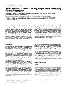
FIG. 1. Reconstitution of microtubule cold stability. A group of polypeptides isolated from microtubule protein by passage through a DEAEcellulose ion exchange column was used to reconstitute microtubule cold stability. (Left) Cold-labile microtubule protein was purified (three cycles) from bovine brain. The DEAE-cellulose first eluate fraction was added in a ratio of 0.05:1. Assembly in MEM buffer at 30TC was monitored by turbidity measurement at 350 nm; 0.1 mM GTP was added to the warm solution at time 0 to initiate polymerization. After assembly, the solutions were chilled to 000 for 10 min and then rewarmed for another assembly cycle. Podophyllotoxin (25 MM) was added and the solutions were chilled again and rewarmed to establish the cold-stable level. The plot shows cold-labile microtubule protein alone (---) or with first eluate material (-). Final protein concentration was 1.0 mg/ml. (Right) Lanes: A, polyacrylamide gel of polypeptides contained in the first eluate fraction; B, purified whole cold-stable microtubule protein; C, purified stabilizing fraction in the first eluate fraction, phosphorylated prior to the column run. Phosphorylation conditions were: 0.5 AuM calmodulin, 100 ,uM Ca2 , and 200 ,LM ATP. The eluate fraction contained approximately 8% ofthe total protein applied to the column. Asterisks, previously designated STOPs. To compare purified cold-stable to cold-labile microtubule proteins, see Job et al. (10); for comparison with X protein, see Pirollet et al. (11).
published procedures (10, 12, 24) with the exception that two different tissue homogenate methods were used as described in Results. The material was either used directly for disassembly assays or depolymerized through shearing by rapid and repeated passage at 0C through a syringe fitted with a 26-gauge needle (10). The disassembled cold-stable microtubule proteins were centrifuged at 40,000 x g for 30 min at 5YC, and the supernatant was chromatographed on a column of DEAE-cellulose for the separation of the STOPs as described (11). Preparation of STOPs. Purified cold-stable microtubules from eight rat brains were disassembled by shearing and incubation at 0C. The disassembled material (ca. 1.0 ml containing 10 mg of protein) was passed through a 1.5-ml DEAE-cellulose column; 0.35-ml fractions were collected at a flow rate of 0.35 ml/ 10 min. The peak flow-through fractions containing the STOPs (ca. 0.4 mg/ml) were collected. These fractions were added directly to cold-labile microtubule preparations in experiments designed to restore cold stability. Turbidimetric Measurement of the Assembled State. Assays for the assembly and disassembly of microtubules were performed on 0.4- or 0.8-ml samples in micro or semimicro cuvettes. Changes in optical density were followed at 350 nm and 30'C by using a Varian Cary 219 recording spectrophotometer equipped with a constant-temperature chamber and a "cell programmer for rapid successive assay of five samples. Assembly was initiated by addition of GTP (final concentration, 0.1 mM) to samples previously adjusted for base line in the spectrophotometer.
Other Methods. Cold-stable material was routinely examined by electron microscopy according to Margolis and Wilson (5) to confirm that it did consist of microtubules. Slab gel electrophoresis was run in 8% polyacrylamide/0. 1% sodium lauryl sulfate, according to Schier-Neiss et al. (25). Gels were stained with Coomassie blue R. Free Ca2+ concentration was determined with a computer program kindly supplied by R. Stein-
hardt (University of California, Berkeley) using the binding constants of Steinhardt et al. (26). RESULTS Microtubule-associated proteins that confer cold stability were isolated from rat brain. Restoration of cold stability was observed on addition of the active fraction in low ratio to bovine brain purified (three cycles) cold-labile microtubule protein (Fig. 1 Left). The results are similar to those found (11) with STOPs derived from sheep brain. The polypeptides associated with the active fraction are compared to whole microtubule protein in Fig. 1 Right. Phosphorylation of the active fraction prior to isolation by column chromatography had minimal effect on the active fraction's column elution profile. The first eluted fraction (containing 50 ,ig of protein in 350 1.d) from the DEAE-cellulose column lost its cold-stabilizing properties upon trypsin digestion (0.4 Ag of trypsin for 30 min at 30'C, pH 6.75, followed by a 4-fold molar excess of soybean trypsin inhibitor). Controls in which the trypsin inhibitor was added before trypsin were not affected. The reaction likewise was unaffected by incubation with a large excess (1 unit each) of RNase A or micrococcal nuclease for 15 min at 300C and pH 6.75. By contrast, boiling the fraction for 2 min totally abolished the restoration of cold stability. From these data, one can conclude that proteins in the DEAE-cellulose column first eluate fraction are directly implicated in the microtubule-stabilizing process. Effect of Calmodulin and ATP on Cold-Stable Microtubules. Micromolar concentrations of Ca2+-calmodulin have been shown to labilize cold-stable microtubules (24), an effect that could not be abolished by the presence of excess phenothiazines (24), compounds known to bind to calmodulin (27-30). We describe here a separate effect of Ca2+-calmodulin that is observed at even lower (submicromolar) concentrations of the
Biochemistry: job et at
3896
Proc. Natl. Acad. Sci. USA 80 (1983) --
A
--
origin
-
-0 u
-a 200
(_b] -__
100 F 0
Qua
B
_ -- -
80 V
.--
---
d).
-are
e
--
116.5
94
60 F
..
-....t
*
g
..;
- 68 40 F 20
hi
,-v .....
....
.I.
0
)
..
20
43
)fwg
...
1.
40
60
) -----
--- 'front
>
Time, min
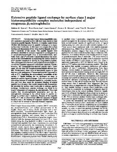
FIG. 2. Effect of ATP on cold-stable microtubules. Purified cold-stable microtubules were prepared and assayed as reported (24). (Left) Disassembly was assayed by turbidity measurement at 350 nm relative to a buffer blank. All samples contained 25 AuM PLN to prevent ATP-dependent reassembly; experiments were performed in MEM buffer at 300C. The 100% value represents turbidity in the absence of any additive. The turbidity of untreated controls did not vary with time. Curve a, control with PLN only. At 5 min the following additions were made: b, 9.0 ,uM free calcium, 0.5 AM calmodulin; c, 0.2 mM GTP; d, 0.2 mM adenylyl imidodiphosphate; e, 9.0 1LM free calcium, 0.5 /iM calmodulin, 0.2 mM adenylyl imidodiphosphate; f, 0.2 mM ATP; g, 9.0 jLM free calcium, 0.2 mM ATP; h, as g but plus 0.5 1LM calmodulin, 25 ,M trifluoperazine; i, 9.0 ,uM free calcium, 0.5 tuM calmodulin, 0.2 mM ATP. (Right) Autoradiograph of 32P-labeled STOPs. Cold-stable microtubules were purified and sheared at 0C. The supernatant from this disassembled preparation was incubated at 30'C for 15 min with 0.2 mM [Y-32P]ATP (500 cpm/pmol) in the absence (lane A) or presence (lane B) of 9.0 AM free calcium and 0.5 /uM calmodulin. The phosphorylated samples were then chilled to 0C and passed through DEAE-cellulose columns. The first eluate fractions were subjected to polyacrylamide gel electrophoresis; each lane contained 50 pg of protein. Asterisk, 56-kilodalton STOP that appears to be the major substrate of the Ca2+-calmodulin-activated kinase.
regulatory protein and that is totally dependent on the presence of ATP. Addition of 0.2 mM ATP alone to cold-stable microtubules resulted in a slow but complete depolymerization of the material after an initial decrease in optical density varying from 5% to 15% from experiment to experiment (Fig. 2 Left, curve f). The
rate
of
disassembly (t1/2
20
min)
was
not
affected
by
micromolar concentrations of Ca2+ (curve g). However, when both calmodulin and Ca2+ were added together with ATP, the disassembly was accelerated approximately 3-fold (curve i) with t1/2 6 min. Adenylyl imidodiphosphate, a nonhydrolyzable analog of ATP, added at 0.2 mM was without effect after the initial decrease. GTP (200 ttM) likewise did not induce disassembly in either the presence or absence of Ca2+ (curve c) whereas addition of 10 ,uM trifluoperazine totally abolished this Ca2+-calmodulin response (curve h). The rate of the ATP-dependent depolymerization could neither be augmented nor reduced by the following additions: cAMP or cGMP (10 /iM), the catalytic subunit of cAMP-dependent protein kinase or of cGMP-dependent protein kinase at 10 ,ug/ ml in the presence of excess cGMP or of purified beef heart myosin light chain kinase or phosphorylase kinase at 10 Ag/ml. Likewise, the pure heat-stable inhibitor of cAMP-dependent protein kinase (2.5 ,g/ml) (15) and myosin light chain (1 mg/ ml) were without effect. Control experiments were performed to confirm that the above measurements truly represent a reduction in the level of coldstable microtubules. For this, purified cold-stable microtubules were exposed to ATP in the presence or absence of Ca2+calmodulin; at various times, aliquots were removed and chilled, and their turbidity was measured quickly upon rewarming. The time course of cold-labilization observed by this approach was nearly identical to that obtained at 30'C in the presence of PLN when measured with the same protein preparation at the same time (data not shown). Polyacrylamide Gel Analysis of Protein Phosphorylation. In order to determine the pattern of phosphorylation of micro-
tubule proteins, disassembled cold-stable microtubule protein incubated with [y-32P]ATP and the product was passed through a DEAE-cellulose column (Fig. 2 Right). A number of polypeptides emerging in the first eluate were phosphorylated under these conditions. When the phosphorylation reaction was carried out in the presence of calcium and calmodulin, one major radioactive band appeared (marked by an asterisk) and phosphorylation of some of the other proteins seemed to be either slightly enhanced or suppressed. Trifluoperazine, which specifically counteracts the destabilization due to ATP-calmodulin (Fig. 2, curve h), blocked the labeling of that particular 56-kilodalton protein as well as that of the other polypeptides whose phosphorylation was calmodulin dependent. was
Response of Cold-Stable Microtubules to Calmodulin-De-
pendent Phosphorylation Only. All results discussed so far have been obtained with rat brain tissue that was initially homogenized with a motor-driven mortar and pestle. A more gentle procedure using a glass/glass loosely fitted Dounce homogenizer yielded somewhat different results. ATP (200 /.LM) alone had no effect on purified cold-stable microtubules obtained from such preparations (Fig. 3 Left), but when the material was exposed to both ATP and Ca2-calmodulin (0.5 uM), rapid disaggregation resulted. The reaction was totally inhibited by 50 tkM trifluoperazine. At the concentration used, calmodulin had no measurable effect on cold-stable microtubules in the absence of ATP. After the above reaction was carried out in the presence of [y-32P]ATP, the STOPs were isolated. Gel autoradiographs of the material obtained showed that two predominant proteins of 56 and 72 kilodaltons became phosphorylated (along with three minor ones) (Fig. 3 Right). Simultaneous Phosphorylation and Release of Stabilizing
Polypeptides. In order to test whether the cold-stabilizing proteins are released or remain bound upon phosphorylation, coldstable microtubules (prepared by the vigorous homogenization procedure) were exposed to 0.5 /LM Ca2+-calmodulin and 50
Biochemistry: job et aL
Proc. NatL. Acad. Sci. USA 80 (1983) A
0.4 A B
C
B
3897
D
-mm-
C
0.3
-
200
-
116.5
E CD LO)
cM 0.2 Co
0 0.1
-7
mck.... .: 9:
r:
-
94
-
68
-
43
1.
L-
Time, min FIG. 3. Effect of gentle homogenization on cold-stable microtubule phosphorylation and lability to ATP. Response of purified (two cycles) cold-stable microtubules (isolated by Dounce homogenization) assayed by turbidity as in Fig. 2. To the sample protein (2.5 mg/ml), was added at time 0: 200 pM ATP alone (curve A); 200 pM ATP, 9.0 p.M free calcium, 0.5 A.M calmodulin, 25 p.M trifluoperazine (curve B); or same as B but without trifluoperazine (curve C). Podophyllotoxin (25 yXM) was present and the assay was performed at 30'C in MEM buffer containing 10 A.M free calcium. (Right) Gel autoradiograph showing the phosphorylation pattern of the stabilizing fraction isolated by DEAE-cellulose chromatography. Lanes: A, with ATP only; B, with ATP, calmodulin, and trifluoperazine; C, with ATP and calmodulin. Twenty micrograms of protein was loaded in each lane.
AM [y-32P]ATP. After 10 min at 30TC, the remaining cold-stable microtubules were separated from the disaggregated material by centrifugation at 00C. Those proteins corresponding to the stabilizing fraction (from the DEAE-cellulose column) were phosphorylated in the absence of calmodulin only upon release from microtubules (Fig. 4). The calmodulin-dependent phosphorylation appears to occur to a minor extent on several polypeptides regardless of their association with microtubules. DISCUSSION
Evidence has been presented that a subset of polypeptides associated with cold-stable microtubules can restore cold stability to purified cold-labile material obtained from beef brain. These polypeptides, designated STOPs, can be separated by chromatography on DEAE-cellulose; they emerge from the column in the first eluate fraction. It is not yet clear how many participate in generating cold-stability, but we have found that they do represent subunits of a structural complex (unpublished data). We have reported previously that cold-stable microtubules are rendered cold-labile by exposure to ATP (12). Unexpected differences in response to phosphorylation resulted from changes in the homogenization procedure. Provided that microtubules are isolated by vigorous homogenization of brain tissue, most of the polypeptides of the stabilizing fraction are phosphorylated during microtubule cold-labilization. Under these conditions, there appears to be a strong correlation between phosphorylation of the stabilizing polypeptides and microtubule labilization, although it is not known which of the individual phosphorylated species is responsible. By contrast, with gentle glass/glass homogenization, the subsequently purified cold-stable microtubules did not respond to ATP alone, nor was substantial phosphorylation of the stabilizing fraction apparent. However, in the presence of cal-
FIG. 4. Phosphorylation of STOPs upon release from microtubules. To compare phosphorylation of microtubules and of released protein, purified cold-stable microtubules were incubated at 30'C in MEM buffer with 50 pM [y-32P]ATP. At 10 min the samples were chilled, 25 A.M podophyllotoxin was added, and the samples were centrifuged for 30 min at 200,000 x g. The supernatant and the cold-stable microtubule pellets were rinsed with cold buffer and used for gel analysis. The autoradiograph shows the supernatant protein sample incubated with ATP only (lane A) or with 10 pM free Ca2l and 0.5 p.M calmodulin (lane B). Lanes C and D are pellets corresponding to lanes A and B, respectively. Each lane contained 50 pg of protein. Bracket indicates those radioactive bands seen primarily in the supernatant samples.
modulin, microtubules became labile concurrently with phosphorylation of two major bands in the stabilizing fraction. This effect occurred at concentrations of calmodulin that by themselves had no direct influence on cold stability (24). Trifluoperazine abolishes both the ATP-dependent cold-labilization of microtubules and the phosphorylation of the stabilizing polypeptides. The effect observed only in the presence of calmodulin is important. It shows, first, that there is a good correlation between phosphorylation of stabilizing polypeptides and cold labilization and second, that the labilizing effect occurs in parallel with the phosphorylation of only two major polypeptides. Either of these two polypeptides is a potential candidate for a direct involvement in the stabilizing mechanism. The reason for loss of the calmodulin-independent kinase activity remains to be determined: perhaps a calmodulin-independent kinase is released from the particulate fraction by vigorous homogenization. Surprisingly, the addition of a number of exogenous calmodulin-dependent kinases had no influence on either the extent of protein phosphorylation or the rate of microtubule cold-labilization, including purified phosphorylase kinase which contains bound calmodulin as one of its subunits (31, 32) or myosin light chain kinase (33-35). Perhaps the calmodulin-dependent kinase operative in this system is one of a small family of such enzymes recently described in brain (36, 37). It is
interesting
to note
that
a
protein band of Mr
50,000
is appreciably less phosphorylated when the phosphorylation reaction is performed in the presence of Ca2+-calmodulin. A possible explanation is that this protein is specifically dephosphorylated by a calmodulin-dependent phosphatase (38). It does not appear to be involved in the cold-stabilizing process because cold stability is not affected by its phosphorylation (Fig.
3).
3898
Biochemistry: job et aL
STOPs migrate on gels in the region of the microtubule-associated r protein cluster (Fig. 1) (39-41). Furthermore, both the factors that render microtubules cold-stable and the T proteins bind to calmodulin affinity columns (41). However, we have recently shown that the X proteins and STOPs do not correspond on gels, nor do they overlap in function (11). That is, isolated r protein did not induce cold stability even when added at high concentration, and the STOPs did not promote microtubule polymerization. The absence of phosphorylated STOPs from intact cold-stable microtubules indicates either that they dissociate from microtubules upon phosphorylation or that they serve as substrates only when they become exposed at the end of the polymer. It should be of importance to the physiology of the cell (unless one is dealing with a remarkable coincidence) that a calmodulin-containing microtubule insertion site appears to occur in the pericentriolar region of the mitotic apparatus (42). Such a region could function in the mitotic spindle by regulating the reeling in and disassembly of cold-stable microtubules (3). It should be emphasized that, although the microtubule regulatory system described here could be useful to mitotic apparatus function, it remains to be determined whether polypeptides similar to those found in brain extracts are indeed present in microtubules of dividing cells. The STOPs and their associated kinases can be looked upon as a system for the physiological fine tuning of microtubule assembly in response to calcium levels or other transient signals. In vitro, this system alters dramatically the kinetic activity of microtubules and does so with economy because it functions through a substoichiometric mechanism. The change in kinetic behavior can be profound although the assembled state of microtubules may change very little. Thus, in the intact cell, the effect can be both subtle and substantial. The authors acknowledge the expert technical assistance of Mr. Dorr Tippens. This work was supported by Grants GM 28189 and AM 07902 from the National Institutes of Health, a grant from the Muscular Dystrophy Association, and starting funds from Institut National de la Sante et de la Recherche Medicale and the Ministry of Research and Technology, France. 1. Snyder, J. A. & McIntosh, J. R. (1976) Annu. Rev. Biochem. 45, 699-720. 2. Justin, P. (1978) Microtubules (Springer, New York). 3. Margolis, R. L. & Wilson, L. (1981) Nature (London) 293; 705712. 4. Satir, P. (1974) in Cilia and Flagella, ed. Sleight, M. A. (Academic, New York), Vol. 7, pp. 131-142. 5. Margolis, R. L. & Wilson, L. (1978) Cell 13, 1-8. 6. Karr, T. L. & Purich, D. L. (1979) J. Biol. Chem. 254, 10,885-
10,888. 7. Bergen, L. G. & Borisy, G. G. (1980) J. Cell Biol. 84, 141-150.
Proc. Natl. Acad. Sci. USA 80 (1983) 8. Grisham, L. M. (1976) Dissertation (Stanford Univ., Stanford, CA). 9. Webb, B. C. & Wilson, L. (1980) Biochemistry 19, 1993-1998. 10. Job, D., Rauch, C. T., Fischer, E. H. & Margolis, R L. (1982) Biochemistry 21, 509-515. 11. Pirollet, F., Job, D., Fischer, E. H. & Margolis, R. L. (1983) Proc. Natl Acad. Sci. USA 80, 1560-1564. 12. Margolis, R. L. & Rauch, C. T. (1981) Biochemistry 20, 4451-4458. 13. Watterson, D. M., Harrelson, W. G., Jr., Kelle, P. M., Sharief, F. & Vanaman, T. C. (1976) J. Biol Chem. 251, 4501-4513. 14. Peters, K. A., Demaille, J. G. & Fischer, E. H. (1977) Biochemistry 16, 5691-5697. 15. Demaille, J. G., Peters, K. A. & Fischer, E. H. (1977) Biochemistry 16, 3080-3086. 16. Demaille, J. G., Peters, K. A., Strandjord, T. P. & Fischer, E. H. (1978) FEBS Lett. 86, 113-116. 17. Glass, D. B. & Krebs, E. G. (1979)J. Biochem. 254, 9728-9738. 18. Krebs, E. G., Love, D. S., Bratvold, G. E., Trayser, K. A., Meyer, W. L. & Fischer, E. H. (1964) Biochemistry 3, 1022-1033. 19. Cohen, P. (1973) Eur. J. Biochem. 34, 1-14. 20. Perrie, W. T., Smillie, L. B. & Perry, S. V. (1973) Biochem.J. 128, 105P-106P. 21. Hiratsuka, T. (1980) Biochim. Biophys. Acta 625, 369-373. 22. Margolis, R. L. & Wilson, L. (1979) Cell 18, 673-679. 23. Asnes, C. F. & Wilson, L. (1979) Anal. Biochem. 98, 64-73. 24. Job, D., Fischer, E. H. & Margolis, R. L. (1981) Proc. Natl Acad. Sci. USA 78, 4679-4682. 25. Schier-Neiss, G., Lai, M. H. & Morris, N. R. (1978) Cell 15, 639647. 26. Steinhardt, R., Zucker, R. & Shatten, G. (1977) Dev. Biol 58, 158196. 27. Cheung, W. Y. (1980) Science 207, 19-27. 28. Klee, C. B., Crouch, T. H. & Richman, P. G. (1980) Annu. Rev. Biochem. 49, 489-515. 29. Levin, R. M. & Weiss, B. (1977) Mol Pharmacol. 13, 690-697. 30. Vincenzi, F. F. (1981) Cell Calcium 2, 387-409. 31. Shenolikar, S., Cohen, P. T. W., Cohen, P., Nairn, A. C. & Perry, S. V. (1979) Eur. J. Biochem. 100, 329-337. 32. Cohen, P. (1980) in Calcium and Cell Function, ed. Cheung, W. Y. (Academic, New York), Vol. 1, pp. 184-198. 33. Dabrowska, R., Aromatorio, D., Sherry, J. M. F. & Hartshorne, D. J. (1978) Biochemistry 17, 253-258. 34. Yagi, K., Yazawa, M., Kakiuchi, S., Ohshima, M. & Uenishi, K. (1978)J. Biol. Chem. 253, 1338-1340. 35. Adelstein, R. S. & Klee, C. B. (1980) in Calcium and Cell Function, ed. Cheung, W. Y. (Academic, New York), Vol. 1, pp. 167179. 36. Yamauchi, T. & Fujisawa, H. (1980) FEBS Lett. 116, 141-144. 37. Kennedy, M. B. & Greengard, P. (1981) Proc. Natl Acad. Sci. USA 78, 1293-1297. 38. Stewart, A. A., Ingebritsen, T. S., Manalan, A., Klee, C. B. & Cohen, P. (1982) FEBS Lett. 137, 80-84. 39. Weingarten, M. D., Lockwood, A. H., Hwo, S.-Y. & Kirschner, M. W. (1975) Proc. Nati Acad. Sci. USA 72, 1858-1862. 40. Cleveland, D. W., Spiegelman, B. M. & Kirschner, M. W. (1979)
J. Biol Chem. 254, 12,670-12,678.
41. Sobue, K., Fujita, M., Muramoto, Y. & Kakiuchi, S. (1981) FEBS
Lett. 132, 137-140.
42. Welsh, M. J., Dedman, J. R., Brinkley, B. R. & Means, A. R. (1979)
J. Cell Biol. 81, 624-634.