Dec 3, 2012 - and the superior temporal sulcus (STS). ... The connection between the OFA and STS is thought to be ..... lOFA, 74%; rSTS, 79%; lSTS, 29%).
Intra- and interhemispheric connectivity between face-selective regions in the human brain Jodie Davies-Thompson and Timothy J. Andrews
J Neurophysiol 108:3087-3095, 2012. First published 12 September 2012; doi: 10.1152/jn.01171.2011 You might find this additional info useful... This article cites 47 articles, 15 of which you can access for free at: http://jn.physiology.org/content/108/11/3087.full#ref-list-1 Updated information and services including high resolution figures, can be found at: http://jn.physiology.org/content/108/11/3087.full
This information is current as of December 3, 2012.
Journal of Neurophysiology publishes original articles on the function of the nervous system. It is published 24 times a year (twice monthly) by the American Physiological Society, 9650 Rockville Pike, Bethesda MD 20814-3991. Copyright © 2012 the American Physiological Society. ESSN: 1522-1598. Visit our website at http://www.the-aps.org/.
Downloaded from http://jn.physiology.org/ at University of York on December 3, 2012
Additional material and information about Journal of Neurophysiology can be found at: http://www.the-aps.org/publications/jn
J Neurophysiol 108: 3087–3095, 2012. First published September 12, 2012; doi:10.1152/jn.01171.2011.
Intra- and interhemispheric connectivity between face-selective regions in the human brain Jodie Davies-Thompson1,2 and Timothy J. Andrews1 1
Department of Psychology and York Neuroimaging Centre, University of York, York, United Kingdom; and 2Department of Ophthalmology and Visual Sciences, University of British Columbia, Vancouver, British Columbia, Canada
Submitted 21 December 2011; accepted in final form 10 September 2012
connectivity; fusiform face area; occipital face area; superior temporal sulcus; amygdala; inferior frontal gyrus MODELS OF FACE PERCEPTION propose a network of regions in the brain that are involved in different aspects of face processing. These regions have been subdivided into a core and an extended system (Haxby et al. 2000; Ishai 2008). The core system comprises regions in the occipital and temporal lobes, such as the occipital face area (OFA), the fusiform face area (FFA), and the superior temporal sulcus (STS). The OFA is proposed to have a feedforward projection to both the STS and the FFA. The connection between the OFA and STS is thought to be important in processing dynamic changes in the face that are important for social interactions, whereas the connection between the OFA and FFA is important for the representation of invariant facial characteristics that are used for recognition
(Andrews and Ewbank 2004; Hoffman and Haxby 2000; Winston et al. 2004). The extended face-processing system includes regions such as the amygdala, inferior frontal gyrus, intraparietal sulcus, orbitofrontal cortex, and anterior temporal regions (Fairhall and Ishai 2007; Haxby et al. 2000). However, the way that the core regions interact with the extended regions is not fully understood. One model suggests that the core regions interact with the extended regions through two parallel routes: one from the FFA to the anterior temporal lobe, and another from the STS to the amygdala and other regions in the extended system (Haxby et al. 2000). However, another model suggests that the flow of information between the core and extended systems is mediated primarily through the FFA (Fairhall and Ishai 2007; Ishai 2008). The first objective of this study was to examine which regions in the brain respond to faces in a large population of participants. Although many studies have shown that regions in the core system are face selective, it is not clear whether all regions in the extended system are face selective or are merely recruited by the face-processing system (Berman et al. 2010; Ishai 2008; Wiggett and Downing 2008). Our second objective was to determine how these face-selective regions are connected. To examine the functional connectivity between regions, we removed the stimulus driven activity that was used to define the location of face-selective regions in the first part of this study and correlated the remaining or residual time courses between face regions (see Norman-Haignere et al. 2012). This method is slightly different from psychophysiological interactions (PPI) in that PPI looks at modulation in the activity of the stimulus driven activity, whereas this approach examines the modulation of the residual activity. Therefore, this can be thought of as an extension of resting state connectivity in which correlations between regions, independent of a response to stimuli, are examined (Biswal et al. 1995; Margulies et al. 2010). The benefit of this technique over resting state analysis is that it allows additional information (connectivity) to be extracted from a standard functional magnetic resonance imaging (fMRI) experiment. Furthermore, this approach allows correlations between the time courses as a function of condition to be examined and thereby provide further support for functional connectivity between regions (Friston et al. 1997; Hampson et al. 2004). METHODS
Participants Address for reprint requests and other correspondence: T. J. Andrews, Dept. of Psychology, Univ. of York, York YO10 5DD, UK (e-mail: t.andrews @psych.york.ac.uk). www.jn.org
Data were collected from 72 participants (44 females; mean age 25 yr) who had taken part in previous fMRI experiments (Andrews et al.
0022-3077/12 Copyright © 2012 the American Physiological Society
3087
Downloaded from http://jn.physiology.org/ at University of York on December 3, 2012
Davies-Thompson J, Andrews TJ. Intra- and interhemispheric connectivity between face-selective regions in the human brain. J Neurophysiol 108: 3087–3095, 2012. First published September 12, 2012; doi:10.1152/jn.01171.2011.—Neuroimaging studies have revealed a number of regions in the human brain that respond to faces. However, the way these regions interact is a matter of current debate. The aim of this study was to use functional MRI to define faceselective regions in the human brain and then determine how these regions interact in a large population of subjects (n ⫽ 72). We found consistent face selectivity in the core face regions of the occipital and temporal lobes: the fusiform face area (FFA), occipital face area (OFA), and superior temporal sulcus (STS). Face selectivity extended into the intraparietal sulcus (IPS), precuneus (PCu), superior colliculus (SC), amygdala (AMG), and inferior frontal gyrus (IFG). We found evidence for significant functional connectivity between the core face-selective regions, particularly between the OFA and FFA. However, we found that the covariation in activity between corresponding face regions in different hemispheres (e.g., right and left FFA) was higher than between different face regions in the same hemisphere (e.g., right OFA and right FFA). Although functional connectivity was evident between regions in the core and extended network, there were significant differences in the magnitude of the connectivity between regions. Activity in the OFA and FFA were most correlated with the IPS, PCu, and SC. In contrast, activity in the STS was most correlated with the AMG and IFG. Correlations between the extended regions suggest strong functional connectivity between the IPS, PCu, and SC. In contrast, the IFG was only correlated with the AMG. This study reveals that interhemispheric as well as intrahemispheric connections play an important role in face perception.
3088
FUNCTIONAL CONNECTIVITY BETWEEN FACE REGIONS
2010a, 2010b; Davies-Thompson et al. 2009). All observers were right-handed and had normal or corrected-to-normal vision. Written consent was obtained for all participants, and the study was approved by the York Neuroimaging Centre Ethics Committee. Stimuli
Imaging Parameters The experiment was carried out using a GE 3 Tesla HD Excite MRI scanner at the York Neuroimaging Center at the University of York. An 8-channel, phased-array head coil (GE, Milwaukee, WI) tuned to 127.4 MHz was used to acquire MRI data from the whole brain. A gradient-echo echo-planar image (EPI) sequence was used to collect data from 38 contiguous axial slices (TR ⫽ 3 s, TE ⫽ 25 ms, field of view ⫽ 28 ⫻ 28 cm, matrix size ⫽ 128 ⫻ 128, voxel size ⫽ 2.1875 ⫻ 2.1875 mm, slice thickness ⫽ 3 mm). These were coregistered onto a T1-weighted anatomic image (1 ⫻ 1 ⫻ 1 mm) from each participant. To improve registrations, a T1-weighted image was taken in the same plane as the EPI slices.
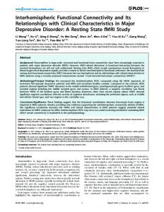
Fig. 1. Examples of stimuli from face and non-face stimulus conditions. From top to bottom: faces, bodies, places, objects, and scrambled images. Face images were taken from the Psychological Image Collection at Stirling (http://www.pics.psych.stir.ac.uk).
J Neurophysiol • doi:10.1152/jn.01171.2011 • www.jn.org
Downloaded from http://jn.physiology.org/ at University of York on December 3, 2012
There were five stimulus conditions: faces, bodies, inanimate objects, places, and scrambled images of the former categories. Figure 1 shows examples of the stimuli used. Face images were taken from the Psychological Image Collection at Stirling (http://www.pics.psych.stir.ac.uk). Face images varied in identity, sex, viewpoint (frontal, ¾ view), and expression (neutral, happy, speaking). Body images were taken from a body image collection at Bangor (http://www.bangor.ac.uk/⬃pss811/) and contained clothed male and female bodies without heads in a variety of postures. Images of places consisted of a variety of unfamiliar indoor scenes, houses and buildings, city scenes, and natural landscapes. Stimuli in the object condition consisted of 40 images of different inanimate objects including tools, ornaments, and furniture. Fourier-scrambled images were created by randomizing the phase of each two-dimensional frequency component in the original image while keeping the power of the components constant. Ten images from each of the four stimulus categories were scrambled for this condition. All images (⬃8° ⫻ 8°) were presented in grayscale and were back-projected onto a screen located inside the magnetic bore, ⬃57 cm from participants’ eyes. Images from each stimulus condition were presented in blocks. Within each block, an image was presented for
700 ms, followed by a 200-ms fixation cross. There were 10 images in each block, resulting in a block length of 9 s. Stimulus blocks were separated by a 9-s gray screen with a central fixation cross. Each condition was repeated 4 times in a counterbalanced design, resulting in a total of 20 stimulus blocks per scan. Participants were required to monitor all images for the presence of a red dot that was superimposed on one or two images in each block. Participants were required to respond, with a button press, as soon as they saw the image containing the target. The target could appear in any location on the image and was counterbalanced across conditions. We found no effect of stimulus condition on reaction times [F(4,160) ⫽ 1.52, P ⫽ 0.20] or percent correct [F(4,160) ⫽ 0.84, P ⫽ 0.50], suggesting that subjects were not significantly faster or more accurate at responding to the target in any of the conditions.
FUNCTIONAL CONNECTIVITY BETWEEN FACE REGIONS
fMRI Analysis
superior colliculus (rSC), and the right precuneus (rPCu). An ROI size of 27 voxels (0.4 cm3) was chosen to capture the peak voxels at the individual level while also allowing for slight variation in the location of face-selective regions at the group level. Furthermore, previous findings suggest 0.3– 0.8 cm3 to be optimal for detecting differences between stimulus conditions in the core face-selective regions (Fox et al. 2008). Connectivity between face-selective regions. To assess functional connectivity between regions, we first removed any stimulus-driven activity, because two regions will appear highly correlated if both are driven by the stimulus in parallel through a common input. As such, this analysis with stimulus-driven activity removed is orthogonal to the whole brain general linear model (GLM) analysis. The stimulusdriven activity was removed through two steps (Fig. 2A). First, the stimulus-driven activation as modeled in the GLM analysis was removed, resulting in a residual time series response for each participant (see Norman-Haignere et al. 2012 for a similar approach). Second, to capture any remaining stimulus-driven response that might not be fully accounted for by the hemodynamic model, the residual time series response from each region was averaged across all participants. The group average residual time series was then used as an additional regressor and the first-level analysis repeated. This generated a second residual time course. A group average of the new residuals revealed no consistent response across participants (Fig. 2A). Correlations between the second residual time courses were then run for each pair of regions in each participant (Fig. 2B). To determine whether the regions share a selective variance, we conducted partial correlations between face-selective regions of interest, entering the residual time course of a control region (PPA) as a random variable. Pearson’s r correlation values were then converted to Fisher’s z values (Zr) before being entered into statistical tests. Finally, to determine how the correlations between the residual time courses of the core face-selective regions were affected by the stimulus category that was being viewed, we extracted the time courses when each stimulus
Fig. 2. Correlating residual activity between face-selective regions. A: the general linear model (GLM) removes the stimulus-driven activity from the time course. The residual activity was then averaged across participants to ensure there was no remaining stimulus-driven activity. MR, magnetic resonance. B: the residuals were then entered into a correlation analysis to determine functional connectivity between pairs of face-selective regions. J Neurophysiol • doi:10.1152/jn.01171.2011 • www.jn.org
Downloaded from http://jn.physiology.org/ at University of York on December 3, 2012
Preprocessing. Statistical analysis of the fMRI data was carried out using FEAT (http://www.fmrib.ox.ac.uk/fsl; Smith et al. 2004). The initial 9 s of data from each scan were removed to minimize the effects of magnetic saturation, and slice-time correction was applied. Motion correction was followed by spatial smoothing [Gaussian, full-width at half-maximum ⫽ 6 mm] and temporal high-pass filtering (cutoff, 0.01 Hz). The functional data were transformed onto a high-resolution T1-anatomic image before being coregistered onto a standard brain (ICBM152). Face-selective regions. Regressors for each condition in the GLM were convolved with a gamma hemodynamic response function, and four face-selective contrasts were run on each participant: 1) faces ⬎ scrambled, 2) faces ⬎ places, 3) faces ⬎ objects, 4) faces ⬎ bodies. Individual participant data were then entered into a higher level group analysis using a mixed-effects design (FLAME, http://www.fmrib.ox. ac.uk/fsl). Contrasts were resel corrected for multiple comparisons (P ⬍ 0.05). Core regions of interest (ROIs) were defined individually for each participant in EPI space by taking a 27-voxel mask around the peak of each ROI from the average statistical map (P ⬍ 0.001, uncorrected), and the responses in each ROI were averaged across the voxels. We also defined the parahippocampal place area (PPA) as a control, non-face-selective region using the contrast places ⬎ faces. This region was defined by the individual level. Because it was not always possible to reliably define regions from the extended system at the individual level, these regions were defined from a higher level group analysis (P ⬍ 0.05, resel corrected) using a mixed-effects design (FLAME, http://www.fmrib.ox.ac.uk/fsl). To determine how these regions interact with the core face-selective regions, 27-voxel masks were drawn at the peak of each ROI at the group level and transformed back into the EPI coordinates for each participant. These regions included the right and left amygdala (AMG), the right intraparietal sulcus (rIPS), the right inferior frontal gyrus (rIFG), the right
3089
3090
FUNCTIONAL CONNECTIVITY BETWEEN FACE REGIONS
condition was presented (for example, all blue data points in Fig. 2 from the face condition) and entered these data into a correlation. To take into account the delay of the MR response, we removed a data point (1 TR) from the start and the end of each stimulus block.
RESULTS
Face-Selective Regions
Fig. 3. Average face-selective statistical map thresholded at P ⬍ 0.05 (corrected for multiple comparisons). A: location of core face-selective regions (FFA, fusiform face area; OFA, occipital face area; STS, superior temporal sulcus) across subjects in a whole brain analysis. B: location of other regions showing face selectivity (IFG, inferior frontal gyrus; IPS, intraparietal sulcus; PCu, precuneus; SC, superior colliculus; AMG, amygdala).
J Neurophysiol • doi:10.1152/jn.01171.2011 • www.jn.org
Downloaded from http://jn.physiology.org/ at University of York on December 3, 2012
Figure 3 shows regions in the brain that were more responsive to faces compared with other non-face objects (P ⬍ 0.05, resel corrected for multiple comparisons). The peak locations of the core and extended regions are shown in Table 1. The core face-selective regions, FFA, OFA, and STS, can be clearly identified in Fig. 3A. Recent studies using high-resolution fMRI have reported that there may be more than one faceselective patch along the fusiform gyrus (Pinsk et al. 2009; Weiner and Grill-Spector 2010). However, due to anatomic variability across subjects and their spatial proximity, these regions are unlikely to be differentiated in a group analysis. In addition to the core regions, a number of other faceselective regions were identified. Figure 3B shows the location of six regions showing a greater response to faces than the other object categories. These were bilateral amygdala (lAMG, rAMG), rIPS, rIFG, rSC, and rPCu. At the group level, the proportion of significant face-selective voxels was greater in the right hemisphere (84% of all face-selective voxels, 77.6 cm3) compared with the left hemisphere (16%, 14.4 cm3).
Connectivity Between Face-Selective Regions Correlations between core regions. Next, we determined the connectivity between face-selective regions in the core system. To do this, face-selective regions were defined for each participant (%participants: rFFA, 94%; FFA, 82%; rOFA, 72%; lOFA, 74%; rSTS, 79%; lSTS, 29%). Figure 4 shows the mean correlations between the residual time courses of the core face-selective regions. First, we examined connectivity between corresponding face-selective regions in the left and right hemisphere. We found strong interhemispheric correlations for each face-selective region, FFA (Zr ⫽ 0.63), OFA (Zr ⫽ 0.71), and STS (Zr ⫽ 0.49). To determine whether these were significantly higher than the intrahemispheric correlations, we compared the correlation between the corresponding left and right regions (i.e.. lFFA-rFFA) with the averaged withinhemisphere correlations (i.e., lFFA-lOFA, lFFA-lSTS, rFFArOFA, rFFA-rSTS). Interhemispheric correlations were significantly higher than the intrahemispheric correlations for the FFA [Zr ⫽ 0.63 ⬎ 0.37; t(55) ⫽ 7.88, P ⬍ 0.001], OFA [Zr ⫽ 0.71 ⬎ 0.38; t(41) ⫽ 7.81, P ⬍ 0.001], and STS [Zr ⫽ 0.49 ⬎ 0.20; t(19) ⫽ 7.86, P ⬍ 0.001]. Next, we examined the intrahemispheric correlations between the core face-selective regions. One-sampled t-tests showed significant correlations (compared with 0, P ⬍ 0.001 corrected for multiple comparisons) between the residual time courses of all the core regions: OFA-FFA (left: Zr ⫽ 0.53; right: Zr ⫽ 0.46), OFA-STS (left: Zr ⫽ 0.17; right: Zr ⫽ 0.12), FFA-STS (left; Zr ⫽ 0.27; right: Zr ⫽ 0.18). Paired-samples t-tests showed no differences between intrahemispheric corre-
FUNCTIONAL CONNECTIVITY BETWEEN FACE REGIONS
Table 1. Peak MNI coordinates of face-selective (and control) regions from the averaged face contrast Coordinates Region Core system FFA OFA STS Extended system AMG
x
y
z
R L R L R L
41 ⫺39 38 ⫺36 53 ⫺55
⫺54 ⫺55 ⫺82 ⫺82 ⫺49 ⫺55
⫺24 ⫺23 ⫺16 ⫺18 3 6
R L R R R R
20 ⫺20 42 50 6 4
⫺6 ⫺8 ⫺70 22 ⫺34 ⫺64
⫺18 ⫺20 40 22 ⫺2 26
R L
29 ⫺27
⫺50 ⫺53
⫺13 ⫺12
Core face-selective regions and the control (parahippocampal, PPA) region were defined at the individual level; extended regions were defined at the group level and transformed back to the individual level. FFA, fusiform face area; OFA, occipital face area; STS, superior temporal sulcus; AMG, amygdala; IPS, intraparietal sulcus; IFG, inferior frontal gyrus; SC, superior colliculus; PCu, precuneous.
lations in the right and left hemisphere [OFA-FFA: t(34) ⫽ 1.77, P ⫽ 0.09; OFA-STS: t(10) ⫽ 0.89, P ⫽ 0.39; FFA-STS: t(17) ⫽ 0.77, P ⫽ 0.45]. Next, we compared the strength of the correlations between pairs of regions. We found significantly greater correlations between the OFA-FFA compared with both the OFA-STS [left: t(13) ⫽ 5.01, P ⬍ 0.001; right: t(40) ⫽ 7.63, P ⬍ 0.001] and FFA-STS [left: t(13) ⫽ 3.31, P ⬍ 0.01; right: t(40) ⫽ 5.37, P ⬍ 0.05]. The correlations between the FFASTS were significantly greater than those for the OFA-STS in the right hemisphere [t(40) ⫽ 3.02, P ⬍ 0.005], but not in the left hemisphere [t(13) ⫽ 1.97, P ⫽ 0.07]. To validate our functional connectivity analysis, we performed a separate analysis to ensure that all stimulus-driven activity was removed from the residual time series. Rather than calculating correlations between ROIs within participants, correlations in this control analysis were calculated between random pairs of participants, e.g., OFA (participant 1)-FFA (participant 2). Unlike the positive values generated by the within-participant correlations (Fig. 4A), none of the control correlations across participants were significantly different from 0 (Fig. 4B).
To determine how the correlations between the residual time courses of the core face-selective regions were affected by the stimulus category that was being viewed, we repeated the correlation on different segments of the time course (Fig. 5). A 2 ⫻ 5 ANOVA (hemisphere, condition) for the OFA-FFA revealed an effect of condition [F(4,136) ⫽ 4.41, P ⬍ 0.005] but no effect of hemisphere [F(1,34) ⫽ 2.61, P ⫽ 0.12] or an interaction [F(4,136) ⫽ 0.47, P ⫽ 0.76]. In the right hemisphere, a 1 ⫻ 5 ANOVA for OFA-FFA showed an effect of condition [F(4,196) ⫽ 9.74, P ⬍ 0.001], which was caused by increased correlations when faces (Zr ⫽ 0.68) were presented as compared with all other conditions [bodies: Zr ⫽ 0.48, t(49) ⫽ 4.11, P ⬍ 0.001; objects: Zr ⫽ 0.37, t(49) ⫽ 5.01, P ⬍ 0.001; places: Zr ⫽ 0.43, t(49) ⫽ 4.09, P ⬍ 0.001; scrambled: Zr ⫽ 0.38, t(49) ⫽ 5.26, P ⬍ 0.001]. In the left hemisphere, there was also an effect of condition for OFA-FFA [F(4,176) ⫽ 2.67, P ⬍ 0.05], which was caused by higher correlations when faces were presented (Zr ⫽ 0.65) as compared with bodies [Zr ⫽ 0.52, t(44) ⫽ 2.29, P ⬍ 0.05] and scrambled images [Zr ⫽ 0.45, t(44) ⫽ 3.52, P ⬍ 0.001], but not relative to objects [Zr ⫽ 0.53, t(44) ⫽ 2.00, P ⫽ 0.05] or places [Zr ⫽ 0.53, t(44) ⫽ 1.84, P ⫽ 0.07]. There were no significant effects of condition on the correlations between residual time courses of the OFA-STS [F(4,40) ⫽ 0.22, P ⫽ 0.92] or the FFA-STS [F(4,68) ⫽ 1.09, P ⫽ 0.37]. To determine whether the increased correlations during face blocks could be due to the residuals being greater in face relative to non-face blocks, we compared the absolute summed residuals during face blocks with the absolute summed residuals during non-face blocks. Paired-samples t-tests (across subjects) showed no difference between the size of the residuals when faces were viewed compared with non-face blocks for any region (P value range: 0.27– 0.98, no corrections). Correlations between core regions and extended regions. Despite the significant face selectivity shown in the group analysis (see Fig. 3), only 21% of the extended regions could be identified at the individual participant level (%participants: rAMG, 19%; lAMG, 15%; rIPS, 25%; rIFG, 32%; rSC, 8%; rPCu, 28%). Accordingly, the extended regions were defined from masks defined from the group analysis and transformed back into the EPI coordinates for each participant. Figure 6 shows the correlations between the core regions (FFA, OFA, STS) and the extended regions (AMG, IPS, IFG, SC, PCu). A two-way ANOVA (core, extended) revealed that there was a significant effect of core region [F(2,80) ⫽ 47.47, P ⬍ 0.001] and a significant effect of extended region [F(4,160) ⫽ 94.77, P ⬍ 0.001] on the correlations between regions. The effect of Fig. 4. A: average correlations (Pearson’s r transformed into Fisher’s z) in the residual time courses between core face-selective regions within participants. This shows significant interhemispheric correlations between corresponding face regions (lFFA-rFFA, lOFA-rOFA, lSTS-rSTS, where “l” is left and “r” is right) and strong intrahemispheric correlations between the OFA and FFA. B: average correlations in the residual time courses between core face-selective regions across participants. The absence of significant correlations between regions in this analysis shows that the correlations in A are specific to each individual and do not reflect any global trend.
J Neurophysiol • doi:10.1152/jn.01171.2011 • www.jn.org
Downloaded from http://jn.physiology.org/ at University of York on December 3, 2012
IPS IFG SC PCu Control region PPA
Hemisphere
3091
3092
FUNCTIONAL CONNECTIVITY BETWEEN FACE REGIONS
Fig. 5. Effect of stimulus condition on the average correlation between the residual time courses of different core face-selective regions. Correlations between the OFA-FFA were significantly increased when faces were presented.
Fig. 6. Average correlations (Pearson’s r transformed into Fisher’s z) in the residual time courses between the core regions and extended regions. This shows strong correlations between the OFA or FFA and the IPS, PCu, and SC. In contrast, the STS was more strongly correlated with the AMG and IFG.
0.001]. FFA-IPS correlations were greater for faces (Zr ⫽ 0.61) compared with bodies [Zr ⫽ 0.51, t(67) ⫽ 2.10, P ⬍ 0.05], objects [Zr ⫽ 0.44, t(67) ⫽ 3.23, P ⬍ 0.005], places [Zr ⫽ 0.50, t(67) ⫽ 2.21, P ⬍ 0.05], and scrambled images [Zr ⫽ 0.45, t(67) ⫽ 3.78, P ⬍ 0.001]. There was no influence of condition on the STS-IPS correlations. The PCu showed a significant effect of core region [F(2,80) ⫽ 31.74, P ⬍ 0.001]. This was due to larger correlations between FFA-PCu (Zr ⫽ 0.52) compared with the OFA-PCu [Zr ⫽ 0.41; t(49) ⫽ 3.44, P ⫽ 0.001] and STS-PCu [Zr ⫽ 0.14; t(54) ⫽ 9.77, P ⬍ 0.001]. The correlation between the OFAPCu was also greater than that for STS-PCu [t(41) ⫽ 6.00, P ⬍ 0.001]. We also found a significant effect of condition [F(4,160) ⫽ 3.13, P ⬍ 0.05]. The effect of condition was due to higher correlations for faces (Zr ⫽ 0.42) and bodies (Zr ⫽ 0.43) compared with objects (Zr ⫽ 0.36) and places (Zr ⫽ 0.35). There was no interaction between core region and condition [F(8,320) ⫽ 0.73, P ⫽ 0.67]. The SC showed a significant effect of core region [F(2,80) ⫽ 56.83, P ⬍ 0.001]. This was due to larger correlations between the FFA-SC compared with the OFA-SC [Zr ⫽ 0.78 ⬎ 0.50; t(49) ⫽ 5.31, P ⬍ 0.001] and STS-SC [Zr ⫽ 0.78 ⬎ 0.12; t(54) ⫽ 11.78, P ⬍ 0.001]. The correlation between the OFA-SC was also greater than STS-SC [Zr ⫽ 0.48 ⬎ 0.12; t(41) ⫽ 8.65, P ⬍ 0.001]. We also found a significant effect of condition [F(4,160) ⫽ 4.44, P ⬍ 0.005]. The effect of condition was due to higher correlations for faces (Zr ⫽ 0.54) compared with bodies (Zr ⫽ 0.51), objects (Zr ⫽ 0.49), places (Zr ⫽ 0.49), and scrambled images (Zr ⫽ 0.48). There was no interaction between core region and condition [F(8,320) ⫽ 0.51, P ⫽ 0.85]. The AMG showed a significant effect of core region [F(2,80) ⫽ 7.08, P ⬍ 0.001]. This was due to higher correlations between the STS-AMG [Zr ⫽ 0.22; t(41) ⫽ 3.94, P ⬍ 0.001] and FFA-AMG [Zr ⫽ 0.18; t(49) ⫽ 3.16, P ⬍ 0.005] compared with the OFA-AMG (Zr ⫽ 0.10). There was no difference in the correlations between STS-AMG and FFAAMG [t(54) ⫽ 1.35, P ⫽ 0.18]. There was a significant effect of condition [F(4,160) ⫽ 3.42, P ⬍ 0.05]. The effect of condition was due to higher correlations for faces (Zr ⫽ 0.21)
J Neurophysiol • doi:10.1152/jn.01171.2011 • www.jn.org
Downloaded from http://jn.physiology.org/ at University of York on December 3, 2012
core region was due to higher correlations with the OFA (Zr ⫽ 0.37) and FFA (Zr ⫽ 0.37) compared with the STS (Zr ⫽ 0.17). The effect of the extended regions was due to higher correlations in the IPS (Zr ⫽ 0.49), PCu (Zr ⫽ 0.36), and SC (Zr ⫽ 0.45) compared with the AMG (Zr ⫽ 0.16) and IFG (Zr ⫽ 0.04). There was also an interaction between core and extended regions [F(8,320) ⫽ 73.40, P ⬍ 0.001]. To determine how each core region interacts with each extended region and to determine how these interactions were influenced by stimulus condition, a two-way ANOVA (core, condition) was used for each extended region. The IPS showed a significant effect of core region [F(2,80) ⫽ 98.26, P ⬍ 0.001]. This was due to larger correlations between OFA-IPS compared with the FFA-IPS [Zr ⫽ 0.92 ⬎ 0.47; t(49) ⫽ 7.74, P ⬍ 0.001] and STS-IPS [Zr ⫽ 0.92 ⬎ 0.10; t(41) ⫽ 15.46, P ⬍ 0.001]. The correlation between the FFA-IPS was also greater than that for STS-IPS [Zr ⫽ 0.47 ⬎ 0.10; t(54) ⫽ 9.91, P ⬍ 0.001]. We also found a significant effect of condition [F(4,160) ⫽ 3.63, P ⬍ 0.01] and an interaction between core region and condition [F(8,320) ⫽ 1.97, P ⬍ 0.05]. OFA-IPS correlations were higher for faces (Zr ⫽ 1.06) compared with places [Zr ⫽ 0.93; t(51) ⫽ 2.43, P ⬍ 0.05] and scrambled images [Zr ⫽ 0.84; t(51) ⫽ 4.29, P ⬍
FUNCTIONAL CONNECTIVITY BETWEEN FACE REGIONS
DISCUSSION
The aim of this study was to determine which areas in the human brain respond selectively to faces and to determine the functional connectivity between these regions in a large population of participants. We found evidence for functional connectivity between all the core face-selective regions. However, we found that corresponding core regions in different
Fig. 7. Average correlations (Pearson’s r transformed into Fisher’s z) in the residual time courses between the extended regions across all subjects. This shows significant correlations between the IPS, PCu, and SC.
hemispheres were more connected with each other than with core regions in the same hemisphere. Our results also suggest that there is marked variability in the connectivity between the core and extended regions. The OFA and FFA showed stronger connectivity with the IPS, PCu, and SC. In contrast, the STS showed more functional connectivity with the AMG and IFG. Face-Selective Regions The locations of the core face-processing regions (FFA, OFA, and STS) were consistent with those described in previous studies (Andrews and Ewbank 2004; Berman et al. 2010; Downing et al. 2006; Fox et al. 2009; Hoffman and Haxby 2000; Kanwisher et al. 1997). We also found significant face selectivity in the right IFG (see also Chan and Downing 2011; Scalaidhe et al. 1999; Tsao et al. 2008; Vignal et al. 2000) and the right IPS. Although the role of these areas in face processing remains unclear, they have both been implicated in models of attentional control (Corbetta and Shulman 2002) and in the mirror neuron system (Iacoboni and Dapretto 2006). The AMG is known to be involved in the perception of facial expressions (Breiter et al. 1996; Morris et al. 1996; Phillips et al. 1998; Vuilleumier et al. 2001), but it has not been clear whether this region is face selective. Our results clearly show that the AMG has a significant selective response to faces. We also found other regions that showed face-selective responses: the PCu (a region on the medial surface of the parietal lobe). This region overlaps with a region that has been referred to as posterior cingulate cortex (Gschwind et al. 2012). Again it is not clear what role the PCu plays in face processing, but it has been associated with memory and visual imagery (Cavanna and Trimble 2006) and in retrieving episodic memories associated with faces (Gobbini and Haxby 2007). Finally, we found significant face-selective activity in the SC. This region is known to play an important role in orienting movements of the head and eyes (Sparks 1999), so it is possible that this selectivity may reflect planning or execution of eye movements associated with face images (Yarbus 1967). Connectivity Between Face-Selective Regions Models of face processing propose that the OFA has a feedforward projection to the FFA and STS (Haxby et al. 2000; Ishai 2008). Our results provide clear support for a functional connection between the OFA and FFA. To further establish the functional nature of the connectivity between the OFA and FFA, we determined whether the correlations between the residual time courses in these regions were influenced by the stimulus that was presented. We found an increased correlation between the OFA and FFA when participants viewed faces compared with any other stimuli, providing further support for a face-selective connection between these regions. The evidence for functional connectivity between the OFA and FFA is consistent with a recent diffusion tensor imaging (DTI) study showing strong connectivity between these regions (Gschwind et al. 2012). Although these results provide support for a functional connection between the OFA and FFA, this does not rule out the possibility that the FFA receives input from other sources. For example, prosopagnosic patients, with lesions that affect the OFA, continue to show activity in the FFA (Rossion et al. 2003; Steeves et al. 2006).
J Neurophysiol • doi:10.1152/jn.01171.2011 • www.jn.org
Downloaded from http://jn.physiology.org/ at University of York on December 3, 2012
and bodies (Zr ⫽ 0.20) compared with objects (Zr ⫽ 0.13) and places (Zr ⫽ 0.15). There was no interaction between core region and condition [F(8,320) ⫽ 0.64, P ⫽ 0.75]. The IFG showed a significant effect of core region [F(2,80) ⫽ 33.56, P ⬍ 0.001]. This was due to larger correlations between STS-IFG compared with the FFA-IFG [Zr ⫽ 0.22 ⬎ ⫺0.01; t(54) ⫽ 8.68, P ⬍ 0.001] and OFA-IFG [Zr ⫽ 0.22 ⬎ ⫺0.06; t(41) ⫽ 6.15, P ⬍ 0.001]. The FFA-IFG and OFA-IFG correlations were not significantly greater than 0. There was no effect of condition [F(4,160) ⫽ 0.36, P ⫽ 0.84] and no interaction between core region and condition [F(8,320) ⫽ 0.70, P ⫽ 0.69]. Correlations between extended regions. Figure 7 shows the correlations between residual time courses in the extended regions. One-sampled t-tests showed significant correlations (compared with 0, P ⬍ 0.005) between the residual time courses of all extended regions, with the exception of correlations between the IFG and the IPS, PCu, and SC. The highest correlations between the extended regions were between the PCu-SC [Zr ⫽ 0.68; t(71) ⫽ 26.26, P ⬍ 0.001], PCu-IPS [Zr ⫽ 0.45; t(71) ⫽ 17.58, P ⬍ 0.001], and IPS-SC [Zr ⫽ 0.52; t(71) ⫽ 21.15, P ⬍ 0.001]. The only region that showed a significant correlation with the IFG was the AMG [Zr ⫽ 0.09; t(71) ⫽ 5.74, P ⬍ 0.001]. The AMG also showed significant correlations with the IPS [Zr ⫽ 0.13; t(71) ⫽ 7.00, P ⬍ 0.001], the PCu [Zr ⫽ 0.19; t(71) ⫽ 7.68, P ⬍ 0.001], and the SC [Zr ⫽ 0.16; t(71) ⫽ 6.70, P ⬍ 0.001]. Next, we determined whether the pattern of correlations was affected by the stimulus condition using a one-way ANOVA (condition). There was an effect of condition for correlations between the PCu and the AMG [F(4,284) ⫽ 3.04, P ⬍ 0.05]. This was due to the correlations being greater when faces were being viewed compared with places [Zr ⫽ 0.24 ⬎ 0.10, t(71) ⫽ 3.27, P ⬍ 0.005]. There were no other significant effects of stimulus condition between the other extended regions.
3093
3094
FUNCTIONAL CONNECTIVITY BETWEEN FACE REGIONS ACKNOWLEDGMENTS We thank Heidi Baseler, Thomas Busigny, and members of the Human Vision and Eye Movement Laboratory (University of British Columbia) for helpful comments on the manuscript. GRANTS This work was supported by Wellcome Trust Grant WT087720MA. J. Davies-Thompson was supported by an Economic and Social Research Council UK studentship. DISCLOSURES No conflicts of interest, financial or otherwise, are declared by the authors. AUTHOR CONTRIBUTIONS J.D.-T. and T.J.A. conception and design of research; J.D.-T. and T.J.A. performed experiments; J.D.-T. analyzed data; J.D.-T. and T.J.A. interpreted results of experiments; J.D.-T. and T.J.A. prepared figures; J.D.-T. and T.J.A. drafted manuscript; J.D.-T. and T.J.A. edited and revised manuscript; J.D.-T. and T.J.A. approved final version of manuscript. REFERENCES Andrews TJ, Clarke A, Pell P, Hartley T. Selectivity for low-level features of objects in the human ventral stream. Neuroimage 49: 703–711, 2010a. Andrews TJ, Davies-Thompson J, Kingstone A, Young AW. Internal and external features of the face are represented holistically in face-selective regions of visual cortex. J Neurosci 30: 3544 –3552, 2010b. Andrews TJ, Ewbank MP. Distinct representations for facial identity and changeable aspects of faces in the human temporal lobe. Neuroimage 23: 905–913, 2004. Berman MG, Park J, Gonzalez R, Polk TA, Gehrke A, Knaffla S, Jonides J. Evaluating functional localizers: the case of the FFA. Neuroimage 50: 56 –71, 2010. Biswal B, Yetkin FZ, Haughton VM, Hyde JS. Functional connectivity in the motor cortex of resting human brain using echo-planar MRI. Magn Reson Med 34: 537–541, 1995. Breiter HC, Etcoff NL, Whalen PJ, Kennedy WA, Rauch SL, Buckner RL, Strauss MM, Hyman SE, Rosen BR. Response and habituation of the human amygdala during visual processing of facial expressions. Neuron 17: 875– 887, 1996. Cavanna AE, Trimble MR. The precuneus: a review of its functional anatomy and behavioural correlates. Brain 129: 564 –583, 2006. Chan AW, Downing PE. Faces and eyes in human lateral prefrontal cortex. Front Hum Neurosci 5: 51, 2011. Corbetta M, Shulman GL. Control of goal-directed and stimulus-driven attention in the brain. Nat Rev Neurosci 3: 201–215, 2002. Cordes D, Haughton VM, Arfanakis K, Wendt GJ, Turski PA, Moritz CH, Quigley MA, Meyerand ME. Mapping functionally related regions of brain with functional connectivity MR imaging. AJNR Am J Neuroradiol 21: 1636 –1644, 2000. Davies-Thompson J, Gouws A, Andrews TJ. An image-dependent representation of familiar and unfamiliar faces in the human ventral stream. Neuropsychologia 47: 1627–1635, 2009. Downing PE, Chan AW. Y, Peelen MV, Dodds CM, Kanwisher N. Domain specificity in visual cortex. Cereb Cortex 16: 1453–1461, 2006. Ethofer T, Gshwind M, Vuilleumier P. Processing social aspects of human gaze: a combined fMRI-DTI study. Neuroimage 55: 411– 419, 2011. Fairhall SI, Ishai A. Effective connectivity within the distributed cortical network for face perception. Cereb Cortex 17: 2400 –2406, 2007. Fox CJ, Iaria G, Barton JJ. Defining the face processing network: optimisation of the functional localiser in fMRI. Hum Brain Mapp 30: 1637–1651, 2009. Friston KJ, Buechel C, Fink GR, Morris J, Rolls E, Dolan RJ. Psychophysiological and modulatory interactions in neuroimaging. Neuroimage 6: 218 –229, 1997. Gobbini MI, Haxby JV. Neural systems for recognition of familiar faces. Neuropsychologia 45: 32– 41, 2007. Gshwind M, Pourtois G, Schwartz S, Van De Ville D, Vuilleumier P. White matter connectivity between face-responsive regions in the human brain. Cereb Cortex 22: 1564 –1576, 2012.
J Neurophysiol • doi:10.1152/jn.01171.2011 • www.jn.org
Downloaded from http://jn.physiology.org/ at University of York on December 3, 2012
We also found evidence for a functional connection between the OFA and STS and between the FFA and STS (see also Fairhall and Ishai 2007; Li et al. 2010; TurkBrowne et al. 2010). However, the correlations between the residual time courses of the OFA-STS and FFA-STS were not significantly increased when participants viewed faces compared with other non-face objects. A reason for the absence of face-selective connectivity between the STS and the other core face regions could be the face images that were used in this study. It is likely that functional connectivity with the STS would be affected by face images if they are more salient for social communication (see Ethofer et al. 2011). Although models of face processing propose that information from the core face-selective regions is relayed to an extended face-processing network, they differ on which areas are functionally connected (Haxby et al. 2000; Ishai 2008). We found significant differences in the functional connectivity between the core and extended face-selective regions. The residual time courses from the OFA and FFA correlated most with the IPS, PCu, and SC. In contrast, the residual time courses of the STS correlated more with the AMG and IFG (see Ethofer et al. 2011). We also found evidence for significant functional connectivity between regions in the extended network. The residual time courses in the IPS, PCu, and SC all showed significant correlations. In contrast, the IFG was only significantly correlated with the AMG. Together, these results suggest that the OFA and FFA are involved in a network involving the IPS, PCu, and SC. In contrast, the STS shares functional connections with the IFG and AMG. Future studies using DTI are necessary to determine whether these functional connections are based on direct structural links (see Gschwind et al. 2012). Models of face processing have focused on the intrahemispheric connections between regions. However, we found high correlations between the time courses of response between corresponding face-selective regions in the left and right hemisphere. Indeed, the correlation in response was greater between corresponding face regions in different hemispheres than between different face regions in the same hemisphere. These data fit with other studies that have shown highly correlated responses between equivalent regions in each hemisphere (Biswal et al. 1995; Cordes et al. 2000; Kleinschmidt et al. 1994; Lowe et al. 1998; Nir et al. 2006; Salvador et al. 2005). This functional connectivity is likely to be mediated by the corpus callosum, because damage to this commissure dramatically reduces correlated magnetic resonance activity across the hemispheres (Quigley et al. 2003). The implication of these findings is that models of face processing should take account of interhemispheric as well as intrahemispheric connections. In conclusion, we found evidence for functional connectivity between the core face-selective regions. However, we found that the functional connectivity between corresponding face regions in different hemispheres was greater than the connectivity between face regions within a hemisphere. We also found evidence for functional connectivity between face-selective regions in the core and extended system. However, the degree of connectivity varied between regions. In summary, these results provide a framework for understanding how different regions in the brain interact to process information in faces.
FUNCTIONAL CONNECTIVITY BETWEEN FACE REGIONS
Rossion B, Caldara R, Seghier M, Schuller AM, Lazeyras F, Mayer E. A network of occipito-temporal face-sensitive areas besides the right middle fusiform gyrus is necessary for normal face processing. Brain 126: 2381– 2395, 2003. Salvador R, Suckling J, Coleman MR, Pickard JD, Menon D, Bullmore E. Neurophysiological architecture of functional magnetic resonance images of human brain. Cereb Cortex 15: 1332–1342, 2005. Scalaidhe SP, Wilson FA, Goldman-Rakic PS. Face-selective neurons during passive viewing and working memory performance of rhesus monkeys: evidence for intrinsic specialization of neuronal coding. Cereb Cortex 9: 459 – 475, 1999. Smith SM, Jenikinson M, Woolrich MW, Beckmann CF, Behrens TE, Johansen-Berg H, Bannister PR, De Luca M, Drobnjak I, Flitney DE, Niazy RK, Saunders J, Vickers J, Zhang Y, De Stefano N, Brady JM. Advances in functional and structural MR image analysis and implementation as FSL. Neuroimage 23, Suppl 1: S208 –S219, 2004. Sparks DL. Conceptual issues related to the role of the superior colliculus in the control of gaze. Curr Opin Neurobiol 9: 698 –707, 1999. Steeves JK, Culham JC, Duchaine BC, Pratesi CC, Valyear KF, Schindler I, Humphrey GK, Milner AD, Goodale MA. The fusiform face area is not sufficient for face recognition: evidence from a patient with dense prosopagnosia and no occipital face area. Neuropsychologia 44: 594 – 609, 2006. Tsao DY, Schweers N, Moeller S, Freiwald WA. Patches of faceselective cortex in the macaque frontal lobe. Nat Neurosci 11: 877– 879, 2008. Turk-Browne NB, Norman-Haignere SV, McCarthy G. Face-specific resting functional connectivity between the fusiform gyrus and posterior superior temporal sulcus. Front Hum Neurosci 4: 1–15, 2010. Vignal JP, Chauvel P, Halgren E. Localised face processing by the human prefrontal cortex: stimulation-evoked hallucinations of faces. Cogn Neuropsychol 17: 281–291, 2000. Vuilleumier P, Armony JL, Driver J, Dola RJ. Effects of attention and emotion on face processing in the human brain: an event-related fMRI study. Neuron 30: 829 – 841, 2001. Weiner KS, Grill-Spector K. Sparsely-distributed organization of face and limb activations in human ventral temporal cortex. Neuroimage 52: 1559 – 1573, 2010. Wiggett AJ, Downing PE. The face network: overextended? (Comment on: “Let’s face it: it’s a cortical network” by Alumit Ishai). Neuroimage 40: 420 – 422, 2008. Winston JS, Henson RNA, Fine-Goulden MR, Dolan RJ. fMRI-adaptation reveals dissociable neural representations of identity and expression in face perception. J Neurophysiol 92: 1830 –1839, 2004. Yarbus AL. Eye Movements and Vision. New York: Plenum, 1967.
J Neurophysiol • doi:10.1152/jn.01171.2011 • www.jn.org
Downloaded from http://jn.physiology.org/ at University of York on December 3, 2012
Hampson M, Olson IR, Leung HC, Skudlarski P, Gore JC. Changes in functional connectivity of human MT/V5 with visual motion input. Neuroreport 15: 1315–1319, 2004. Haxby JV, Hoffman EA, Gobbini MI. The distributed human neural system for face perception. Trends Cogn Sci 4: 223–233, 2000. Hoffman EA, Haxby JV. Distinct representations of eye gaze and identity in the distributed human neural system for face perception. Nat Neurosci 3: 80 – 84, 2000. Iacoboni M, Dapretto M. The mirror neuron system and the consequences of its dysfunction. Nat Rev Neurosci 7: 942–951, 2006. Ishai A. Let’s face it: it’s a cortical network. Neuroimage 40: 415– 419, 2008. Kanwisher N, McDermott J, Chun MM. The fusiform face area: a module in extrastriate cortex specialised for face perception. J Neurosci 17: 4302– 4311, 1997. Kleinschmidt A, Merbold KD, Hanicke W, Steinmetz H, Frahm J. Correlational imaging of thalamocortical coupling in the primary visual pathway of the human brain. J Cereb Blood Flow Metab 14: 952–957, 1994. Li J, Liu J, Liang J, Zhang H, Zhao J, Rieth CA, Huber DE, Li W, Shi G, Ai L, Tian J, Lee K. Effective connectivities of cortical regions for top-down face processing: a dynamic causal modelling study. Brain Res 1340: 40 –51, 2010. Lowe MJ, Mock BJ, Sorenson JA. Functional connectivity in single and multislice echoplanar imaging using resting-state fluctuations. Neuroimage 7: 119 –132, 1998. Margulies DS, Bottger J, Long X, Yating L, Kelly C, Schafer A, Goldhahn D, Abbushi A, Milham MP, Lohmann G, Villringer A. Resting developments: a review of fMRI post-processing methodologies for spontaneous brain activity. MAGMA 23: 289 –307, 2010. Morris JS, Frith CD, Perrett DI, Rowland D, Young AW, Calder AJ, Dolan RJ. A differential neural response in the human amygdala to fearful and happy facial expressions. Nature 383: 812– 815, 1996. Nir Y, Hasson U, Levy I, Yeshurun Y, Malach R. Wisespread functional connectivity and fMRI fluctuations in human visual cortex in the absence of visual stimulation. Neuroimage 30: 1313–1324, 2006. Norman-Haignere SV, McCarthy G, Chun MM, Turk-Browne NB. Category-selective background connectivity in ventral visual cortex. Cereb Cortex 22: 391– 402, 2012. Phillips ML, Young AW, Scott SK, Calder AJ, Andrew C, Giampietro V, Williams SE, Bullmore ET, Brammer M, Gray JA. Neural responses to facial and vocal expressions of fear and disgust. Proc Biol Sci 265: 1809 –1817, 1998. Pinsk MA, Arcaro M, Weiner KS, Kalkus JF, Inati SJ, Gross CG, Kastner S. Neural representations of faces and body parts in macaque and human cortex: a comparative FMRI study. J Neurophysiol 101: 2581–2600, 2009. Quigley M, Cordes D, Turski P, Moritz C, Haughton V, Seth R, Meyerand ME. Role of the corpus callosum in functional connectivity. AJNR Am J Neuroradiol 24: 208 –212, 2003.
3095