of April 27, 2018. This information is current as and L-Selectin Expression. Requires Intercellular Adhesion Molecule 1. The Cutaneous Reverse Arthus Reaction.
The Cutaneous Reverse Arthus Reaction Requires Intercellular Adhesion Molecule 1 and L-Selectin Expression This information is current as of April 27, 2018.
Yuko Kaburagi, Minoru Hasegawa, Tetsuya Nagaoka, Yuka Shimada, Yasuhito Hamaguchi, Kazuhiro Komura, Eriko Saito, Koichi Yanaba, Kazuhiko Takehara, Takafumi Kadono, Douglas A. Steeber, Thomas F. Tedder and Shinichi Sato
References
This article cites 56 articles, 34 of which you can access for free at: http://www.jimmunol.org/content/168/6/2970.full#ref-list-1
Why The JI? Submit online. • Rapid Reviews! 30 days* from submission to initial decision • No Triage! Every submission reviewed by practicing scientists • Fast Publication! 4 weeks from acceptance to publication *average
Subscription Permissions Email Alerts
Information about subscribing to The Journal of Immunology is online at: http://jimmunol.org/subscription Submit copyright permission requests at: http://www.aai.org/About/Publications/JI/copyright.html Receive free email-alerts when new articles cite this article. Sign up at: http://jimmunol.org/alerts
The Journal of Immunology is published twice each month by The American Association of Immunologists, Inc., 1451 Rockville Pike, Suite 650, Rockville, MD 20852 Copyright © 2002 by The American Association of Immunologists All rights reserved. Print ISSN: 0022-1767 Online ISSN: 1550-6606.
Downloaded from http://www.jimmunol.org/ by guest on April 27, 2018
J Immunol 2002; 168:2970-2978; ; doi: 10.4049/jimmunol.168.6.2970 http://www.jimmunol.org/content/168/6/2970
The Cutaneous Reverse Arthus Reaction Requires Intercellular Adhesion Molecule 1 and L-Selectin Expression1 Yuko Kaburagi,* Minoru Hasegawa,* Tetsuya Nagaoka,* Yuka Shimada,* Yasuhito Hamaguchi,* Kazuhiro Komura,* Eriko Saito,* Koichi Yanaba,* Kazuhiko Takehara,* Takafumi Kadono,† Douglas A. Steeber,† Thomas F. Tedder,† and Shinichi Sato2*
T
he formation and local deposition of immune complexes (IC)3 induces an acute inflammatory response with significant tissue injury. IC injury is implicated in a variety of human diseases, including vasculitis syndrome, systemic lupus erythematosus, rheumatoid arthritis, Goodpasture’s syndrome, and glomerulonephritis (1). The classical experimental model for ICmediated tissue injury is the Arthus reaction, which is characterized by edema, hemorrhage, and neutrophil infiltration (2). Cellbound Fc receptors for IgG (Fc␥Rs) play a central role in the initiation of IC-triggered inflammation (3, 4). Specifically, the FcR type III (Fc␥RIII, CD16) on mast cells initiates the cutaneous Arthus reaction since mast cell-deficient Kitw/KitW-v mice and Fc␥RIII-deficient (Fc␥RIII⫺/⫺) mice exhibit substantially reduced inflammation (5–9). C5aR expression is also required for normal neutrophil influx and edema formation during the cutaneous Arthus reaction (10). Although Fc␥R and C5aR are codominant receptors in the initiation of a cutaneous Arthus reaction, few stud-
*Department of Dermatology, Kanazawa University Graduate School of Medical Science, Kanazawa, Japan; and †Department of Immunology, Duke University Medical Center, Durham, NC 27710 Received for publication October 16, 2001. Accepted for publication January 11, 2002. The costs of publication of this article were defrayed in part by the payment of page charges. This article must therefore be hereby marked advertisement in accordance with 18 U.S.C. Section 1734 solely to indicate this fact. 1 This work was supported by a Grant-in-Aid from the Ministry of Education, Science, and Culture of Japan (to S.S.) and National Institutes of Health Grants CA54464 and CA81776 (to T.F.T.). 2 Address correspondence and reprint requests to Dr. Shinichi Sato, Department of Dermatology, Kanazawa University Graduate School of Medical Science, 13-1 Takaramachi, Kanazawa, Ishikawa 920-8641, Japan. E-mail address: s-sato@ med.kanazawa-u.ac.jp 3
Abbreviation used in this paper: IC, immune complex.
Copyright © 2002 by The American Association of Immunologists
ies have addressed the contribution of leukocyte accumulation to the effector phase of the reaction. Leukocyte recruitment into inflammatory sites is achieved using distinct constitutive or inducible families of cell adhesion molecules (11–13). L-selectin (CD62L) which primarily mediates leukocyte capture and rolling on the endothelium is constitutively expressed by most leukocytes (14, 15). In vitro, L-selectin binds several glycosylated mucin-like proteins expressed by high endothelial venules (15). Cytokine-inducible ligands for L-selectin have also been described for peripheral endothelial cells, but their identity remains unknown (16 –18). L-selectin⫺/⫺ mice demonstrate decreased trauma- and TNF-␣-induced rolling of leukocytes, decreased leukocyte recruitment into an inflamed peritoneum, decreased delayed-type hypersensitivity responses, delayed rejection of allogeneic skin transplants, and resistance to LPS -induced septic shock (19 –25). ICAM-1 (CD54) is constitutively expressed at low levels by endothelial cells and is rapidly up-regulated during inflammation, resulting in increased leukocyte-endothelial cell adhesion (26). Leukocytes express 2 integrins, including LFA-1 (CD11a/CD18), which interact with ICAM-1. ICAM-1-2 integrin interactions promote leukocyte rolling, but also mediate firm adhesion and the transmigration of leukocytes at sites of inflammation (13, 27). ICAM-1⫺/⫺ mice have significantly reduced numbers of infiltrating neutrophils during peritonitis, reduced susceptibility to LPS-induced septic shock, delayed skin wound repair, and impaired delayed-type hypersensitivity reactions, although allogeneic skin graft rejection is normal (20, 28 –30). Recent studies using L-selectin/ICAM-1⫺/⫺ mice demonstrate a direct role for ICAM-1 in leukocyte rolling as the frequency of rolling leukocytes in L-selectin⫺/⫺ mice treated with TNF-␣ is decreased significantly by the additional loss of ICAM-1 expression (27). Furthermore, the loss of both L-selectin and ICAM-1 0022-1767/02/$02.00
Downloaded from http://www.jimmunol.org/ by guest on April 27, 2018
The deposition of immune complexes (IC) induces an acute inflammatory response with tissue injury. IC-induced inflammation is mediated by inflammatory cell infiltration, a process highly regulated by expression of multiple adhesion molecules. To assess the role of L-selectin and ICAM-1 in this pathogenetic process, the cutaneous reverse passive Arthus reaction was examined in mice lacking L-selectin (L-selectinⴚ/ⴚ), ICAM-1 (ICAM-1ⴚ/ⴚ), or both (L-selectin/ICAM-1ⴚ/ⴚ). Edema and hemorrhage, which peaked 4 and 8 h after IC challenge, respectively, were significantly reduced in L-selectinⴚ/ⴚ, ICAM-1ⴚ/ⴚ, and L-selectin/ICAM-1ⴚ/ⴚ mice compared with wild-type littermates. In general, edema and hemorrhage were more significantly inhibited in ICAM-1ⴚ/ⴚ mice than in L-selectinⴚ/ⴚ mice, but were most significantly reduced in L-selectin/ICAM-1ⴚ/ⴚ mice compared with ICAM-1ⴚ/ⴚ or L-selectinⴚ/ⴚ mice. Decreased edema and hemorrhage correlated with reduced neutrophil and mast cell infiltration in all adhesion molecule-deficient mice, but leukocyte infiltration was most affected in L-selectin/ICAM-1ⴚ/ⴚ mice. Reduced neutrophil and mast cell infiltration was also observed for all mutant mice in the peritoneal Arthus reaction. Furthermore, cutaneous TNF-␣ production was inhibited in each deficient mouse, which paralleled the reductions in cutaneous inflammation. These results indicate that ICAM-1 and L-selectin cooperatively contribute to the cutaneous Arthus reaction by regulating neutrophil and mast cell recruitment and suggest that ICAM-1 and L-selectin are therapeutic targets for human IC-mediated disease. The Journal of Immunology, 2002, 168: 2970 –2978.
The Journal of Immunology
Materials and Methods Mice L-selectin⫺/⫺ mice were produced as described previously (19). ICAM1⫺/⫺ mice (28) expressing residual amounts of ICAM-1 splice variants in the thymus and spleen but not in other organs, including skin (36), were obtained from The Jackson Laboratory (Bar Harbor, ME). Mice lacking both L-selectin and ICAM-1 were generated as described elsewhere (27). All mice were healthy, fertile, and did not display evidence of infection or disease. All mice were backcrossed between 5 and 10 generations onto the C57BL/6 genetic background. Mice used for experiments were 12–16 wk old. Age-matched wild-type littermates and C57BL/6 mice (The Jackson Laboratory) were used as controls with equivalent results so all control results were pooled. All mice were housed in a specific pathogen-free barrier facility and screened regularly for pathogens. All studies and procedures were approved by the Committee on Animal Experimentation of Kanazawa University School of Medicine.
Reverse passive Arthus reactions For cutaneous Arthus reactions, mice anesthetized by inhalation of diethyl ether were shaved on their dorsal skin and wiped with 70% alcohol. Rabbit IgG anti-chicken egg albumin Abs (60 g/30 l; Cappel, Aurora, OH) were injected intradermally with a 29-gauge needle, followed immediately thereafter by i.v. injection of chicken egg albumin (20 mg/kg; Sigma-Aldrich, St. Louis, MO) (10). The intradermal injection of purified polyclonal rabbit IgG (60 g/30 l; Sigma-Aldrich) followed by i.v. installation of chicken egg albumin served as a control. The solution of chicken egg albumin contained 1% Evans blue dye (Sigma-Aldrich). Tissues were harvested 4 or 8 h later and assessed for edema, hemorrhage, and numbers of infiltrating neutrophils and mast cells. The peritoneal reverse passive Arthus reaction was initiated by the i.v. injection of chicken egg albumin at 20 mg/kg, followed immediately by the i.p. injection of 800 g of rabbit IgG anti-chicken egg albumin Ab or control purified rabbit polyclonal IgG in a volume of 400 l. Four or 8 h later, the peritoneum was exposed by a midline abdominal incision, and 5 ml of ice-cold PBS containing 0.1% BSA was injected into the peritoneal cavity via a 27-gauge needle. Cells in the recovered lavage fluid were centrifuged onto glass slides and stained with Giemsa for light microscopic examination to quantify neutrophil and mast cell numbers.
tissue section was weighed. The amount of hemorrhage was assessed 8 h after IC challenge by direct macroscopic measurement of the purpuric spot. The diameter of the major and minor axis of the blue spot was averaged for analysis.
Histological examination and immunohistochemical staining Tissues were fixed in 3.5% paraformaldehyde and then paraffin embedded. Six-micrometer sections were stained using H&E for neutrophil evaluation and toluidine blue for mast cell staining. Extravascular neutrophils and mast cells were counted in the entire section. For immunohistochemistry, tissue sections of skin biopsies were acetone fixed and then incubated with 10% normal rabbit serum in PBS (10 min, 37°C) to block nonspecific staining. Sections were then stained with rat mAbs specific for mouse ICAM-1 (Beckman Coulter, Miami, FL) as described previously (30). Rat IgG (Southern Biotechnology Associates, Birmingham, AL) was used as a control for nonspecific staining. Sections were then incubated sequentially (20 min, 37°C) with biotinylated rabbit anti-rat IgG secondary Abs (Vectastain avidin-biotin complex method; Vector Laboratories, Burlingame, CA), then HRP-conjugated avidin-biotin complexes (Vectastain ABC method; Vector Laboratories). Sections were finally developed with 3,3⬘diaminobenzidine tetrahydrochloride and hydrogen peroxide and counterstained with methyl green.
Flow cytometric analysis After lavage fluid recovery, it was immediately placed on ice to inhibit the endoproteolytic release of cell surface L-selectin. Isolated peritoneal lavage cells (0.5 ⫻ 106) were stained using predetermined optimal concentrations of either anti-c-Kit-FITC Ab (CD117, clone 2B8; BD PharMingen, San Diego, CA) or anti-Gr-1-FITC Ab (clone RB6-8C5; BD PharMingen) plus either anti-CD18-PE Ab (clone C71/16; Beckman Coulter) or antiL-selectin-PE Ab (clone MEL-14; Beckman Coulter) for 20 min at 4°C as described elsewhere (37, 38). Cells were washed and analyzed on a FACScan flow cytometer (BD PharMingen) by gating on c-Kit-positive mast cells or Gr-1-positive granulocytes. Positive and negative populations of cells were determined using unreactive isotype-matched mAbs (Beckman Coulter) as controls for background staining.
RT-PCR and real-time PCR Total RNA was isolated from frozen skin tissues using a RNA PCR kit according to the manufacturer’s instructions (Promega, Madison, WI). RNA yield and purity were determined by spectrophotometry. RNA was then reverse transcribed into cDNA and amplified. Amplification was performed in a PCR thermal cycler MP (Takara, Kusatsu, Japan) for 30 cycles of denaturation at 94°C for 30 s, annealing at 60°C for 45 s, and extension at 72°C for 60 s. The final extension was performed for 10 min and then for 5 min at 5°C. The sense primer for mouse TNF-␣ was 5⬘-AGC CCA CGT AGC AAA CCA CCA A-3⬘ and the antisense primer was 5⬘-ACA CCC ATT CCC TTC ACA GAG CAA T-3⬘ (Bex, Tokyo, Japan). The sense primer for -actin was 5⬘-GTG GGG CGC CCC AGG CAC CA-3⬘ and the antisense primer was 5⬘-GCT CGG CCG TGG TGG TGA AGC-3⬘ (Bex). The PCR products were electrophoresed on a 2% agarose gel and stained with ethidium bromide. Real-time PCR was performed as previously described (39). Briefly, RNA was isolated and reverse transcribed as described above. The level of TNF-␣ mRNA was determined by real-time PCR using the predeveloped TaqMan probe and primers to TNF-␣ (Applied Biosystems, Foster City, CA) on a model 7700 Applied Biosystems Prism Sequence Detector (Applied Biosystems). The 18S rRNA endogenous control (Applied Biosystems) was used to normalize RNA. The TNF-␣ mRNA level in wild-type littermates was used as the calibrator. Real-time RT-PCR assays were conducted in triplicate for each sample.
Statistical analysis The Mann-Whitney U test was used for determining the level of significance of differences in sample means and Bonferroni’s test was used for multiple comparisons.
Quantitation of edema and hemorrhage Edema was evaluated by two methods 4 h after IC challenge (10). First, the diameter of extravascular Evans blue dye on the reverse side of the injection site was measured directly. Evans blue dye binds to serum proteins and thereby can be used to quantify alterations in vascular permeability. The diameter of the major and minor axis of the blue spot was averaged for analysis. Second, the injection area or a control site was removed using a disposable sterile 6-mm punch biopsy (Maruho, Osaka, Japan), and each
Results Edema and hemorrhage in the cutaneous reverse passive Arthus reaction Cutaneous inflammation induced by an Arthus reaction can be separated into two distinct responses: edema, which reaches a maximum at 3– 4 h after IC challenge, and hemorrhage, which peaks in
Downloaded from http://www.jimmunol.org/ by guest on April 27, 2018
expression reduces leukocyte recruitment into sites of inflammation beyond what is observed with loss of either receptor alone (30, 31). Therefore, L-selectin and ICAM-1 mediate optimal leukocyte accumulation during inflammation through overlapping as well as synergistic functions. Neutrophils and mast cells are the primary effector cells in the cutaneous Arthus reaction (1, 7, 8, 32). Despite this, studies investigating the role of adhesion molecules mediating IC-induced neutrophil migration are limited. P-selectin expression is detected along vessel walls before neutrophil accumulation in the rat cutaneous Arthus reaction, with neutrophil accumulation inhibited by mAb against P-selectin (33). Similarly, pretreatment with antiCD18 mAb inhibits neutrophil influx in the cutaneous Arthus reaction of rat and rabbit (34, 35). Although a critical role for Lselectin and ICAM-1 has been demonstrated in various inflammatory models (27, 30, 31), it is unknown whether L-selectin and ICAM-1 contribute to the cutaneous Arthus reaction by mediating leukocyte recruitment in vivo. For this purpose, we analyzed inflammation induced by IC in mice lacking either L-selectin, ICAM-1, or both receptors. The results demonstrate that ICAM-1 and L-selectin cooperatively contribute to IC-induced skin injury by regulating the accumulation of mast cells and neutrophils.
2971
2972
Leukocyte infiltration in the cutaneous Arthus reaction Extravascular neutrophils were assessed in skin tissue sections after 4 and 8 h of IC formation (Figs. 2A and 3). Neutrophil numbers were significantly reduced in L-selectin⫺/⫺ (33–34% decrease,
p ⬍ 0.05), ICAM-1⫺/⫺ (43–54%, p ⬍ 0.05), and L-selectin/ ICAM-1⫺/⫺ mice (54%, p ⬍ 0.005) compared with wild-type mice. The added loss of L-selectin in ICAM-1⫺/⫺ mice did not dramatically affect neutrophil accumulation compared with ICAM1⫺/⫺ mice at either time point. Mast cell numbers were also assessed in skin tissue sections stained with toluidine blue (Figs. 2A and 4). Before IC challenge, there were no significant differences in mast cell numbers between mutant and wild-type littermates. By contrast, 4 h after IC challenge, mast cell numbers were significantly reduced in L-selectin⫺/⫺ (43% decrease, p ⬍ 0.0001), ICAM-1⫺/⫺ (52%, p ⬍ 0.0001), and L-selectin/ICAM-1⫺/⫺ (60%, p ⬍ 0.0001) mice compared with wild-type littermates. Similar results were obtained after 8 h of IC formation. Mast cell numbers did not significantly increase in L-selectin/ICAM-1⫺/⫺ mice after IC challenge: the additional loss of ICAM-1 in L-selectin⫺/⫺ mice resulted in significantly reduced mast cell numbers relative to L-selectin⫺/⫺ mice after both 4 and 8 h ( p ⬍ 0.05), while mast cell accumulation was significantly diminished in L-selectin/ICAM1⫺/⫺ mice compared with ICAM-1⫺/⫺mice ( p ⬍ 0.01) after 8 h. Thus, the loss of either L-selectin or ICAM-1 expression significantly reduced leukocyte accumulation, but the combined loss of L-selectin and ICAM-1 led to greater reductions in leukocyte accumulation than the loss of each molecule alone. Leukocyte infiltration in the peritoneal Arthus reaction The i.p. injection of Ab with the i.v. injection of Ag elicits a reverse passive Arthus reaction characterized by leukocyte influx into the peritoneal cavity (1). After 4 h of IC challenge, neutrophil numbers in the peritoneal cavity were significantly reduced in L-selectin⫺/⫺ (67% decrease, p ⬍ 0.05), ICAM-1⫺/⫺ (91%, p ⬍ 0.05), and L-selectin/ICAM-1⫺/⫺ mice (67%, p ⬍ 0.01) relative to wild-type littermates (Fig. 2B). After 8 h, neutrophil influx remained significantly inhibited in both ICAM-1⫺/⫺ (67%, p ⬍ 0.02) and L-selectin/ICAM-1⫺/⫺ mice (53%, p ⬍ 0.01) compared with wild-type littermates. Mast cell recruitment was also significantly reduced in L-selectin⫺/⫺ (42% decrease, p ⬍ 0.01), ICAM1⫺/⫺ (64%, p ⬍ 0.0001), and L-selectin/ICAM-1⫺/⫺ mice (68%, p ⬍ 0.0001) after 4 h of IC challenge relative to wild-type littermates (Fig. 2B). Similar differences were detected after 8 h. The
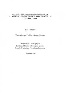
FIGURE 1. Edema and hemorrhage in the cutaneous reverse passive Arthus reaction. Mice were injected intradermally with rabbit IgG anti-chicken egg albumin Ab, followed by systemic chicken egg albumin and 1% Evans blue dye. Dorsal skins were harvested from mutant mice and wild-type (WT) littermates after 4 or 8 h. Edema was evaluated as the diameter of the Evans blue spot (A) as well as the wet weight of a 6-mm punch biopsy (B). Wild-type littermates that received an intradermal injection of polyclonal rabbit IgG followed by i.v. installation of chicken egg albumin served as controls. Hemorrhage after 8 h was assessed as the diameter of the purpuric spot (C). Edema and hemorrhage were significantly inhibited in L-selectin⫺/⫺, ICAM-1⫺/⫺, and L-selectin/ICAM-1⫺/⫺ mice compared with wild-type littermates for all panels (p ⬍ 0.005). Horizontal bars indicate mean values for each group of mice.
Downloaded from http://www.jimmunol.org/ by guest on April 27, 2018
intensity at 8 h (3). Therefore, edema and hemorrhage were evaluated 4 and 8 h after IC challenge, respectively, in L-selectin⫺/⫺, ICAM-1⫺/⫺, and L-selectin/ICAM-1⫺/⫺ mice compared with wild-type littermates. When edema was assessed by measuring the diameter of Evans blue dye in the extravascular space, edema was significantly reduced in L-selectin⫺/⫺ (31% decrease, p ⬍ 0.0001), ICAM-1⫺/⫺ (43%, p ⬍ 0.0001), and L-selectin/ICAM1⫺/⫺ mice (51%, p ⬍ 0.0001) compared with wild-type littermates (Fig. 1A). ICAM-1⫺/⫺ mice exhibited significant inhibition of dye vascular leak when compared with L-selectin⫺/⫺ mice ( p ⬍ 0.01), whereas the loss of both ICAM-1 and L-selectin resulted in a significant further reduction of dye vascular leak relative to the Lselectin loss alone ( p ⬍ 0.001). Similar results were obtained when edema was evaluated as the wet weight of skin biopsies from the site of IC formation (Fig. 1B). No edema was detected in mutant mice or their wild-type littermate controls following intradermal injection of rabbit polyclonal IgG with systemic chicken egg albumin (Fig. 1A and data not shown). Thus, L-selectin loss reduced the early cellular response characterized by edema, with ICAM-1 deficiency inhibiting edema beyond that found with Lselectin deficiency. Hemorrhage was macroscopically quantitated after 8 h by measuring the size of the purpuric spot. Hemorrhage was significantly inhibited in L-selectin⫺/⫺ (45% decrease, p ⬍ 0.002), ICAM-1⫺/⫺ (48%, p ⬍ 0.0005), and L-selectin/ICAM-1⫺/⫺ mice (64%, p ⬍ 0.0001) compared with wild-type littermates (Fig. 1C). L-selectin/ ICAM-1⫺/⫺ mice exhibited significantly reduced hemorrhage compared with both ICAM-1⫺/⫺ ( p ⬍ 0.05) and L-selectin⫺/⫺ mice ( p ⬍ 0.05). Hemorrhage was not detected in mutant mice or their wild-type littermate controls following intradermal injection of rabbit polyclonal IgG with systemic chicken egg albumin (Fig. 1C and data not shown). Therefore, the combined loss of L-selectin and ICAM-1 resulted in a greater inhibition of edema and hemorrhage than the loss of each adhesion molecule alone.
ICAM-1 AND L-SELECTIN IN THE ARTHUS REACTION
The Journal of Immunology
2973
additional loss of ICAM-1 in L-selectin⫺/⫺ mice led to significantly reduced mast cell numbers compared with L-selectin⫺/⫺ mice after 4 and 8 h ( p ⬍ 0.05). By contrast, there was no leukocyte influx in mutant mice or their control littermates following i.p. injection of rabbit polyclonal IgG with systemic chicken egg albumin (data not shown). Thus, the effect of the loss of each adhesion molecule on leukocyte recruitment in the peritoneal Arthus reaction was similar to that observed in the cutaneous Arthus reaction. TNF-␣ production IC stimulate the production and release of TNF-␣ from infiltrating leukocytes (8, 10, 32, 40), which is detected 1–5 h after the initiation of a peritoneal Arthus reaction but not after 6 h (40). To assess the involvement of TNF-␣ in the cutaneous Arthus reaction, TNF-␣ mRNA levels were examined in the skin after 4 h by RTPCR (Fig. 5A) and were quantitated by real-time PCR (Fig. 5B). TNF-␣ mRNA levels were up-regulated in skin from wild-type and in each adhesion molecule-deficient mouse after 4 h (Fig. 5A). However, TNF-␣ mRNA levels were significantly decreased in L-selectin⫺/⫺, ICAM-1⫺/⫺, and L-selectin/ICAM-1⫺/⫺ mice relative to their wild-type littermates ( p ⬍ 0.0001, Fig. 5B). ICAM1⫺/⫺ and L-selectin/ICAM-1⫺/⫺ mice exhibited similar TNF-␣ mRNA levels that were significantly lower than those of L-selectin⫺/⫺ mice ( p ⬍ 0.001). By contrast, TNF-␣ production was not
detected after 4 h in mice injected with control Ab (Fig. 5A and data not shown). Thus, reduced cutaneous inflammatory responses in each adhesion molecule-deficient mouse correlated with reduced TNF-␣ gene transcription. Expression of L-selectin and CD18 on peritoneal mast cells Reduced Arthus reaction-induced mast cell accumulation in adhesion molecule-deficient mice suggests a role for L-selectin and ICAM-1 in mast cell recruitment. Therefore, mouse peritoneal mast cells expressing c-Kit (38, 41) were analyzed for cell surface L-selectin and/or CD18 (2 integrin) expression by flow cytometry. Significant L-selectin expression on the surface of c-Kit-positive mast cells from wild-type mice was detected when compared with mast cells from L-selectin⫺/⫺ mice (Fig. 6A) or staining using an unreactive isotype-matched control mAb (data not shown). CD18 was also expressed on mast cells from wild-type mice compared with staining using an unreactive isotype-matched mAb (Fig. 6A). As a positive control, granulocytes expressed significant levels of both L-selectin and CD18 (Fig. 6B). Deficiency of ICAM-1 did not influence L-selectin or CD18 expression by mast cells or granulocytes (data not shown). Similarly, the loss of Lselectin did not alter CD18 expression on either of these cell populations (data not shown). Thus, L-selectin and CD18 were both highly expressed on the surface of mouse peritoneal mast cells.
Downloaded from http://www.jimmunol.org/ by guest on April 27, 2018
FIGURE 2. Arthus reaction-induced recruitment of neutrophils and mast cells in the skin (A) and the peritoneum (B) from mutant and wildtype littermates at 4 and 8 h after IC challenge. Numbers of neutrophils and mast cells per skin section were determined by counting H&E- and toluidine blue-stained skin sections, respectively. The peritoneal reverse passive Arthus reaction was induced by the i.v. injection of chicken egg albumin, followed immediately by the i.p. injection of rabbit IgG anti-chicken egg albumin Ab. Cells in the recovered lavage fluid were then centrifuged onto glass slides and stained with Giemsa to quantify neutrophil and mast cell numbers. All values represent the mean ⫾ SEM of results obtained from 5 to 10 mice in each group. Statistical analysis is provided in Results.
2974
ICAM-1 AND L-SELECTIN IN THE ARTHUS REACTION
ICAM-1 expression ICAM-1 expression on various types of cells, including keratinocytes and fibroblasts, is induced by stimulation with proinflammatory cytokines in vitro (26, 42). Thus, the loss of ICAM-1 expression on fibroblasts and keratinocytes may contribute to the reduced inflammation observed in ICAM-1⫺/⫺ mice. To assess this, cutaneous ICAM-1 expression during the Arthus reaction was examined immunohistochemically. In normal skin, ICAM-1 was detected exclusively on endothelial cells (Fig. 7A), with up-regulated ICAM-1 expression by endothelial cells 4 h after IC induction (Fig. 7B). However, ICAM-1 expression could not be accurately assessed on endothelial cells after 8 h since large numbers of inflammatory cells infiltrating the vessel wall obscured ICAM-1 staining (Fig. 7C). ICAM-1 expression was not detected on keratinocytes, fibroblasts, or infiltrating inflammatory cells after 4 or 8 h (Fig. 7, B and C, and data not shown). ICAM-1 expression was not detected in either the intact or inflamed skin from ICAM-1⫺/⫺
mice (data not shown). In addition, the loss of L-selectin expression did not affect ICAM-1 expression in either the intact or inflamed skin (data not shown). Thus, ICAM-1 was predominantly expressed on cutaneous endothelial cells.
Discussion In the present study, the loss of ICAM-1, L-selectin, or both significantly inhibited edema and vascular hemorrhage after IC challenge (Fig. 1). The extent that L-selectin and/or ICAM-1 deficiencies influenced inflammation varied at different time points, with each cell type, and in different tissues (Figs. 1 and 2). This suggests that the repertoire of functional adhesion molecules mediating ICmediated inflammation changes during the course of inflammation and is influenced by tissue site. Edema and hemorrhage were generally inhibited to a larger extent by ICAM-1 deficiency after IC formation when compared with L-selectin deficiency (Fig. 1).
Downloaded from http://www.jimmunol.org/ by guest on April 27, 2018
FIGURE 3. Histologic tissue sections showing neutrophil infiltration in the skin of mutant and wild-type littermates at 4 h (A) and 8 h (B) after IC challenge. Neutrophils were revealed by H&E staining. Original magnification, ⫻100.
The Journal of Immunology
2975
Nonetheless, the added loss of L-selectin in ICAM-1⫺/⫺ mice further diminished inflammatory responses relative to ICAM-1-deficiency alone (Fig. 1). This is consistent with the finding that Lselectin and ICAM-1 function cooperatively to mediate optimal leukocyte rolling as well as to recruit leukocytes into inflammatory sites (27, 30, 31). In addition to reduced inflammation in adhesion molecule-deficient mice, neutrophil and mast cell accumulation in the skin and peritoneum were significantly reduced during Arthus reactions (Figs. 2– 4). Similarly, reduced cutaneous inflammatory responses correlated with a significant decrease in TNF-␣ mRNA levels within skin at sites of IC deposition (Fig. 5). Taken together, these results demonstrate that ICAM-1 and L-selectin cooperatively contribute to the development of the cutaneous Arthus reaction by regulating neutrophil and mast cell accumulation at the inflammatory site. L-selectin regulates lymphocyte migration into lymph nodes across high endothelial venules and is involved in leukocyte-en-
dothelial cell interactions at peripheral sites of inflammation (15). L-selectin⫺/⫺ mice exhibit impaired peripheral inflammatory responses including delayed-type hypersensitivity responses and rejection of allogeneic skin transplants (20, 21, 31). However, whether the observed reduction in Ag-induced inflammation is due to disruption of early events in Ag sensitization or later effector phases has been controversial. L-selectin deficiency does not inhibit the in vivo generation of effector T cells able to mount in vitro proliferative responses to Ag or the generation of effector cytotoxic T cell responses in skin allograft recipients (20, 31, 43). In addition, intravital microscopy studies have shown that leukocyte rolling is reduced in L-selectin⫺/⫺ mice in the cremasteric microvasculature during Ag challenge at this site (44). These results suggest that L-selectin contributes directly to leukocyte rolling in the peripheral vasculature during immune responses to Ag (44). However, a requirement for L-selectin during the Ag sensitization
Downloaded from http://www.jimmunol.org/ by guest on April 27, 2018
FIGURE 4. Histologic tissue sections showing mast cell accumulation in the skin of mutant and wild-type littermates at 4 h (A) and 8 h (B) after IC challenge. Mast cells (arrows) were detected as cells with metachromatic staining of granules in toluidine blue-stained sections. Original magnification, ⫻75.
2976
ICAM-1 AND L-SELECTIN IN THE ARTHUS REACTION
FIGURE 5. TNF-␣ mRNA expression in the skin of mutant and wildtype littermates at 4 h after IC challenge. Total RNA was isolated from frozen skin tissues, reverse transcribed into cDNA, and then amplified using TNF-␣ and -actin primers. The PCR products were electrophoresed on an agarose gel and stained with ethidium bromide. Representative mRNA expression of TNF-␣ and -actin is shown (A). The amount of TNF-␣ mRNA was measured by real-time PCR and normalized to the 18S rRNA endogenous control (B). The TNF-␣ mRNA level in wild-type littermates was used as the calibrator. All values represent the mean ⫾ SEM of results obtained with five mice of each group. The sample mean for each group of mutant mice was significantly different from that of wild-type littermates (p ⬍ 0.0001).
phase of immune responses could not be fully excluded since these experimental systems required systemic Ag sensitization before Ag challenge. Catalina et al. (23) have shown that effector functions, including leukocyte entry into inflammatory sites of the skin, are intact in L-selectin⫺/⫺ mice, while the major defect in responsiveness is due to a lack of T cell sensitization in draining lymph nodes. In studies by Xu et al. (22), leukocyte recruitment into the skin was reduced within the first 4 days of sensitization in L-selectin⫺/⫺ mice, but not after 9 days of sensitization. Based on this, Xu et al. (22) concluded that the impaired migration of naive T cells into the draining lymph nodes impaired Ag sensitization in L-selectin⫺/⫺ mice, rather than the subsequent impairment of leukocyte recruitment into the skin. However, the finding that L-se-
FIGURE 7. ICAM-1 expression in skin from wild-type mice during a cutaneous passive Arthus reaction. ICAM-1 expression in normal skin (A) and in inflamed skin after 4 h (B) and 8 h (C) of IC challenge was assessed by immunohistochemistry using anti-ICAM-1 Abs. Sections were counterstained with methyl green. Original magnification, ⫻100.
lectin deficiency significantly reduced leukocyte accumulation during the passive cutaneous and peritoneal Arthus reactions excludes the Ag sensitization process (Figs. 2– 4). Thus, reduced Aginduced cellular hypersensitivity in L-selectin⫺/⫺ mice results from interrupted leukocyte-endothelial cell interactions and not from reduced immune responses. Furthermore, these results fully confirm the existence of a peripheral vascular endothelial ligand for L-selectin. Mast cells derive from bone marrow progenitors that migrate through the circulation into tissues where they proliferate and mature (45). Mature mast cells exist exclusively within tissues, although mast cell numbers increase at sites of inflammation (46). Since mature mast cells from the blood accumulate in the CNS within 1–2 h in response to altered physiological conditions (47), it is possible that mature mast cells also migrate from the circulation into inflamed sites. Consistent with this, peritoneal mast cells expressed significant levels of both L-selectin and CD18 (Fig. 6). Furthermore, Arthus reaction-induced mast cell accumulation in both skin and the peritoneum was substantially reduced in mice lacking L-selectin, ICAM-1, or both adhesion molecules (Fig. 2). It is unlikely that mast cells migrated from surrounding tissues into sites of inflammation since ICAM-1 expression was only detected on the endothelium during IC-induced inflammation (Fig. 7), and mast cell numbers increased rapidly in the peritoneal cavity of
Downloaded from http://www.jimmunol.org/ by guest on April 27, 2018
FIGURE 6. Expression of L-selectin and CD18 on peritoneal mast cells and granulocytes. Peritoneal cells from untreated wild-type mice were stained with either anti-c-Kit-FITC Ab or anti-Gr-1-FITC Ab plus either anti-CD18-PE Ab or anti-L-selectin-PE Ab. Flow cytometric analysis was performed by gating on c-Kit-positive mast cells or Gr-1-positive granulocytes. Representative histograms are shown for expression of L-selectin or CD18 on mast cells (A) or granulocytes (B). The dashed lines for Lselectin expression represent staining obtained using L-selectin⫺/⫺ mice. The dashed lines for CD18 expression represent control staining using an unreactive isotype-matched mAb. These results represent those obtained with five wild-type mice.
The Journal of Immunology
References 1. Kohl, J., and J. E. Gessner. 1999. On the role of complement and Fc ␥-receptors in the Arthus reaction. Mol. Immunol. 36:893. 2. Arthus, M. 1903. Injections re´ pete´ es de serum de cheval chez le lapin. V. R. Soc. Biol. 55:817. 3. Sylvestre, D. L., and J. V. Ravetch. 1994. Fc receptors initiate the Arthus reaction: redefining the inflammatory cascade. Science 265:1095. 4. Sylvestre, D., R. Clynes, M. Ma, H. Warren, M. C. Carroll, and J. V. Ravetch. 1996. Immunoglobulin G-mediated inflammatory responses develop normally in complement-deficient mice. J. Exp. Med. 184:2385. 5. Baumann, U., J. Kohl, T. Tschernig, K. Schwerter-Strumpf, J. S. Verbeek, R. E. Schmidt, and J. E. Gessner. 2000. A codominant role of Fc␥RI/III and C5aR in the reverse Arthus reaction. J. Immunol. 164:1065. 6. Hazenbos, W. L., J. E. Gessner, F. M. Hofhuis, H. Kuipers, D. Meyer, I. A. Heijnen, R. E. Schmidt, M. Sandor, P. J. Capel, M. Daeron, et al. 1996. Impaired IgG-dependent anaphylaxis and Arthus reaction in Fc␥RIII (CD16) deficient mice. Immunity 5:181. 7. Zhang, Y., B. F. Ramos, and B. A. Jakschik. 1991. Augmentation of reverse Arthus reaction by mast cells in mice. J. Clin. Invest. 88:841. 8. Zhang, Y., B. F. Ramos, and B. A. Jakschik. 1992. Neutrophil recruitment by tumor necrosis factor from mast cells in immune complex peritonitis. Science 258:1957. 9. Sylvestre, D. L., and J. V. Ravetch. 1996. A dominant role for mast cell Fc receptors in the Arthus reaction. Immunity 5:387. 10. Hopken, U. E., B. Lu, N. P. Gerard, and C. Gerard. 1997. Impaired inflammatory responses in the reverse Arthus reaction through genetic deletion of the C5a receptor. J. Exp. Med. 186:749. 11. Butcher, E. C. 1991. Leukocyte-endothelial cell recognition: three (or more) steps to specificity and diversity. Cell 67:1033. 12. Ley, K., and T. F. Tedder. 1995. Leukocyte interactions with vascular endothelium: new insights into selectin-mediated attachment and rolling. J. Immunol. 155:525. 13. Springer, T. A. 1995. Traffic signals on endothelium for lymphocyte recirculation and leukocyte emigration. Annu. Rev. Physiol. 57:827. 14. Tedder, T. F., D. A. Steeber, A. Chen, and P. Engel. 1995. The selectins: vascular adhesion molecules. FASEB J. 9:866. 15. Tedder, T. F., X. Li, and D. A. Steeber. 1999. The selectins and their ligands: adhesion molecules of the vasculature. Adv. Mol. Cell Biol. 28:65. 16. Spertini, O., F. W. Luscinskas, M. A. Gimbrone Jr., and T. F. Tedder. 1992. Monocyte attachment to activated human vascular endothelium in vitro is mediated by leukocyte adhesion molecule-1 (L-selectin) under non-static conditions. J. Exp. Med. 175:1789. 17. Spertini, O., F. W. Luscinskas, G. S. Kansas, J. M. Munro, J. D. Griffin, M. A. Gimbrone, Jr., and T. F. Tedder. 1991. Leukocyte adhesion molecule-1 (LAM-1, L-selectin) interacts with an inducible endothelial cell ligand to support leukocyte adhesion. J. Immunol. 147:2565. 18. Brady, H. R., O. Spertini, W. Jimenez, B. M. Brenner, P. A. Marsden, and T. F. Tedder. 1992. Neutrophils, monocytes and lymphocytes bind to cytokineactivated kidney glomerular endothelial cells through L-selectin (LAM-1) in vitro. J. Immunol. 149:2437. 19. Arbones, M. L., D. C. Ord, K. Ley, H. Radich, C. Maynard-Curry, D. J. Capon, and T. F. Tedder. 1994. Lymphocyte homing and leukocyte rolling and migration are impaired in L-selectin (CD62L) deficient mice. Immunity 1:247. 20. Tang, M. L. K., L. P. Hale, D. A. Steeber, and T. F. Tedder. 1997. L-selectin is involved in lymphocyte migration to sites of inflammation in the skin: delayed rejection of allografts in L-selectin-deficient mice. J. Immunol. 158:5191. 21. Tedder, T. F., D. A. Steeber, and P. Pizcueta. 1995. L-selectin deficient mice have impaired leukocyte recruitment into inflammatory sites. J. Exp. Med. 181:2259. 22. Xu, J., I. S. Grewal, G. P. Geba, and R. A. Flavell. 1996. Impaired primary T cell responses in L-selectin-deficient mice. J. Exp. Med. 183:589. 23. Catalina, M. D., M. C. Carroll, H. Arizpe, A. Takashima, P. Estess, and M. H. Siegelman. 1996. The route of antigen entry determines the requirement for L-selectin during immune responses. J. Exp. Med. 184:2341. 24. Kunkel, E. J., and K. Ley. 1996. Distinct phenotype of E-selectin-deficient mice. E-selectin is required for slow leukocyte rolling in vivo. Circ. Res. 79:1196. 25. Ley, K. E., D. Bullard, M. L. Arbones, R. Bosse, D. Vestweber, T. F. Tedder, and A. L. Beaudet. 1995. Sequential contribution of L- and P-selectin to leukocyte rolling in vivo. J. Exp. Med. 181:669. 26. Dustin, M. L., R. Rothlein, A. K. Bhan, C. A. Dinarello, and T. A. Springer. 1986. Induction by IL-1 and interferon-␥: tissue distribution, biochemistry, and function of a natural adherence molecule (ICAM-1). J. Immunol. 137:245. 27. Steeber, D. A., M. A. Campbell, A. Basit, K. Ley, and T. F. Tedder. 1998. Optimal selectin-mediated rolling of leukocytes during inflammation in vivo requires intercellular adhesion molecule-1 expression. Proc. Natl. Acad. Sci. USA 95:7562. 28. Sligh Jr., J. E., C. M. Ballantyne, S. S. Rich, H. K. Hawkins, C. W. Smith, A. Bradley, and A. L. Beaudet. 1993. Inflammatory and immune responses are impaired in mice deficient in intercellular adhesion molecule 1. Proc. Natl. Acad. Sci. USA 90:8529. 29. Xu, H., J. A. Gonzalo, Y. St. Pierre, I. R. Williams, T. S. Kupper, R. S. Cotran, T. A. Springer, and J.-C. Guiterrez-Ramos. 1994. Leukocytosis and resistance to septic shock in intercellular adhesion molecule 1-deficient mice. J. Exp. Med. 180:95. 30. Nagaoka, T., Y. Kaburagi, Y. Hamaguchi, M. Hasegawa, K. Takehara, D. A. Steeber, T. F. Tedder, and S. Sato. 2000. Delayed wound healing in the absence of intercellular adhesion molecule-1 or L-selectin expression. Am. J. Pathol. 157:237.
Downloaded from http://www.jimmunol.org/ by guest on April 27, 2018
wild-type mice following inflammation (Fig. 2B). Although the exact routes of mast cell migration into inflamed foci were not determined in this study, the results suggest that L-selectin and ICAM-1 regulate mast cell recruitment from the circulation. Mature peritoneal mast cells expressed CD18 and L-selectin (Fig. 6). Consistent with this finding, Mac-1 (CD11b/CD18) is expressed by immature mast cells derived from mouse bone marrow and mature peritoneal mast cells (41). CD18 expression is also detected on mast cells in normal human skin, and human mast cell lines express CD18 (48). With regard to L-selectin expression, a previous study has shown that immature mast cells derived from mouse bone marrow do not express L-selectin (49). A second study has shown that L-selectin is not expressed on mature primary mast cells isolated from human lung and uterus (50). However, tissue fragments were first treated with collagenase in the second study and the isolated mast cells were then cultured for at least 24 h. L-selectin is rapidly lost from the cell surface of leukocytes following cellular activation (15). Furthermore, incubating lymphocytes overnight at 4°C can also result in the complete loss of L-selectin from the cell surface (51). Therefore, it is likely that L-selectin expression was endoproteolytically released from the cell surface during mature mast cell isolation. Nonetheless, the present study reveals that mature mast cells freshly isolated from the peritoneal cavity express significant levels of L-selectin. ICAM-1 is expressed on many types of cells, with its expression up-regulated by proinflammatory cytokines in vitro (26). Augmented ICAM-1 expression is also observed on keratinocytes, fibroblasts, and infiltrating leukocytes in the inflamed skin in vivo (52, 53). Furthermore, ICAM-1 expression by fibroblasts may mediate neutrophil migration through fibroblast layers within tissues (54, 55), and ICAM-1 expressed on lung epithelial cells supports the adhesion and retention of neutrophils (56). Therefore, it is possible that ICAM-1 expression on cells other than endothelial cells might be involved in the migration or retention of leukocytes within the perivascular area of inflamed skin during the Arthus reaction. However, our finding that ICAM-1 was expressed exclusively by skin endothelium in the Arthus reaction (Fig. 7) excludes this possibility. Moreover, this finding suggests that endothelial ICAM-1 expression primarily mediates leukocyte accumulation during the Arthus reaction. Two dominant pathways contribute to initiation of the cutaneous Arthus reaction: a Fc␥RIII-dependent pathway and a complementdependent pathway using the C5aR (5, 6, 9). The loss of Fc␥RIII results in a 60% reduction in edema formation and neutrophil recruitment compared with wild-type mice, whereas C5aR deficiency results in a 30 –50% reduction (5, 10). The present study demonstrates that edema, hemorrhage, and neutrophil accumulation are inhibited by 30 – 40% with L-selectin deficiency, 40 –50% with ICAM-1 deficiency, and 50 – 60% with the combined loss of L-selectin and ICAM-1 (Figs. 1 and 2A). Thus, blocking the function of L-selectin, ICAM-1, or both adhesion molecules inhibits the Arthus reaction to a similar extent as blocking either the C5aR or Fc␥RIII pathways. Thus, cell adhesion molecules, including L-selectin and ICAM-1, may play a critical role in Arthus reaction initiation, in addition to its progression. This suggests that L-selectin and ICAM-1 are potential therapeutic targets for human ICmediated diseases such as vasculitis syndrome and some collagen diseases. However, it should be noted that genetic deficiency of L-selectin or ICAM-1 in the development of Arthus reaction was a fundamentally different situation than the selective inhibition of the function of these adhesion molecules during the course of an IC-mediated disease.
2977
2978
44. Kanwar, S., D. A. Steeber, T. F. Tedder, M. J. Hickey, and P. Kubes. 1999. Overlapping roles for L-selectin and P-selectin in antigen-induced immune responses in the microvasculature. J. Immunol. 162:2709. 45. Kirshenbaum, A. S., S. W. Kessler, J. P. Goff, and D. D. Metcalfe. 1991. Demonstration of the origin of human mast cells from CD34⫹ bone marrow progenitor cells. J. Immunol. 146:1410. 46. Ishizaka, T., and K. Ishizaka. 1975. Biology of immunoglobulin E. Molecular basis of reaginic hypersensitivity. Prog. Allergy 19:60. 47. Silverman, A. J., A. K. Sutherland, M. Wilhelm, and R. Silver. 2000. Mast cells migrate from blood to brain. J. Neurosci. 20:401. 48. Weber, S., M. Babina, G. Feller, and B. M. Henz. 1997. Human leukaemic (HMC-1) and normal skin mast cells express 2- integrins: characterization of 2-integrins and ICAM-1 on HMC-1 cells. Scand. J. Immunol. 45:471. 49. Sriramarao, P., W. Anderson, B. A. Wolitzky, and D. H. Broide. 1996. Mouse bone marrow-derived mast cells roll on P-selectin under conditions of flow in vivo. Lab. Invest. 74:634. 50. Wimazal, F., M. Ghannadan, M. R. Muller, A. End, M. Willheim, P. Meidlinger, G. H. Schernthaner, J. H. Jordan, W. Hagen, H. Agis, et al. 1999. Expression of homing receptors and related molecules on human mast cells and basophils: a comparative analysis using multi-color flow cytometry and toluidine blue/immunofluorescence staining techniques. Tissue Antigens 54:499. 51. Tedder, T. F., M. D. Cooper, and L. T. Clement. 1985. Human lymphocyte differentiation antigens HB-10 and HB-11. II. Differential production of B cell growth and differentiation factors by distinct helper T cell subpopulations. J. Immunol. 134:2989. 52. Matsunaga, T., I. Katayama, H. Yokozeki, and K. Nishioka. 1996. ICAM-1 expression on keratinocytes in mechanically-injured skin of a patient with atopic dermatitis. J. Dermatol. Sci. 12:219. 53. Majewski, S., N. Hunzelmann, J. P. Johnson, C. Jung, C. Mauch, H. W. L. Ziegler-Heitbrock, G. Riethmuller, and T. Krieg. 1991. Expression of intercellular adhesion molecule-1 (ICAM-1) in the skin of patients with systemic sclerosis. J. Invest. Dermatol. 97:667. 54. Morzycki, W., and A. C. Issekutz. 1991. Tumor necrosis factor-␣ but not interleukin-1 induces polymorphonuclear leukocyte migration through fibroblast layers by a fibroblast-dependent mechanism. Immunology 74:107. 55. Shang, X. Z., and A. C. Issekutz. 1998. Contribution of CD11a/CD18, CD11b/ CD18, ICAM-1 (CD54) and -2 (CD102) to human monocyte migration through endothelium and connective tissue fibroblast barriers. Eur. J. Immunol. 28:1970. 56. Tosi, M. F., J. M. Stark, C. W. Smith, A. Hamedani, D. C. Gruenert, and M. D. Infeld. 1992. Induction of ICAM-1 expression on human airway epithelial cells by inflammatory cytokines: effects on neutrophil-epithelial cell adhesion. Am. J. Respir. Cell. Mol. Biol. 7:214.
Downloaded from http://www.jimmunol.org/ by guest on April 27, 2018
31. Steeber, D. A., M. L. K. Tang, N. E. Green, X.-Q. Zhang, J. E. Sloane, and T. F. Tedder. 1999. Leukocyte entry into sites of inflammation requires overlapping interaction between the L-selectin and ICAM-1 pathways. J. Immunol. 163: 2176. 32. Ramos, B. F., Y. Zhang, and B. A. Jakschik. 1994. Neutrophil elicitation in the reverse passive Arthus reaction: complement-dependent and -independent mast cell involvement. J. Immunol. 152:1380. 33. Ohnishi, M., H. Koike, N. Kawamura, S. J. Tojo, M. Hayashi, and S. Morooka. 1996. Role of P-selectin in the early stage of the Arthus reaction. Immunopharmacology 34:161. 34. Rote, W. E., E. Dempsey, S. Maki, G. P. Vlasuk, and M. Moyle. 1996. The role of CD11/CD18 integrins in the reverse passive Arthus reaction in rat dermal tissue. J. Leukocyte Biol. 59:254. 35. Nourshargh, S., M. Rampart, P. G. Hellewell, P. J. Jose, J. M. Harlan, A. J. Edwards, and T. J. Williams. 1989. Accumulation of 111In-neutrophils in rabbit skin in allergic and non- allergic inflammatory reactions in vivo. Inhibition by neutrophil pretreatment in vitro with a monoclonal antibody recognizing the CD18 antigen. J. Immunol. 142:3193. 36. King, P. D., E. T. Sandberg, A. Selvakumar, P. Fang, A. L. Beaudet, and B. Dupont. 1996. Novel isoforms of murine intercellular adhesion molecule-1 generated by alternative RNA splicing. J. Immunol. 154:6080. 37. Sato, S., A. S. Miller, M. Inaoki, C. B. Bock, P. J. Jansen, M. L. K. Tang, and T. F. Tedder. 1996. CD22 is both a positive and negative regulator of B lymphocyte antigen receptor signal transduction: altered signaling in CD22-deficient mice. Immunity 5:551. 38. Gommerman, J. L., D. Y. Oh, X. Zhou, T. F. Tedder, M. Maurer, S. J. Galli, and M. C. Carroll. 2000. A role for CD21/CD35 and CD19 in responses to acute septic peritonitis: a potential mechanism for mast cell activation. J. Immunol. 165:6915. 39. Abraham, C., and J. Miller. 2001. Molecular mechanisms of IL-2 gene regulation following costimulation through LFA-1. J. Immunol. 167:5193. 40. Heller, T., J. E. Gessner, R. E. Schmidt, A. Klos, W. Bautsch, and J. Kohl. 1999. Cutting edge: Fc receptor type I for IgG on macrophages and complement mediate the inflammatory response in immune complex peritonitis. J. Immunol. 162:5657. 41. Rosenkranz, A. R., A. Coxon, M. Maurer, M. F. Gurish, K. F. Austen, D. S. Friend, S. J. Galli, and T. N. Mayadas. 1998. Impaired mast cell development and innate immunity in Mac-1 (CD11b/CD18, CR3)-deficient mice. J. Immunol. 161:6463. 42. Middleton, M. H., and D. A. Norris. 1995. Cytokine-induced ICAM-1 expression in human keratinocytes is highly variable in keratinocyte strains from different donors. J. Invest. Dermatol. 104:489. 43. Steeber, D. A., N. E. Green, S. Sato, and T. F. Tedder. 1996. Humoral immune responses in L-selectin deficient mice. J. Immunol. 157:4899.
ICAM-1 AND L-SELECTIN IN THE ARTHUS REACTION