Synaptic rearrangements in developing muscle were studied by visualizing individual neuromuscular junctions in the ster- nomastoid muscle of living neonatal ...
The Journal
/II viva Observations of Pre- and Postsynaptic Transition from Multiple to Single Innervation Neuromuscular Junctions Rita J. Balice-Gordon Department
of Neuroscience,
February
1993,
73(2):
834-855
Changes during the at Developing
and Jeff W. Lichtman
of Anatomy and Neurobiology,
Washington
University
Synaptic rearrangements in developing muscle were studied by visualizing individual neuromuscular junctions in the sternomastoid muscle of living neonatal mice as they underwent the transition from multiple to single innervation. Vital staining of ACh receptors (AChRs) with rhodamine-conjugated a-bungarotoxin showed that while junctions were still multiply innervated (usually by two motor axons), regions of the postsynaptic membrane within each junction became depleted of receptors. Usually, several small postsynaptic,areas lost AChRs in succession. In these areas, AChRs already in the membrane rapidly disappeared compared to a low level of receptor turnover elsewhere in the junction. Moreover, there was no evidence of new AChRs being inserted into these areas. Within each postsynaptic area undergoing ACRR depletion, the intensity of receptor staining decreased gradually over l-2 d. In some junctions, it appeared that AChRs were migrating away from areas being depleted of receptors. The depletion of AChRs from some sites in combination with the spreading apart of the entire receptor-rich area due to muscle fiber growth accounts for the transformation from plaque-like to branched receptor distributions at developing neuromuscular junctions. Vital staining of presynaptic motor nerve terminals at junctions whose postsynaptic AChRs were also stained showed that motor nerve terminals were lost from the same areas that were depleted of receptors postsynaptically. Postsynaptic areas began to be depleted of AChRs before there was any obvious loss of membrane or intracellular staining in the overlying nerve terminal. Only when a single innervating axon remained at a junction did loss of motor nerve terminals and underlying AChRs largely cease. That former synaptic areas could at later times be identified as uninnervated regions within a junction indicates that synapse elimination during development leaves an indelible mark on synaptic structure. These observations suggest that the withdrawal of a motor axon from a neuromuscular junction occurs as a consequence of the stepwise elimination of all of its synapses with that muscle fiber. These results also suggest that an imReceived July 15, 1992; revised Aug. 21, 1992; accepted Aug. 28, 1992. We thank A. Brown, H. Colman, S. Culican, K. Fischer, C. Nelson, M. Rich, S. Tumey, and P. van Mier for helpful discussions and C. Chua, J. Fiez, L. Hedayati, and S. Turney for technical assistance. This work was supported by grants from the NIH and MDA to J.W.L. and the Harry Zimmerman Postdoctoral Fellowship of the MDA to R.B.-G. Correspondence should be addressed to Rita J. Balice-Gordon, Ph.D., Department of Anatomy and Neurobiology, Washington University School of Medicine, 660 South Euclid Avenue, Box 8108, St. Louis, MO 63110. Copyright 0 1993 Society for Neuroscience 0270-6474/93/130834-22$05.00/O
School of Medicine, St. Louis, Missouri 63110
portant aspect of synaptic competition leading to axon withdrawal is the precocious loss of AChRs beneath the nerve terminals of the axon that will be eliminated. A similar early loss of AChRs beneath one axon’s synapses has been shown to occur during synapse elimination in reinnervated adult muscle (Rich and Lichtman, 1989a). Thus, changes in the postsynaptic membrane beneath the nerve terminals of one motor axon may instigate the removal of those terminals, suggesting that the postsynaptic cell plays an important role in competitive synaptic reorganization. [Key words: synapse elimination, competition, neuromuscular junction, ACh receptors, cr-bungarotoxin, nerve terminals, fluorescent dyes, vital dyes, motor neuron, axon, muscle fiber]
Throughout the developing nervous system, competitive interactions between different neurons result in extensive and permanent changesin the pattern of synaptic connections. For example, in the developing visual system, inputs driven by the two eyes are initially convergent, innervating the same target cells in primary visual cortex (Hubel and Wiesel, 1963). After birth, however, theseinputs apparently competefor domination of the samecortical neurons. As a result, inputs driven by the two eyes segregateonto different cortical neurons by the elimination of synaptic connectionsestablishedearlier (LeVay et al., 1980). This rearrangement results in the formation of ocular dominance columnsthat are permanently maintained. Because the processof segregationis profoundly affectedwhen the visual experience of the two eyesis unequal, the neural activity of the competing inputs appearsto have an important influence on the outcome (Hubel and Wiesel, 1970). Activity-driven synaptic rearrangementsmay alsobe involved in other long-term changes in the nervous system, for example, in learning and memory. Few detailsare known about the mechanismof synaptic competition. The complexity and inaccessibilityof synaptic circuitry in the CNS have made it difficult to study this phenomenonat the level of individual synapses.However, a similar competitive reorganization of synapsesalso occurs in the more accessible periphery, in both muscle(Redfem, 1970; Brown et al., 1976; seeJansenand Fladby, 1990, for recent review) and autonomic ganglia (Lichtman, 1977; seePurves and Lichtman, 1980, for review). In developing mammalianmuscle,for example, several different motor axons convergeat the sameneuromuscularjunction on each muscle fiber. During the first weeks after birth, somemotor axon branchesare eliminated, pruning eachmotor unit and leaving each musclefiber innervated by a singlemotor neuron. This processis commonly called synapseelimination.
The Journal
We have studied synapse elimination by repeatedly observing the same junctions over time in the sternomastoid muscle of living mice. In this muscle, most junctions are innervated by two axons at birth but by only one axon 2 weeks later (Steinbach, 198 1). One of our aims was to determine whether axon withdrawal occurs abruptly, with the sudden demise of all of an axon’s synapses, as has been suggested by some experiments (Rosenthal and Taraskevitch, 1977). Alternatively, axon withdrawal may be the outcome of a protracted competition between two axons that leads to the gradual loss of territory by one axon. Our evidence suggests that withdrawal of the losing motor axon from a junction is the culmination of a gradual process of synaptic loss. Another aim was to study the role that the postsynaptic muscle fiber might play in this process. Previous work in adult muscle showed that after muscle nerve crush and reinnervation, terminal sprouts that transiently multiply innervated junctions regressed over time (Rich and Lichtman, 1989a). As sprouts were eliminated, postsynaptic ACh receptors (AChRs) were observed to be lost from the postsynaptic membrane at former sites of synaptic contact. In fact, AChR loss preceded nerve terminal loss. We were interested to learn if a similar relation held for postsynaptic sites that lose nerve terminals during development. Steinbach (1981) and Slater (1982a,b) have noted that the AChR distribution within neuromuscular junctions changes dramatically during the first few postnatal weeks when multiple innervation is being eliminated. Whereas at birth the AChRs in each neuromuscular junction are typically arranged as ovalshaped plaques with only hints of inhomogeneities in receptor density, by 2-3 weeks postnatal the AChR areas have been transformed into a more mature pattern with regions of high and very low receptor density. The high-density areas become the sites of the primary junctional folds that are overlain by nerve terminal branches, while the low-density sites are not innervated. Although these workers did not view the same junctions multiple times, they argued that the transition from plaque to mature forms was accomplished by a disappearance of AChRs from areas within the plaques. This idea was supported by the presence of “transitional” junctions in early postnatal ages in which AChR-rich plaques contained one or more small holes with low receptor density (i.e., “perforated plaques”; NystrBm, 1968). Could it be that during development AChR-rich areas are lost as part of the synapse elimination process? In the present work, we determined that postsynaptic changes in AChR density do occur at sites of nerve terminal elimination. Moreover, postsynaptic AChR depletion precedes the loss of overlying nerve terminals as previously found in reinnervated adult junctions, arguing for the importance of the postsynaptic cell as an intermediary in synaptic competition during normal development. A portion of these results have been reported in abstract form (Balice-Gordon and Lichtman, 1989, 1990a).
Materials
and Methods
Neonatal mice were obtained from breeding colonies established in our animal care facility (CFlB strain initially obtained from HarlanSprague-Dawley, Inc.; C57B16J strain from Jackson Laboratories). The date a copulatory plug was found was designated embryonic day 0 (EO), and the date of birth was designated postnatal day 0 (PO). Litters were culled to six pups on P4, and pups were weaned on P2 1. Female mice of the C57Bl 65 strain were found to be somewhat more tolerant of manipulation of their pups than CF 1B females. Determination of sex after 2 weeks of age showed that approximately half of the animals used were male. No differences were noted between sexes or between strains of mice relevant to the present experiments.
of Neuroscience,
February
1993,
13(2)
535
Methods for anesthesia, preparation of animals, staining and imaging of neuromuscular junctions, and control procedures are described fully elsewhere (Lichtman et al., 1987; Balice-Gordon and Lichtman, 1990b). The interpretive limitations of these techniques are extensively discussed in Balice-Gordon and Lichtman (199 l), and in this work we have attempted to surmount these by (1) making as many observations as possible, (2) extensive use of control experiments, and (3) using several different vital staining techniques. Only those methods specific to the present experiments are described below. Visualization of motor nerve terminals and AChRs. Pups less than 1 week old were anesthetized bv cooling. (- 10°C for 5-10 min) using a thermoelectric temperature-regulating&ice (a Peltier element driven by a Cambion Bipolar Controller) mounted in the microscope stage. Pups older than 1 week were anesthetized with chloral hydrate (0.60 gm/kg), delivered intraperitoneally with a 27 gauge needle, and were intubated and mechanically respirated (Harvard Instruments rodent ventilator). Motor nerve terminals were stained with a 1 1~ solution of 4-Di-2ASP (Molecular Probes, Inc., Eugene, OR, Magrassi et al., 1987) perfused over the muscle for 2 min. This low dose was used so that most of the dye could be washed away at the end of the experiment, enabling junctions to be restained l-2 d later. Postsynaptic AChRs were usually labeled with a nonblocking dose of rhodamine-conjugated cY-bungarotoxin (RaBTX) prepared according to the method of Ravdin and Axelrod (1977) so that synaptic transmission was only minimally affected (5 &ml for 3 min; see also below). In some experiments, after obtaining imaaes of 4-Di-2-ASP and RaBTX staining. motor nerve terminal membranes were stained with the voltage-sensitive dye RH795 (Molecular Probes, Eugene, OR, Grinvald et al., 1986) as described below. Neuromuscular junctions that were on the superficial surface of superficial muscle fibers were visualized using a 100 x , 1.2 NA water-immersion objective (Leitz NPL Fluotar), which was machined into a conical shape to fit more easily into the neck of a neonatal mouse. Images were obtained using a silicon-intensified target (SIT) video camera (series 66, Dage-MTI, Inc.) and an image processor (Trapix, Recognition Concepts, Inc.) as described previously (Lichtman et al., 1987; Balice-Gordon and Lichtman, 1990b). Axon branches were difficult to image in a single focal plane because they often traveled vertically from nerve branches deep within the muscle. Nonetheless, for each junction studied, as many images were obtained as necessary to determine unambiguously the number and position of the innervating axons. Junctions in which the number of innervating axons could not be determined were not studied further. Because of the curvature of individual muscle fibers and the orientation of the muscle in the mouse, areas within a junction were often in different focal planes. Thus, in some figures, portions of images of different focal planes have been spliced together using the image processor and interactive software. Alignment between motor nerve terminals and underlying acetylcholine receptors was determined after 4-Di-2-ASP staining using images of 4-Di-2-ASP “bleedthrough” into rhodamine filters as previously described (BaliceGordon and Lichtman, 1990b). Once images of selected neuromuscular junctions were obtained, the wound was closed with 8-O silk and painted with a liquid bandage preparation (New&in, MedTech Lab., Inc.). Pups were warmed to room temperature using a temperature-controlled blanket (Harvard Instruments) or an infrared heat lamp. When pups became alert and responsive (cold-anesthetized pups, 5-30 min later; chloral hydrate-anesthetized pups, 6-8 hr later), they were returned to their mothers. Pups requiring a longer time to recover fully from the anesthetic were kept warm and fed drops of milk at 2 hr intervals. Although female mice frequently cannibalized manipulated pups, about 60% survived until the next experiment. One to seven days later, the staining and visualization procedures were repeated. The neuromuscular junctions stained at the first view were found by scanning the muscle surface with a 50 x , 1 .O NA waterimmersion fluorescence objective (Leitz) while referring to the pictures taken in the initial experiment. Observations of neuromuscular junctions were repeated at least once and as often as seven times during the first 3 postnatal weeks. Determination of relative density and turnover of AChRs in postsynaptic areas. To compare the relative density of AChRs in faint and normally staining areas ofjunctions, the output of the SIT video camera was calibrated (Rich and Lichtman, 1989a). We adjusted the gain settings of the video camera and the intensity of the incident light used so that the camera was operating in its linear range when viewing RczBTX staining of postsynaptic AChRs as measured by the image processor.
636
Bake-Gordon
and
Lichtman
* Pre- and
Postsynaptic
Changes
during
Synapse
Elimination
In order to measure the value of the camera gain and offset, a calibration curve was generated by measuring the brightness of the same RorBTXstained postsynaptic AChR areas with a series of neutral-density filters of known transmittance (Rolyn Optics) placed in the incident light path. We then asked how much filtration of the bright areas was necessary to obtain a brightness reading equivalent to that of a faintly stained area within the same junction measured with no filtration. Because these measurements were made in the linear range of the SIT camera signal and the slope (gain) and y-intercept (offset) were measured, this value is an accurate estimate of the relative number of RcuBTX-stained AChRs in faintly stained postsynaptic areas. To determine how AChR areas were lost from junctions, receptors were labeled with a saturating dose of RolBTX (5 &ml for 1 hr) and their intensity recorded digitally as described above. At the second view, typically 6-48 hr later, the remaining original staining with RotBTX was reimaged to examine the distribution of AChRs remaining since the first view. Additional RaBTX was then applied to label new AChRs inserted in the junctions since the first observation and the junctions were digitally photographed again. The area of faintly staining AChR regions or of AChR regions that were lost between views was measured using a digital bit pad (Summagraphics). Vital labeling of motor nerve terminal and axon membranes. Because 4-Di-2-ASP labels only intracellular organelles such as mitochondria (Lichtman et al., 1989), we also screened several different classes of fluorescent dyes for their ability to vitally stain motor nerve terminal membranes. Although several different fluorescent compounds were found that labeled the membranes of axons and nerve terminals without damaging them (R. J. Balice-Gordon and J. W. Lichtman, unpublished observations), voltage-sensitive dicationic styryl pyridinium dyes de-, veloned bv Grinvald and his colleagues (see Grinvald, 1985. for review) suchas RH795 and RH4 14 resulted in bright nerve terminal membrane staining with low background fluorescence. To visualize motor nerve terminal membranes, junctions were stained for 3-5 min with a 50 PM solution of RH795 (Molecular Probes, Eugene, OR, Grinvald et al., 1986) diluted in sterile Ringer’s (Travenol) and filtered through a 0.2~pm-pore filter. RH414 was used in some experiments with similar results. Although these dyes can be used as activitydependent labels (Betz and Bewick, 1992), motor axons were not stimulated in the present experiments. Following staining, excess dye was washed away with Ringer’s for lo-30 min; this washing also diminished nonspecific background staining. Junctions were then visualized with conventional fluorescein optics. Motor axon staining was typically more intense than nerve terminal staining with these compounds, especially when axons were myelinated. Blood vessels, connective tissue cells, and perisynaptic cells were more weakly labeled. As with other dyes, to prevent fading or photodamage to the tissue, the output of a 100 W Hg lamp was attenuated at least 70% with neutral-density filters. RH795 staining had no apparent shortor long-term toxic effects on junctions (see Results). However, concentrations in excess of 100 KM were sufficient to occasionally cause spontaneous contractions of muscle fibers, vasoconstriction (London et al., 1989), and damage to neuromuscular junctions upon illumination. Ideally, developing junctions would have been stained with all three markers (RH795, 4-Di-2-ASP, and RaBTX) and followed over time. This proved difficult because, while RH795 staining was excellent at the first view, at a second view l-2 d later the accumulated background staining from RH795 obscured staining ofthe nerve terminal. At longer intervals (weeks to months), however, the background staining diminished so that restaining was possible (e.g., see Fig. 16).
4-Di-2-ASP staining (aswell as more conventional histological (see Fig. 1 caption; Steinbach, 1981) and physiological techniques (J. Nabekura, H. Colman, and J. W. Lichtman, unpublished observations) showed that in the mousesternomastoid muscle,the transition from multiple to singleinnervation takes place during the first 2 postnatal weeks. Shortly before birth, more than 90% of the neuromuscularjunctions in the stemomastoid musclehave at least two different motor axons innervating the junction (Fig. 1). In a few junctions (~5%) three different motor axons could be seeninnervating the junction. Sevendays after birth, however, only approximately 40% of the junctions are innervated by two motor axons; the remainder are singly innervated. By the end of the second postnatal week, virtually none of the musclefibers are innervated by more than one axon. As hasbeen observed by other workers in other animalsand in other muscles(Steinbach, 1981; Slater, 1982a),at birth (when nearly all of the musclefibers are multiply innervated) AChRs are arranged into bands or branches interposed with areas (when muscle fibers are uniformly singly innervated) AChRs are arranged into bands or branchesinterposed between areas of very low receptor density. To determine whether this change in AChR distribution correlates with the transition from multiple to single innervation at individual junctions, rather than being a fortuitous correlation of two independent events that are both a function of animal age, we studied receptor staining in individual junctions from 6-7-d-old mice in which approximately half of the muscle fibers were multiply innervated. As can be seenin Figure 2, at this age 4-Di-2-ASP staining shows somejunctions receive input from two axons whereasothers receive input from a single axon. Junctions that were multiply innervated on P6-P7 were more plaque-like (having a relatively small percentageof junctional areawith very low receptor density; Fig. 2, junction 3) than junctions that were singly innervated at the sameage, which were more broken up (having a higher percentageof junctional area with very low receptor density; Fig. 2, junction 2). Of approximately 100 junctions from two 7-d-old mice studied in this way, 18 were multiply innervated, 15 were singly innervated, and the rest either were not well positioned (i.e., on their sides; see,e.g., Fig. 2, junction 1) or their axons were oriented in such a way that it was difficult to determine whether they were multiply or singly innervated. The percentageof junctional area that had very low or undetectable levels of AChRs was only 13.3 -t 9.9% in multiply innervated junctions compared to 29.7 -t 14.1%in singly innervated junctions (Student’st test, significant atp < 0.00 1). Thus, individual neuromuscularjunctions must undergo a changein AChR distribution from plaque to branched forms and synapseelimination at roughly the sametime.
Results The results are divided into seven sections. The first section describesa correlation between multiple innervation and the distribution of postsynaptic AChRs. Sections2 and 3 describe AChR depletion from areasof the postsynapticmembranewithin junctions. Sections 4-6 describe the spatial and temporal relationshipbetweennerve terminal elimination and AChR loss. The final section describesthe fate of neuromuscularjunctions once the period of synapseelimination is complete.
2. Postsynaptic areas undergoingAChR lossare commonly seenin developingneuromuscularjunctions To understand how areasof very low AChR density appear in junctions undergoing the transition from plaque to branched forms, we stainedjunctions with a nonblocking doseof RotBTX to label AChRs and then reviewed this staining at 2 d intervals. On P14 and P2 1, additional RaBTX was applied and the junctions viewed again(N = 58junctions from 14 P6-P8-d-old mice; 19junctions viewed three times, 2 1 viewed four times, 7 viewed five times, 10 viewed six times, and 1 viewed seven times). As might be expected,junctions that appearedrelatively broken up into branchesat the initial view showedfewer changes
1. AChR distribution differs at singly and multiply innervated junctions
The Journal of Neuroscience,
g
100
February
1993, 73(2) 837
-
9 ;
80
::us,
80
_I
40
--
20
--
ii 2 2 .o g 3 6 8 ii 8 5 CL
if
iijl
:
:
:
:
:
:
:
04 El7
El9
Pl
P3
:
3 :
P5
: P7
:
:
!;naJu P9
Pll
P13
P15
P17
P19
~~lml P21 P30 P60
P90
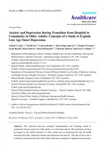
Embryonic / Postnatal Age in Days Figure 1. Most neuromuscular junctions in the mouse sternomastoid muscle are innervated by a single motor axon by 2 weeks of age. Junctions were stained with 4-Di-2-ASP in mice ranging in age from El 7 to P90. Each horizontal line represents the percentage of junctions innervated by more than one axon determined by examining between 17 and 6 1 junctions in each of five to nine animals at each age. Open circles represent mean values. We do not know whether the range of data at any one age represents variation in the rate of synapse elimination between muscles or the relatively small sample sizes that gave rise to each individual value. Other anatomical techniques such as tetranitroblue tetrazolium (TNBT) staining (Jordan et al., 1988) or combined silver/AChE staining (Oda, 1986) in fixed whole-mount preparations showed a similar incidence of multiple innervation at each age examined (for each type of staining, at least 25 junctions were examined from each of five muscles at each age). For example, 4-Di-2-ASP staining showed that 42 + 2 1% of stemomastoid neuromuscular junctions remained multiply innervated on P7, while TNBT or silver/ AChE staining showed that 44 & 13% and 47 f 9% ofjunctions, respectively, were multiply innervated at this age (values not significantly different, Student’s t test). These observations showed that the incidence of multiple innervation can be as reliably determined using vital staining in situ as it can be with conventional staining of fixed tissues removed from the animal.
Figure 2. AChR distribution differs at multiply and singly innervated junctions. Shown are three junctions (1-3) in a stemomastoid muscle from a 6-dold mouse stained with 4-Di-2-ASP (left panel) and RaBTX (right panel). Junction 2 is innervated by one motor axon (arrow), and the AChR distribution at this junction is relatively broken up in appearance, having areas of high AChR density (bright regions) interspersed with areas of relatively low AChR density (dark regions). In contrast, junction 3 is innervated by two motor axons (arrows) and its AChR distribution is relatively homogeneous and plaque-like. Junction I is also multiply innervated, but its AChR distribution was only partially visible due to the curvature of the muscle fiber. Such junctions were not studied further. Scale bar, 20 pm.
The Journal
in AChR distribution than junctions that appeared more plaquelike. Indeed, in 19 junctions that were already broken up at the first view, with more than one-third of the junctional area consisting of very low AChR density, no loss of AChR-rich areas was observed (Fig. 3A). We presume that such junctions underwent loss several days earlier, because nearly all junctions at PO (> 90%) had a plaque-like AChR distribution (see section 4 below; Steinbach, 1981; Slater, 1982a). A few junctions (3 of 58) that appeared to have a moderate degree of branching at the first view lost one or two small AChR-rich areas between P8 and P14 (Fig. 3B). In the remaining junctions we studied (36 of 58), AChRs were distributed into more homogeneous plaques at the first view. In each of these junctions, multiple discrete postsynaptic areas stained with RaBTX on P6 were no longer visible at subsequent views on P8, PI 0, and P12 (Fig. 4). During this time, junctions that showed evidence of loss of previously inserted AChRs from postsynaptic areas lost a mean of 4.3 f 0.5 areas each. Typically, these areas were relatively small, ranging in size from 6 to 58 Mm* (mean, 24 f 10 pmz; N= 167 areas, 39 junctions) compared to a mean junctional area of 3 19 f 43 pm2 on P6-P8 (N = 58 junctions). Because the views from P8 through P12 were made without adding additional RolBTX, these results show that previously inserted AChRs in the postsynaptic membrane are lost from several areas within individual junctions. This loss could mean either that AChRs in these areas undergo more rapid turnover than receptors elsewhere in the junction, in which case insertion of receptors into these areas may also be increased, leading to no net change in receptor density; or that these areas may undergo a net loss of AChRs if the loss of previously inserted receptors is not compensated for by an increased rate of insertion of new receptors into those areas. The junctions were thus restained with RcvBTX on P 14 and again on P2 1 to determine the receptor density in these areas. Every one of the areaswhich lost AChR staining betweenP6 and P12 (167 areasin 39 junctions) could not be restained on P14 or P21 (Fig. 3B, panel P14; Fig. 4, columns P14, P21). Furthermore, between P12 and P14, a few additional AChRrich areaswerelost. The inability to stain AChRs in postsynaptic areasthat lost previously inserted receptorsshowsthat the areas which disappearundergo a net depletion of AChRs, due to loss of previously inserted receptorsthat are not replaced by newly inserted receptors. Examination of successiveviews of eachjunction showedthat postsynaptic areaswithin a junction were depleted of AChRs sequentially rather than simultaneously (Fig. 4). For example, in the junction illustrated in the first row of Figure 4, two small AChR-rich areaspresent on P6 were depleted of receptors by P8. Another area present on PI0 was depleted of receptors by P12. One additional area was depleted of AChRs betweenP12 and P14. In most of thesejunctions, periods of depletion of
of Neuroscience,
February
1993,
13(2)
839
AChR-rich areaswere interspersedwith periods of relative quiescence,indicating a discontinuous process.In the junction illustrated in the first row of Figure 4, no postsynaptic areaswere depleted of AChRs between P8 and PlO. Despite the lossof multiple AChR-rich areaswithin a junction, there was no net decreasein their total AChR-rich area. This cameabout becauseduring the first 2 postnatalweekswhile AChR-rich areaswere being lost, there was substantialgrowth of the junction asa whole. In the example illustrated in the first row of Figure 4, five AChR-rich areastotaling 128 pm2 were lost between P6 and P14 but the total area of the remaining AChR regions increasedfrom 203 pm* on P6 to 3 15 pm2 on P14. A comparableincreasein area was seenin every junction viewed more than once between P6 and P14. However, in none of the junctions we followed during the first weeksafter birth was the steady increasein synaptic area accounted for by the significant addition of new AChR-rich areasthat changedthe shapeof the junctions. Rather, synaptic area increasedby an overall enlargementof the existing postsynaptic territory: not only did AChR-rich areasenlarge, but the spacesbetweenAChR-rich areasalsoenlarged.The ongoing expansion of synaptic and intrasynaptic areaswithin the junction during the first 2 postnatal weeksis analogousto the mode of intercalary enlargementofjunctions in older animals(BaliceGordon and Lichtman, 1990b). Thus, the loss of AChR-rich areasis not compensatedfor by a comparable addition of discrete new areas. One consequenceof this form of generalizedenlargementwas the appearanceof obvious gapsin what formerly appearedto be contiguous AChR-rich areaswhen the junction was more compact (e.g., seeFig. 3B, region marked with arrowheads;see also Fig. 11). The appearanceof gapsas well as the depletion of AChRs from postsynaptic areasdescribedabove contributed to the breaking apart of initially plaque-like receptor distributions. The areasthat were depleted of AChRs, however, were usually easyto distinguish from the gapsthat appeareddue to growth. In many of the areasthat we judged to have been depleted of AChRs, some element in the postsynaptic receptor pattern had disappearedrather than been displaced (seeFig. 11). Another difference betweenAChR areasthat were lost and the gapsthat appearedwas that intermediate views sometimes caught the regionsthat would ultimately be completely depleted of receptorsat a time when they stainedmore faintly with RcvBTX (Fig. 4, first row, small arrows; seealso Fig. 5). In all but one of the junctions we viewed another time after 2 weeksof age,the postsynaptic AChR-rich area remained stable, undergoingno further losses.In the onejunction which did change(illustrated in the last row of Fig. 4), one small area (9 prn2)was lost between 14 and 2 1 d of age. Loss of AChR-rich areasonly rarely occurs in sternomastoidneuromuscularjunctions in normal adult animals (seesection 7 below; Balice-Gordon and Lichtman, 1990b, 1991). On the other hand, loss of
t Figure 3. Junctions with AChR distributions that appeared relatively branched at the first view show little or no loss of receptor areas at subsequent views. Shown are three junctions stained with RolBTX at the first view (left panels) that were restained and rephotographed on two subsequent occasions (middle and right panels). A, More than 30% of the area of these two junctions (top and middle rows) consists of areas of very low or undetectable AChR density (dark regions; see, e.g. top left panel, arrows). Comparison of AChR-rich areas in each view shows that junctions such as these that were already broken up at the first view often did not show evidence of loss of AChR-rich regions at subsequent views. B, Shown is a junction that is relatively branched at the first view. Subsequent views show that AChRs have been depleted from the upper right region of the junction (arrow). The remaining regions have spread apart during this time, and gaps that were present in the original pattern have become more evident (arrowheads). Scale bar, 20 pm.
840
Bake-Gordon
and
Lichtman
l
Pre- and
Postsynaptic
Changes
during
Synapse
Elimination
Figure 4. Junctions with relatively plaque-like AChRdistributionslosemultipleAChR-richareasin succession duringthe first 2 postnatalweeks. The&t panel in each row illustrates a neuromuscular junction in a sternomastoid muscle from a P6 mouse in which postsynaptic AChRs were stained with RcBTX. Adjacent panels show the same junction rephotographed on P8, PlO, and P12 without restaining. It was necessary to restain junctions with RaBTX on P 14 and P2 1 because turnover of existing AChRs resulted in junctional staining being too dim to image. Thus, junctions were restained with RolBTX and rephotographed on P 14 and again on P2 1. In each row, comparison of adjacent panels shows that small AChRrich areas within the junction disappear one after the other (arrows).In the junction in thefirst row, one AChR area present on PI0 (arrow) appears fainter on PI2 (smaN arrow) and is no longer present on P14. In most junctions, loss of AChR-rich areas ceased by 2 weeks after birth. In the
junction illustratedin the bottomrow,onesmallspotof stainingwaslostbetweenPI4 (arrow)andP21.Note that the principalmeansofjunctional growth is the expansion of remaining AChR-rich
areas and the spaces between them. Scale bar, 10 pm.
AChR-rich areasis commonly observedin adult neuromuscular junctions undergoing synapse elimination following reinnervation (Rich and Lichtman, 1989a). 3. Depletion of AChRs from eachpostsynaptic area is gradual To determine how AChRs are depleted from a postsynaptic area, receptors already inserted in the muscle fiber membrane were labeledwith RaBTX at one view and the stainingintensity wasmeasuredat later times (N = 258 junctions, 39 mice; mean ageat first view, 7 f 0.5 d, at secondview, l-4 d later, mean, 3 -t 0.8 d). As with someof the junctions from 1-week-old mice that were viewed three or more times describedin section 2, in
this seriesof experiments, in which junctions from 1-week-old mice were viewed twice, about half (134 of 258) showed no evidence of lossof AChR-rich areas.Thesejunctions are likely to have undergone loss at an earlier time (seealso section 4 below). In someof theseiunctions (104 of 258), AChR-rich areasthat stained as brightly as the rest of the junction at the first view were no longer present at the second view several days later (Fig. 5A). In somejunctions (N= 20), we observedfaintly stained AChR areas at the first view (Fig. 5A, lower arrow; Fig. SB). Thesefaintly stained areaswere completely depletedof AChRs at the secondview. Becausepostsynapticareasthat stainedfaintly with RaBTX were found in junctions the first time that they
The Journal
of Neuroscience,
Figure
5.
February
1993.
i3(2)
841
Postsynaptic areas are grad-
ually depleted of previously inserted AChRs. Two junctions are shown in the process of losing AChR-rich areas. In the left panels, the junction has been stained with RcKBTX. The right panels show the original RolBTX staining rephotographed 2 d later. A, In this junction, postsynaptic AChR-rich areas that on P6 stained more faintly than the rest of the junction (lower arrows) were completely depleted of previously inserted AChRs by P8. Other areas (top arrow) may also have been depleted of previously inserted AChRs in the interval between views. B, In this junction, a postsynaptic AChR-rich area that on P8 stained more faintly than the rest of the junction (lower arrow) was completely depleted of previously inserted AChRs by PIO. Another area that on P8 stained as brightly as the rest of the junction (upper arrow) seems to have been partially displaced upward in the interval between views. This type of change may mean that AChRs are migrating in the plane of the muscle fiber membrane (see Fig. 7). Scale bar, 10 pm.
were viewed, it is unlikely that loss of AChRs was induced by labeling receptors with RaBTX or by other aspects of the viewing procedure. By 14 d of age, faintly stained AChR areas were very rare (Fig. 6) as was evidence of loss of AChR-rich areas. Thus, the data suggest that all faintly stained receptor areas are in the midst of being depleted of AChRs. We retrospectively compared the brightness of AChR-rich areas that 2-3 d later (mean, 2 + 0.3 d) were observed to disappear to the receptor-rich areas in junctions that remained intact (N = 49 junctions, 8 P7 mice, 7 1 areas). Not surprisingly, these brightnesses varied widely. In order to obtain a quantitative approximation of the relative numbers of AChRs in faintly stained compared to brightly stained postsynaptic areas, the output of the SIT camera was calibrated as described in Materials and Methods. Postsynaptic areas stained with RaBTX which are still easily discernable but appear faint were estimated to contain between 3 1% and 86% as much RaBTX staining as parts of the junction which are brightly stained (mean, 58 * 12 %; N = 10 junctions from three P7 mice). The wide range of values suggests that the density of AChRs in areas undergoing receptor depletion changes from the amount initially present in the rest of the junction to ultimately a very low density indistinguishable from background.
To determine how rapidly AChRs are lost from individual postsynaptic areas, muscles were stained with RaBTX and plaque-like junctions were monitored over the courseof 1 d (N = 3 1junctions, 23 P6-P8 mice). In 16junctions, changesin the brightnessof AChR staining were discernablelessthan 24 hr after the first view (Fig. 7A). In four junctions, faint AChR areas were viewed a second time just 6 hr after the first view was made. In each case,a significant loss of staining intensity was discerned. In somejunctions (N = 1l), a portion of the labeled AChRs in areas undergoing loss appeared to be redistributed in the postsynaptic membranein the brief interval between views. In the,junction shown in Figure 7, at the time AChRs were found to have been depleted from the upper portion of the junction, the distribution of labeled receptors had also changed.In particular, postsynaptic regionsthat did not contain RaBTX staining at the first view (Fig. 7B, left panel, arrows) did contain someRL~BTXstaining 17 hr later (Fig. 7B, right panel, arrows). Application of additional RaBTX at the final view showeda similar pattern of lossand changesin overall receptor density and distribution; thus, RcuBTX-labeled AChRs are unlikely to have behaved differently from unlabeled receptorsin the same junctions. Theseobservationsindicate that depletion of AChRs
842
Balice-Gordon
and
Lichtman
l
Pre- and
Figure 6. AChR areas within junctions that stain faintly with RolBTX are commonly observed during the first 2 postnatal weeks and are rare after 14 d ofage. The percentage ofjunctions containing faint AChR areas was determined by examining at least 10 junctions in each of five muscles stained with RolBTX at each age. Faint AChR areas are approximately 30-90% as bright as the rest of the receptor-rich areas and are typically greater than 5 pm2 in size. See Results and Materials and Methods for details. At the time multiple innervation in the muscle is declining the most rapidly (see Fig. I), faint AChR areas are the most prevalent. Bars indicate average values and linesindicate SD.
Postsynaptic
Changes
during
Synapse
Elimination
P
g 20 c
5 E 15 ‘$ f 3 10
5 z 8 ,om p! g a
El7
El9
Pl
P3
from postsynaptic membrane areascan be rapid. In each case studied, once postsynaptic areasbegan to lose AChRs, the remaining receptors in those areasinvariably disappearedcompletely in lessthan 24 hr. 4. Multiply innervated junctions are more likely than singly innervatedjunctions to loseAChR-rich areas Given that multiply innervated junctions are typically plaquelike and that singly innervated junctions are more branched(see section 1 above), lossof AChR-rich areasmay occur during (and perhapsbecauseot) the transition from multiple to single innervation. We thus wanted to study the AChR distribution at individual junctions that we knew were undergoing synapse elimination. As a first step, we observedjunctions stained with 4-Di-2-ASP multiple times to ensurethat this dye could be used to follow changesin junctional innervation over time in living neonatal mice. Junctions from 4-S-d-old mice (mean age, 6 f 0.4 d) were stained with 4-Di-2-ASP, and the number of innervating axons was determined (N = 360 junctions, 74 mice). Junctions were then restained and relocated l-4 d later (mean, 3 -+ 0.3 d later; 291 junctions viewed twice and 32 junctions viewed three times). An additional 37 junctions were unable to be studied at the secondview becausetheir muscle fibers had shifted, altering the orientation of junctions onto their side, which in thesecasespartially obscuredeither the axons or the synaptic sites. In 148 of 360 junctions, 4-Di-2-ASP staining showed two motor axons entering the junctional area at the first view (Fig. 8, left panels).In 116 of these 148junctions, 4-Di-2-ASP staining at the secondview showed that only one axon remained (Fig. 8, right panels). In some cases,a second axon was still
P5
P7
P9
Pll
P13
P15
P17
P19
P21 P30 P60
P90
Embryonic / Postnatal Age in Days
present at the secondview but no longer projected all the way to the junction (Fig. 8, bottom row). In 32 of 148junctions, two motor axons were still presentat the secondview (see,e.g., Fig. 10). However, in each of thesejunctions, only one axon projected to the junction when a third view was made l-4 d later (mean,2 f 0.1 d). In the junctions which were singly innervated at the first view (175 of 360) there wasno evidence at the second view either that the singleaxon withdrew from the junction or that a secondaxon appeared. We compared a group of junctions that several days previously had been stained and viewed with junctions from littermate control animals that had never been previously stained and viewed. No significant differencesin junction size, extent of multiple innervation, or time courseof synapseelimination were observed (comparison by age or weight). For example, 7 d after birth, 41 f 6% of junctions in the control group were innervated by two motor axons (N = 162 junctions, 18 mice). In a group of mice whosejunctions were stained and viewed twice (first view on P4 and secondview on P7), 43 f 4% were multiply innervated on P7 (N = 50 junctions, 7 mice). Thus, axon withdrawal appearedto be unaffected by the staining and viewing procedureswe used. To correlate the depletion of AChRs from postsynaptic areas directly with the transition from multiple to singleinnervation, junctions were stainedboth with 4-Di-2-ASP (to assess the number of innervating motor axons)and with RatBTX and werethen viewed repeatedly (N = 376 junctions, 74 P4-P8 mice; mean ageat first view, 5 + 0.6 d; 3 12junctions viewed twice and 64 junctions viewed three times). 4-Di-2-ASP stainingshowedthat 257 junctions were innervated by two motor axons and 119 were innervated by one motor axon at the first view. We found
Figure 7. Postsynaptic areas are rapidly depleted of previously inserted AChRs. A, Shown is a junction from a P6 mouse (left panel) stained with RolBTX. The right panel shows the same junction 17 hr later. No additional RolBTX had been applied; thus, the labeled AChRs are those that have remained in the junction since the first view. The top one-third of the junction has been depleted of AChRs in this brief interval and thus appears faint. B, The top one-third of the junction is shown at higher magnification. A portion of the labeled AChRs in this region appear to have been redistributed in the postsynaptic membrane. Three regions that did not contain RaBTX staining at the first view did contain some staining 17 hr later (compare regions marked with arrowsin left and right panels).This comparison was made by taking the two images with identical illumination and gain settings of the SIT video camera. For illustration purposes only, the rightpanelshave been enhanced so that the faint areas would be more visible in the photographs. Scale bars, 10 pm.
Original
RocBTX
Remaining 17 hours
RaBTX later
The Journal of Neuroscience,
February
1993, 13(2) 845
Figure 9. Individual junctions undergoing the transition from multiple to single innervation lose postsynaptic AChR-rich areas. Top, Motor nerve terminals and axons at a junction from a PI mouse stained with 4-Di-2-ASP. This junction is multiply innervated by two axons (left panel, arrows). The right panel shows the same junction restained and rephotographed 3 d later, when only one axon remains to singly innervate the junction (right panel, arrow). Bottom, Postsynaptic AChR-rich areas in this junction stained with RotBTX. AChR-rich areas present on P7 (leftpanel, arrow) are no longer present when the junction was revisualized on PlO (right panel, arrow). Scale bar, 10 w-n. ,,
that multiply and singly innervated neuromuscularjunctions differed dramatically in the incidence of loss of postsynaptic AChR-rich areas. In every one of the 257 junctions that 4-Di-2-ASP staining showedwere innervated by two axons at the first view, AChRt
rich areaswere observed to be depleted of receptors when the junctions were revisualized several days later (mean, 3 + 0.5 d later; Fig. 9, bottom panels). In 193 of 257 junctions, one of the two axons initially innervating the junction at the first view was no longer present at the secondview (Fig. 9, top panels).
Figure 8. Visualization of the transition from multiple to single innervation in the sternomastoid muscle of living mice. Top, Motor axons and nerve terminals from a P5 mouse (left panel) vitally labeled with 4-Di-2-ASP. Shown is a portion of the end-plate band containing five superficial neuromuscular junctions; the bright spots of staining are probably intracellular mitochondria within motor axons and nerve terminals. Muscle fibers are more weakly stained, and some myofibrillar striations can be seen. The right panel shows the same area restained and rephotographed 2 d later. The junctions labeled 1 and 2 are shown at higher magnification below. Middle, Shown is a neuromuscular junction from a PS mouse that is innervated by two motor axons that are entwined (left panel, arrows). Two days later, focusing through this region revealed that only one of these axons remains (right panel, arrow). Bottom, Shown is a neuromuscular junction innervated by two parallel motor axons (left panel, arrows). Two days later, one axon has withdrawn from the junction but is still present (right panel, right arrow) adjacent to the preterminal portion of the axon that remains (right panel, left arrow). Scale bars, 10 pm.
846 Bake-Gordon
and Lichtman
l
Pre- and Postsynaptic
Changes
during Synapse
Elimination
Figure 10. Postsynaptic AChR-rich areas are lost prior to axon withdrawal from multiply innervated junctions. A, Motor nerve terminals and axons in a junction from a P5 mouse stained with 4-Di-2-ASP. The junction is innervated by two motor axons (top left panel, arrows). The top right panel shows the postsynaptic AChR-rich areas in this junction stained with RLuBTX. Bottom panels show the same junction restained and rephotographed on P7. Although the junction remains multiply innervated (bottom left panel, arrows), AChRs have been depleted from a small area of the junction in the interval between views (right panels, arrow). B, Motor nerve terminals and axons in a multiply innervated junction from a P6 mouse stained with 4-Di-2-ASP (top left panel). The top right panel shows the postsynaptic AChR-rich areas in this junction stained with RaBTX. Bottom panels show the same junction restained and rephotographed on P8. AChRs have been depleted from the bottom portion of the junction in the interval between views (compare top right panel, arrows, with corresponding area in bottom panel). Scale bar, 10 pm.
both axons remainedat the secondview. Nonetheless,each of thesejunctions also contained AChR-rich
In 64 of 257 junctions,
areas that were depleted
of receptors
between
views (Fig. 10).
Thus, lossof AChR-rich areascan occur prior to the withdrawal I of a motor axon from a junction. In contrast, 116 of 119junctions that appearedsingly innervated at the first view showedno evidenceof depletion of AChRs from postsynaptic areaswhen they were viewed a secondtime severaldays later (mean, 4 + 0.4 d later; Fig. 11). Theseresults show that depletion of AChRs occurs very frequently in junctions undergoing the transition from multiple to singleinnervation but relatively rarely in junctions in which axon withdrawal hasalready taken place.Thus, the largegroup ofjunctions described in sections 2 and 3 which did not undergo loss of AChR-rich areas were most likely already singly innervated prior to the time the first view was made. 5. Motor nerve terminal depleted of AChRs
staining
disappears
over areas
The observation that depletion of AChRs from postsynaptic areasis occurring in junctions losingmultiple innervation raised the obvious question of what is the spatial relationship between
pre- and postsynaptic loss. We first determined that when one axon withdrew from a junction, a change in nerve terminal staining is apparent that is consistent with the elimination of sites of nerve terminal contact. Junctions were stained with 4-Di-2-ASP, and those in which the two axons and their nerve terminals appearedto be at least partially segregatedwithin the junction were selectedfor further study (N = 33 junctions, 12 mice; mean ageat first view, 6 f 0.3 d). Thesejunctions were viewed a secondtime a mean of 2 * 0.9 d later. In 26 of 33 of thesejunctions, as one axon withdrew, motor nerve terminal staining adjacent to that axon also disappeared. In the examples illustrated in Figure 12, several small 4-Di-2ASP-stained spotsnear one axon that were present at the first view (left panels,rightmost arrows)were no longer presentwhen the junctions were restainedand reviewed 2 d later (right panels). That areas in fact lost 4-Di-2-ASP staining in each of these junctions at the secondview suggeststhat the remaining axon did not re-occupy those areas(Fig. 12, right panels). To compare pre- and postsynaptic sites within a junction before and after one axon withdrew, junctions were stainedwith both 4-Di-2-ASP and RcvBTX (N = 124 junctions, 27 mice; mean age at first view, 6 + 0.5 d; secondview, mean of 3 +
The Journal
of Neuroscience,
February
1993,
13(2)
847
Figure I I.
AChR-rich areasin singly innervatedjunctions spreadapart as junctionsgrow but are otherwiseunchanged.Top, Motor nerve terminals in ajunctionfroma P5mouse(leftpanet) stainedwith 4-Di-2-ASP.Thisjunction is singlyinnervatedby onemotor axon (arrow) whosefull lengthis not visible in the sinalefocal planeillustratedhere.The spotsof stakingabove andbelowthis axon are probablymitochondriain Schwanncellsthat begin to ensheathaxonsat theseages.The rightpanelshows thesame junctionrestained and rephotographedon PlO. Bottom, Postsynapticdistribution of AChR-richareasin thisjunctionstained with RczBTX.Thejunctionenlarged by spreading apartof existingpostsynaptic AChR-rich areasin the interval betweenviews.As thisoccurred,gapsbecameapparentin formerlycompactreceptor-richareas.In contrastto multiply innervatedjunctionsundergoingsynapseeliminationat the sameages,no AChR-rich areascompletely disappearedfrom singly innervatedjunctionsin theintervalbetween views.Scale bar, 10Wm. 0.3 d later). In every casewherepostsynaptic areasweredepleted of AChRs, motor nerve terminal staining waslost from the same synaptic areasthat were depleted of AChRs (Fig. 13, uppermost arrow). Depletion of AChRs was not observed from areasof the junction that maintained presynaptic staining. On the other hand, areasthat lost 4-Di-2-ASP staining did not always also lose postsynaptic AChRs (seeFig. 13, arrow-
heads).Because4-Di-2-ASP only stainsintracellular organelles suchas mitochondria (Lichtman et al., 1989),one possibility is that loss of 4-Di-2-ASP staining may merely represent mitochondrial rearrangementswithin the nerve terminal rather than actual lossof a nerve terminal per se. However, in neuromuscular junctions stainedwith RH795 (which intercalatesinto the motor nerve terminal membrane; Grinvald et al., 1986), the
848
Bake-Gordon
and
Lichtman
l
Pre- and
Postsynaptic
Changes
during
Synapse
Elimination
Figure 12. Areas of motor nerve terminal staining disappear during the transition from multiple to single innervation. A, Motor nerve terminals and axons stained with 4-Di-2-ASP in a junction from a P5 mouse multiply innervated by two motor axons (left panel, upper arrows). The right panel shows the same junction restained and rephotographed on PI, when one axon remains (arrow). In addition to the withdrawal of one motor axon from the upper right-hand area of the junction, motor nerve terminal staining has also disappeared from a nearby area (compare left panel, rightmost arrow, with corresponding area in right panel). On PI, mitochondria in an ensheathing Schwann cell are evident surrounding the remaining motor axon. B, Motor nerve terminals and axons stained with 4-Di-2-ASP in a junction from a P6 mouse multiply innervated by two motor axons (left panel, Ieftmost arrows). The right panel shows the same junction restained and rephotographed on P8, when one axon remains (arrow); this axon is ensheathed by a Schwann cell whose mitochondria are evident surrounding the preterminal portion of the axon. Motor nerve terminal staining has disappeared from the top portion of the junction (compare Ieji panel, rightmost arrow, with corresponding area in right panel). Scale bar, 10 pm.
nerve terminal membrane (contiguous with the motor axon) aligned exactly with brightly stained postsynaptic AChR-rich areas at all of the ageswe examined (N = 79 junctions, 30 P4-
P14 mice; seeFigs. 15, 16). Thus, as AChR-rich areasare lost during development, the overlying nerve terminal must alsobe eliminated.
6. AChR depletion begins before the overlying motor nerve terminal is eliminated One question raised by these observations is whether the loss of postsynaptic AChR-rich areasoccurs after and perhapsas a consequenceof motor nerve terminal elimination, or alternatively, whether elimination of motor nerve terminals is a consequenceof postsynapticchanges,including depletion of AChRs. To addressthis question, it was necessaryto determine more precisely when motor nerve terminals disappearedrelative to
when AChRs in the underlying membranebeganto be depleted. By looking at faintly stainedAChR areasthat werein the process of being eliminated, we could ask if they consistently were no longer overlain by nerve terminal staining,which would indicate that loss of AChR staining occurs after the nerve has already vacated those areas.Examination of multiply innervated junctions stainedwith 4-Di-2-ASP and RaBTX that contained faint AChR areas(N= 89junctions, 27 P6-P8 mice), however, showed that AChR loss often occurred prior to loss of motor nerve terminal staining,becausefaint receptor areaswere still overlain by 4-Di-2-ASP staining (78 of 89 junctions; Fig. 14). This indicates that 4-Di-2-ASP staining is not eliminated until after AChRs begin to be depleted postsynaptically. By staining with 4-Di-2-ASP as well as RH79.5 to label the membrane of the nerve terminals, we could determine whether or not the 4-Di2-ASP staining remaining over areasof low AChR density was
The Journal
of Neuroscience,
February
1993,
13(2)
949
Figure 13. Motor nerve terminals are eliminated from junctional areas that lose postsynaptic AChRs. Top, Theleft panel shows motor nerve terminals and axons in a junction from a P6 mouse stained with 4-Di-2-ASP. This iunction is multiply innervated by two axons (lower arrows). The right panel shows the same junction restained and rephotographed on P9, when one axon remains (lower arrow). In addition to the withdrawal of one axon, the pattern of motor nerve terminal staining has changed in the interval between views. One ofthese changes is the loss of 4-Di2-ASP staining from the uvver region of thejunctionlupperarrows). A second change is the loss of staining from the left-hand region of the junction (arrowhead). Bottom, Postsynaptic AChR-rich areas in ais junction stained with RolBTX. The postsynaptic area beneath a site of loss of 4-Di-2-ASP staining has been depleted of AChRs (compare right panel, arrow, with left panel, arrow). Because changes in 4-Di-2-ASP staining did not ilways correspond with changes in AChR staining (compare preand postsynaptic sites marked by arrowheads in top and bottom panels), we also stained junctions with a vital dye that labeled motor nerve terminal membranes (see Fig. 15). Scale bar, 10 pm.
part of the nerve terminal. We compared the pattern of staining of multiply innervated junctions with 4-Di-2-ASP and RH795 at various stages of synapse elimination (N = 154 junctions, 65 P6-P8 mice). In each of the multiply innervated junctions in which postsynaptic AChR areas were stained uniformly brightly with RaBTX (N = 113 junctions), the postsynaptic area was entirely overlain by RH795 stained motor nerve terminals and the RH795 staining enveloped 4-Di-2-ASP staining (Fig. 15, top row). However, in 35 of 41 multiply innervated junctions in which postsynaptic areas stained faintly with RaBTX, the faint AChR areas still had RH795 and 4-Di-2-ASP stained motor nerve terminals over them (Fig. 15, middle row). This result not only confirms that depletion of AChRs can occur before a motor axon entirely withdraws from a junction (see section 4 and Fig. lo), but also shows that depletion of receptors begins before an overlying motor nerve terminal is eliminated from that site. In 6 of 4 1 junctions, faintly stained AChR areas were unoccupied or only partially overlain by nerve terminal staining, suggesting that after AChRs began to disappear from a region, nerve terminal staining is short-lived (Fig. 15, bottom row).
7. Loss of synaptic sites is rare after single innervation
is
established Studiesin which neuromuscularjunctions in other mammalian muscles(Wigston, 1989, 1990; Hill et al., 1991) and in other species(Herrera et al., 1990; Chen et al., 1991) were followed over time have suggestedthat loss.of synaptic sitesmay occur throughout life. Becausejunctions in adult skeletal musclesare assumedto be uniformly singly innervated (at least in mammals), this raisesthe possibility that there are causesother than the presenceof two (or more) competing inputs for pre- or postsynaptic loss.To study further the relative incidence of loss of synaptic regionsfrom junctions in the sternomastoidmuscle after the period of synapseelimination and to understandbetter the cause,we followed junctions stained with RH795, 4-Di-2ASP, and RolBTX during subsequentmaturation (N = 39 junctions, 12 1-month-old mice;junctions viewed at roughly 1month intervals; 26junctions viewed three times, 12viewed four times, 1 viewed five times). In most junctions (35 of 39), no postsynaptic AChR-rich areas or presynaptic motor nerve terminals were observed to be lost when junctions were followed for up
550 Bake-Gordon
and Lichtman
* Pre- and Postsynaptic
Changes
during Synapse
Elimination
Figure 14. Postsynaptic areas begin to be depleted of AChRs before overlying motor nerve terminals are eliminated. Shown are two examples (A, B) of neuromuscular junctions in sternomastoid muscles from P7 mice that were stained with 4-Di-2-ASP (left panels) and RaBTX (middle panels). These junctions are multiply innervated by two motor axons (left panels, arrowheads). Within each junction there are areas of RaBTX staining that are fainter than the remainder of the junction (middle panels, arrows) and that are in the process of being depleted of AChRs. A “bleedthrough” image (see Materials and Methods) of 4-Di-2-ASP staining plus RaBTX staining taken with rhodamine filters shows that these faintly stained areas are overlain by motor nerve terminal staining (compare left, middle, and right panels, arrows). Scale bar, 10 pm. to 7 months (seealsoBalice-Gordon and Lichtman, 1990b,their Figs. 7, 8, 11, 12; Balice-Gordon and Lichtman, 1991, their
Figs. 1, 2). In 4 of 39 junctions, however, synaptic areasdisappearedover the time the junction was observed. Theseareas were typically small, lessthan 40 Mm*in area (compared to an average junctional area of 1296 + 3 19 pm* at 1 month of age). One of these junctions is illustrated in Figure 16. Over a 5 month
interval, three small AChR-rich areaswere observedto be lost from the junction. Between 1 and 4 months of age,a smallgap appearedin the upper left-hand region of this junction (top right panel, arrow) and similarly sized gapsappeared in two other junctions. It is possible,however, that the appearanceof such gapsis an example of spreadingapart of adjacent receptor areas due to growth rather than true loss of a synaptic region (see
Figure 15. Alignment of pre- and postsynaptic regions before, during, and after synapse elimination. Each row illustrates a neuromuscular junction in which motor nerve terminals and axons were stained with 4-Di-2-ASP (left panels), RH795 (middle panels); postsynaptic AChR-rich areas were stained with RaBTX (right panels). Top, Shown is a multiply innervated neuromuscular junction from a P5 mouse in which all of the AChRrich areas are occupied by motor nerve terminal staining. Middle, Shown is a multiply innervated neuromuscular junction from a P7 mouse that contains a postsynaptic area that is partially depleted of AChRs (right panel, arrow). This area stains more faintly than the other AChR-rich areas in the junction. Motor nerve terminals, stained with 4-Di-2-ASP (left panel, arrow) or RH795 (middle panel, arrow) not only arepresentover the brightly stained AChR-rich areas but also are present over the faintly stained area. Observations of junctions such as this one (see also Fig. 14) suggested that AChRs begin to be depleted from a postsynaptic area before the overlying motor nerve terminals are eliminated and the motor axon withdraws from the junction. Bottom, Shown is a junction from a P8 mouse that is innervated by a well-stained axon (left and middle panels). There may also be a faintly stained remnant of a second axon entering the junction above the well-stained axon that could be traced back to an intramuscular nerve branch. Brightly stained areas of AChRs are occupied by motor nerve terminal staining, and a faint AChR area at the top of the junction appears to be unoccupied by nerve terminal staining. This suggests that after AChRs begin to disappear from a region, nerve terminal staining over that region is short-lived. Note the more highly branched AChR distribution in this junction from a P8 mouse, which is typical of singly innervated junctions (compare right panels in each row). Scale bar, 10 pm.
852 Babe-Gordon
and Lichtman
l
Pre- and Postsynaptic
Changes
during Synapse
Elimination
Figure 16. Loss of synaptic areas is only occasionally observed in stemomastoid neuromuscular junctions after single innervation is established in the muscle. Shown are 4-Di-2-ASP staining (left panels), RH795 staining (middle panels), and RL~BTX labeling (right panels) of a junction in a sternomastoid muscle from a l-month-old mouse (Im.) that was restained and rephotographed at 4 months (4m.) and 6 months (6m.) of age. RH795 staining shows the outline of terminal and axon membrane, which completely encloses 4-Di-2-ASP staining. Presynaptic motor nerve
The Journal
section 2). Between 4 and 6 months of age, two other AChRrich areas in this junction have been depleted of receptors (middle left panel, arrows). By viewing the RH795stained nerve terminals at the same time points, it is clear that in this case, as well as a similar loss of a receptor-rich area in one other junction, the loss is related to the myelination of a former nerve terminal branch (bottom middle panel, arrows). These observations show that in the mouse sternomastoid muscle, ongoing loss of pre- or postsynaptic areas in singly innervated junctions is relatively rare after the period of synapse elimination is completed. Moreover, the synaptic sites that remain after synapse elimination is complete are stable for many months. Thus, the synaptic sites that are lost give riseto changes in the junctional branching pattern that are maintained over long intervals. Discussion We have attempted to analyze the changesthat occur in mouse neuromuscularjunctions during early postnatallife when muscle fibers undergothe transition from multiple to singleinnervation. The principal findings are that (1) postsynapticAChRs are commonly depleted from areasof neuromuscularjunctions during early postnatal life but not later. This depletion is much more frequently seen in junctions that are innervated by multiple motor axons than in junctions that are singly innervated. (2) More than one postsynaptic area is depleted of AChRs within eachjunction. These lossesoccur in successionrather than simultaneously. (3) Nerve terminals are eliminated presynaptitally from the sameareasthat are depleted of AChRs postsynaptically. (4) Presynaptic nerve terminal loss does not occur until after postsynaptic AChRs begin to be depleted, and (5) this pre- and postsynapticlosspermanently changesthe branching pattern of a junction. From these results,we conclude that the withdrawal of a motor axon from a neuromuscularjunction occurs as a consequenceof the sequentialelimination of all of its synapseswithin that junction. It appearsthat one axon begins to lose the battle for exclusive innervation of a junction as the postsynaptic areasit occupiesare depletedof AChRs. Thus, the postsynaptic target cell may play an active role in deciding which synapsesare to be maintained and which are to be eliminated. One weaknessin this work is that the quality of the vital presynaptic staining was usually insufficient to identify the axonal source of the presynaptic terminals that were eliminated. We cannot say with certainty that the nerve terminal staining that wasseento disappearalways originated from the axon that ultimately waseliminated. In a few optimally stainedjunctions (seeFig. 14; Fig. 15, middle row), motor nerve terminals over a faintly stainedAChR area could be seento originate from the vicinity of one axon, arguing that the area being lost was associated with that axon. In other work (Balice-Gordon and Lichtman, 1989; Chua et al., 1990; R. J. Balice-Gordon, C. K. Chua, and J. W. Lichtman, unpublishedobservations),we have approachedthis problem more directly by usingnonvital staining techniques,which showthe full extent of eachof the terminal
of Neuroscience,
February
1993,
13(2)
853
arbors at multiply innervated muscle fibers by staining each of them with a different-colored fluorescentdye. That work indicates that prior to axon withdrawal there is a gradual loss of synaptic territory occupied by one axon. The presentresultsare consistentwith this idea, and add to this conclusionby showing that there are a number of sites of postsynaptic lossthat correspondto sitesof motor nerve terminal elimination. However, we still do not know whether someof the synaptic sitesthat are eliminated belong to the axon that will remain. It seemslikely that the losing axon must undergo the majority (perhapsall) of the synaptic loss,which ceasesonly when the losing axon finally withdraws. The low level of synapseloss observed at singly innervated junctions seemsto be due to small changesin the maturing nerve terminal suchasmyelination of former terminal branches. If withdrawal of a motor axon from a neuromuscularjunction is the consequenceof the lossof its synapses,theseresultssuggest that there is a temporal disjunction betweenthesetwo phenomena. This may mean that in certain situations in which the number of axonsinnervating a target celldoesnot change,changes in synaptic input to that cell could occur by the elimination of someof the synapsesof belongingto an axon. Becauseindividual synaptic siteswere lost over short times (lessthan 2 d; seeFigs. 4, 5, 7) this form of synaptic plasticity could be involved in rapid changesthat occur in the nervous system as a result of synaptic competition. The presentresults offer somecluesabout the mechanismof synapseelimination. The fact that postsynaptic sites are lost with suchrapidity is inconsistentwith the idea that postsynaptic lossis merely the consequenceof the lossof the overlying terminal. Vacated postsynaptic sitesin developing neuromuscular junctions have been observedto lose their morphological characteristics,but this occursover longer periods (> 1 week; Slater, 1982b).Moreover, the postsynaptic areasthat will be lost begin to be depletedof AChRs in advance of the structural withdrawal of the overlying nerve terminal. Thus, in both development and reinnervation (Rich and Lichtman, 1989a), lossof AChR-rich areas occurs prior to the structural elimination of the nerve terminal. The early postsynaptic changessuggesta possiblerole for the postsynaptic cell both in eliminating and perhaps also maintaining a presynaptic nerve terminal. Previous work in the sternomastoid and bulbocavemosus muscleshasmade the casethat nerve terminals are adherent to the postsynaptic cell at synaptic sites(Balice-Gordon and Lichtman, 1990b, 1991; Balice-Gordon et al., 1990). This adhesion seemsto be the basis for both the intercalary enlargementof nerve terminals in musclesduring growth as well asthe maintenance of precisealignment of pre- and postsynaptic sitesduring growth. If such adhesion is also present during the first 2 postnatal weeks,then synapseelimination not only dismantles the postsynaptic apparatus, but also must break the adhesive bond the nerve terminal makeswith the postsynapticmembrane (via the basallamina). We do not know what kind of causallink exists between lossof postsynaptic AChR-rich areasand nerve
t terminalswereobservedto alignpreciselywith postsynaptic AChR-richareas.Overthe 5 monthsthisjunctionwasstudied,threesmallpostsynaptic AChR-richareasdisappeared. Between1 monthand 4 monthsof age,a smallgapappearsin the upperleft-handregionof thejunction (top right panel, arrow),probablydueto the spreading apartof adjacentpostsynaptic areasasthe musclefibergrows(Balice-GordonandLichtman,1990b). Between4 monthsand6 monthsof age,two smallreceptor-richareasarelost (middle right panel, arrows)asthe overlyingnerveterminalbranch becamemyelinatedand thus stainedmuch more intensely(bottom middle panel, arrows). Lossof synapticregionsafter singleinnervationis established is far moreinfrequentthan the lossobservedin multiply innervatedjunctionsover the first 2 postnatalweeks.Scalebar, 20pm.
854
Balice-Gordon
and
Lichtman
* Pre- and
Postsynaptic
Changes
during
Synapse
terminal elimination, but these phenomena seem to be closely associated. Loss of postsynaptic AChR-rich areas during development, reinnervation (Rich and Lichtman, 1989a), and muscle degeneration (Rich and Lichtman, 1989b) in each case precedes loss of the overlying presynaptic nerve terminal, whereas the converse is not always true: in adult animals presynaptic nerve terminal loss as a consequence of denervation does not cause postsynaptic areas to disappear (Rich and Lichtman, 1989a). Taken together, these results argue that factors derived from the postsynaptic
cell are necessary to maintain
the over-
lying nerve terminal actively, whereaspresynaptic maintenance of postsynaptic sitesmay require lessactive maintenance. Two questions raised by this work are the following. (1) By what mechanism does the postsynaptic cell signal the nerve terminal adhesion
that it should withdraw? Is it possible that loss of is sufficient to cause nerve terminal loss? (2) How do
postsynaptic AChR-rich areascome to losereceptors?The present work suggestspart of the answerto the secondquestion. We have learned that existing (previously inserted) AChRs are quickly removed from postsynaptic areas undergoing depletion. In views of the same junction obtained over relatively short intervals (hours), AChRs from areasin the processof disappearing occasionally seemed to undergo migration in the membrane between views (see Fig. 7, arrows). More recent efforts using focal labeling with RotBTX (Balice-Gordon and Lichtman,
1990a, and unpublished observations) and fluorescencerecovery after photobleaching
(Brown
et al., 1992; A. P. Brown,
P.
van Mier, and J. W. Lichtman, unpublishedobservations)show that even in adult neuromuscular junctions, postsynaptic AChRs are capable of movement within the postsynaptic membrane. If AChRs were more mobile in areasof the postsynaptic membrane in the processof being depleted of receptors than elsewhere within the junction, then receptors might migrate away from one synaptic site and perhaps be trapped by the remaining synaptic sites. Such a mechanism has been invoked to explain
AChR clustering (Stollberg and Fraser, 1990), and it may turn out that during
synapse elimination
axons are competing
for
AChRs that are “declustering.” Because withdrawal of axons is widespread during neural development, it is possible that the pre- and postsynaptic loss
described here is the basis of analogousphenomena in other parts of the nervous system. If this is the case, then loss of synaptic connections generally may require both an early postsynaptic change, including a rapid removal of postsynaptic molecules, and a secondary presynaptic change, which might include
the breaking of an adhesivebond betweenthe presynaptic nerve terminal
and the postsynaptic
apparatus.
References Balice-Gordon RJ, Lichtman JW (1989) Competing motor nerve terminals and the acetylcholine receptors underlying them are rearranged during synapse elimination. Sot Neurosci Abstr 15: 165. Balice-Gordon RJ, Lichtman JW (1990a) Loss of synaptic sites during competitive synapse elimination is both rapid and saltatory. Sot Neurosci Abstr 16456. Balice-Gordon RJ, Lichtman JW (1990b) In viva visualization of the growth of pre- and postsynaptic elements of mouse neuromuscular junctions. J Neurosci 10:894-908. Balice-Gordon RJ, Lichtman JW (199 1) The plasticity and stability of neuromuscular synapses in living mice. In: Restorative neurology, Vo15, Plasticity of motoneuronal connections, peripheral and central (Wemig A, ed), pp 7 l-84. Berlin: Springer. Balice-Gordon RJ, Breedlove SM, Bernstein S, Lichtman JW (1990) Neuromuscular junctions shrink and expand as muscle fiber size is
Elimination
manipulated: in vivo observations in the androgen-sensitive bulbocavemosus muscle of mice. J Neurosci 10:2660-2671. Betz WJ, Bewick GS (1992) Optical analysis of synaptic vesicle recycling at the frog neuromuscular junction. Science 255:200-203. Brown AP, van Mier P, Lichtman JW (1992) Synaptic plasticity: photobleaching recovery in living animals demonstrates neurotransmitter receptor movement in the postsynaptic membrane. Am Assoc Neurol Surgeons Abstr 60:323-324. Brown MC, Jansen J, Van Essen D (1976) Polyneuronal innervation of skeletal muscle in new-born rats and its elimination during maturation. J Physiol (Lond) 261:387422. Chen L, Folsom DB, Ko C-P (1991) The remodeling of synaptic extracellular matrix and its dynamic relationship with nerve terminals at living frog neuromuscular junctions. J Neurosci 11:2920-2930. Chua CK, Balice-Gordon RJ, Lichtman JW (1990) Differential labeling of terminal arbors of multiple axons innervating the same target cell with DiI and a new lipophilic tracer, 4-Di- 16-ASP. Sot Neurosci Abstr 16: 1004. Grinvald A (1985) Real-time optical mapping of neuronal activity: from single growth cones to the intact mammalian brain. Annu Rev Neurosci 8:263-306. Grinvald A, Lieke E, Frostig RD, Gilbert CD, Wiesel TN (1986) Functional architecture of cortex revealed by optical imaging of intrinsic signals. Nature 324:361-364. Herrera AA, Banner LR, Nagaya N (1990) Repeated in vivo observation of frog neuromuscular junctions; remodelling involves concurrent growth and retraction. J Neurocytol 19:85-99. Hill RR, Robbins NJ, Fang Z-P (1991) Plasticity of pre- and postsynaptic elements of neuromuscular junctions repeatedly observed in living adult mice. J Neurocytol 20: 165-182. Hubel DH, Wiesel TN (1963) Receptive fields of cells in striate cortex of very young, visually inexperienced kittens. J Neurophysiol 28: 1041-1059. Hubel DH, Wiesel TN (1970) The period of susceptibility to the physiological effects of unilateral eye closure in kittens. J Physiol (Lond) 206:419-436. Jansen JKS, Fladby T (1990) The perinatal reorganization of the innervation of skeletal muscle in mammals. Prog Neurobiol 34:39-90. Jordan CL, Letinsky MS, Arnold AP (1988) Synapse elimination occurs late in the hormone-sensitive levator and muscle of the rat. J Neurobiol 19:335-356. LeVay S, Wiesel TN, Hubel DH (1980) The development of ocular dominance columns in normal a‘nd visually deprived monkeys. J Comp Neurol 19: l-5 1. Lichtman JW (1977) The reorganization of synaptic connections in the rat submandibular ganglion during postnatal development. J Physiol (Lond) 273: 155-177. Lichtman JW, Magrassi L, Purves D (1987) Visualization of motor nerve terminals over time in living mice. J Neurosci 7: 12 15-l 222. Lichtman JW, Sunderland WJ, Wilkinson RS (1989) High resolution imaging of synaptic structure with a simple confocal microscope. New Biol 1:75-82. London JA, Cohen LB, Wu J-Y (1989) Optical recordings of the cortical response to whisker stimulation before and after the addition of an epileptogenic agent. J Neurosci 9:2 182-2 190. Magrassi L, Purves D, Lichtman JW (1987) Fluorescent probes that stain living nerve terminals. J Neurosci 7: 1207-l 2 14. Nystrijm B -( 1968) Postnatal development of motor nerve terminals in ‘slow-red’ and ‘fast-white’ cat muscles. Acta Neurol Stand 44:363383. Oda K (1986) Motor innervation and acetylcholine receptor distribution of human extraocular muscle fibres. J Neurol Sci 74: 125-133. Purves D, Lichtman JW (1980) Elimination of synapses in the developing nervous system. Science 2 10: 153-l 57. Ravdin P. Axelrod D (1977) Fluorescent tetramethvl rhodamine derivatives of a-bungarotoxin: preparation, separation, and characterization. Anal Biochem 80:585-592. Redfem PA (1970) Neuromuscular transmission in new-born rats. J Physiol (L&d) 269:70 l-709. Rich MM, Lichtman JW (1989a) In vivo visualization of pre- and postsynaptic changes during synapse elimination in reinnervated mouse muscle. J Neurosci 9:1781-1805. Rich MM, Lichtman JW (1989b) Motor nerve terminal loss from degenerating muscle fibers. Neuron 3:677-688. Rosenthal JL, Taraskevitch PS (1977) Reduction of multiaxonal in-
The Journal
nervation at the neuromuscular junction of the rat during development. J Physiol (Lond) 270:299-3 10. Slater CR (1982a) Postnatal maturation of nerve-muscle junctions in hindlimb muscles of the mouse. Dev Biol 94: 1 l-22. Slater CR (1982b) Neural influence on the postnatal changes in acetylcholine receptor distribution at nerve-muscle junctions in the mouse. Dev Biol 94:23-30. Steinbach JH (198 1) Developmental changes in acetylcholine receptors at rat skeletal neuromuscular junctions. Dev Biol 84:267-276.
of Neuroscience,
February
1993,
13(2)
855
Stollberg J, Fraser SE (1990) Local accumulation of acetylcholine receptors is neither necessary nor sufficient to induce cluster formation. J Neurosci 10:247-255. Wigston DJ (1989) Remodeling of neuromuscular junctions in adult mouse soleus. J Neurosci 9:639-647. Wigston D (1990) Repeated in vivo visualization of neuromuscular junctions in adult mouse lateral gastrocnemius. J Neurosci 10: 17531761.