Zhan et al. BMC Gastroenterology (2015) 15:82 DOI 10.1186/s12876-015-0312-4
RESEARCH ARTICLE
Open Access
Frequent co-occurrence of high-grade dysplasia in large flat colonic polyps (>20 mm) and synchronous polyps Tianzuo Zhan1,2*, Felix Hahn2, Thomas Hielscher3, Johannes Betge1,2, Georg Kähler4, Matthias P. Ebert2 and Sebastian Belle2,4*
Abstract Background: Large colonic polyps are associated with advanced dysplasia, but prevalence and characteristics of synchronous polyps in patients with large flat colonic polyps are poorly investigated. This study aims to characterize clinicopathological features of large flat colonic polyps and their impact on occurrence and characteristics of synchronous polyps. Methods: A total of 802 patients that underwent endoscopic mucosal resection (EMR) of flat colonic polyps >20 mm from 2003 to 2014 in an academic endoscopy unit were retrospectively analyzed for size, location and histology of large polyps and synchronous polyps. Results: Average size of large polyps was 34.1 mm (range 20–150 mm, standard deviation 16.1 mm). Histology included 52.5 % adenomas with low-grade dysplasia (LGD), 26.7 % with high-grade dysplasia (HGD), 9.6 % serrated polyps and 11.2 % adenocarcinomas. The majority of large polyps were localized in the proximal colon (61 %). 72.2 % of adenocarcinomas were found in the distal colon, while 80.5 % of all serrated polyps were detected in the proximal colon. Increase in polyp size, advanced age and location in the distal colon were associated with presence of HGD/ adenocarcinoma in large polyps, as identified by multivariate analysis. Synchronous polyps were detected in 67.2 % of patients undergoing complete colonoscopy during EMR. Presence of HGD/adenocarcinoma in the large polyp, localization of any synchronous polyp in the rectosigmoid colon and occurrence of multiple synchronous polyps were associated with presence of HGD/adenocarcinoma in synchronous polyps. Conclusions: Synchronous polyps are frequently found in patients with large flat colonic polyps. The prevalence of synchronous polyps with high grade dysplasia is highest in patients with large flat polyps containing HGD/ adenocarcinoma. Keywords: Colonic polyp, Colonoscopy, Adenoma, Synchronous polyps, Endoscopic mucosal resection
Background Despite all activities in prevention, colorectal cancer is still one of the leading causes of cancer-associated morbidity and mortality worldwide [1, 2]. The main route of colorectal cancer development is a progression from adenoma with low to high-grade dysplasia to adenocarcinoma [3]. * Correspondence:
[email protected];
[email protected] 1 Division of Signaling and Functional Genomics, German Cancer Research Center (DKFZ), Im Neuenheimer Feld 580, D-69120 Heidelberg, Germany 2 Department of Internal Medicine II, Universitätsmedizin Mannheim, Medical Faculty Mannheim, Heidelberg University, Theodor-Kutzer Ufer 1-3, D-68167 Mannheim, Germany Full list of author information is available at the end of the article
Depending on the site of occurrence, specific histological subtypes and driver mutations can be found. Adenomatous polyps for instance are more frequent in the distal part of the colorectum and usually have mutations in the APC gene, while serrated polyps are predominantly located in the proximal colon and show a high prevalence of BRAF mutations [4, 5]. Screening colonoscopy has been proven to be effective in reducing death from colorectal cancer by detection and removal of early polyps [6]. Colonic polyps are found in approximately 20–49 % of asymptomatic patients undergoing screening colonoscopy, most of which
© 2015 Zhan et al. This is an Open Access article distributed under the terms of the Creative Commons Attribution License (http://creativecommons.org/licenses/by/4.0), which permits unrestricted use, distribution, and reproduction in any medium, provided the original work is properly credited. The Creative Commons Public Domain Dedication waiver (http:// creativecommons.org/publicdomain/zero/1.0/) applies to the data made available in this article, unless otherwise stated.
Zhan et al. BMC Gastroenterology (2015) 15:82
are adenomas [7–9]. Small lesions 20 mm are associated with a high proportion of high-grade dysplasia and carcinoma in situ [12–14]. However, there is a considerable variance in frequency between studies, ranging from 8 % [15] to over 60 % [14]. In addition, some studies described a preference of large flat adenomas to occur in the right colon [12, 16, 17], while others found a predominantly left sided localization [14, 18]. Finally, the correlation between histological subtypes and localization has been described by a few studies, showing that high grade dysplasia and carcinoma in large flat polyps predominantly occur in the left colon [11, 13, 16]. Synchronous polyps are found in approximately 15–36 % of all screening colonoscopies [19, 20]. In patients with colorectal cancer, occurrence of synchronous polyps correlates with incidence of synchronous and metachronous cancer [19]. Furthermore, the incidence of non-invasive and invasive colorectal carcinoma is higher in patients with multiple synchronous polyps [8]. Thus, the occurrence of synchronous polyps and factors associated with the presence of synchronous polyps are clinically relevant. Recently, several studies showed that the presence of large serrated polyps (>10 mm) in the proximal colon is associated with synchronous polyps containing advanced dysplasia [20–22]. Whether this observation applies on large colonic adenomas in general has so far not been sufficiently investigated. We therefore aimed to characterize localization and histopathology of flat colonic polyps >20 mm in a large single center cohort. Our goal was to identify associations between histology and location of large flat polyps with prevalence and characteristics of synchronous polyps.
Methods Ethical approval
This study is part of a project investigating life style associated risk factors for large adenomas and has been approved by the local board of ethics (Medizinische Ethikkomission II, Heidelberg University, identifier: 2013–557 N-MA) and is in accordance with the Treaty of Helsinki. Data collection
The electronic database of the Central Interdisciplinary Endoscopy Unit of Mannheim University Hospital, Heidelberg University, was reviewed for all patients who underwent endoscopic mucosal resection (EMR) from January 2003 to January 2014. All patients with colonic polyps >20 mm in maximal dimension were included in the preliminary review. Polyp size was determined
Page 2 of 8
during endoscopy by comparing polyps with forceps or snares as reference. EMRs of the large colonic polyp by both sigmoidoscopy and colonoscopy were included. Resection was performed by senior endoscopists with at least five years of experience. If not described in the endoscopy report, polyp morphology was determined by post hoc review of endoscopic images. Only patients with flat polyps (Paris classification 0–Is, 0–IIa, 0–IIb, 0–IIc) were included, those with pedunculated polyps were excluded. For patients who underwent consecutive EMRs of several large polyps, only the first resected polyp was included in the analysis. By applying these criteria, we obtained a list of 802 large polyps from 802 unique patients. Localization of large colonic polyps was extracted from the endoscopy report. The area proximal of the splenic flexure was defined as the proximal colon and the descending colon, sigmoid and rectum were defined as the distal colon. Histopathology reports provided by the central pathology department were reviewed for histological subtype and grade of dysplasia. Histological findings were assigned into major groups according to Vienna Classification [23]: adenoma with low-grade dysplasia (LGD), adenoma with high-grade dysplasia (HGD) and adenocarcinoma (both invasive and non-invasive). Serrated polyps include hyperplastic polyps, traditional serrated adenomas and/or sessile serrated adenomas/ polyps [5, 24]. If a large polyp contained more than one histological subtype, the subtype with the highest degree of dysplasia was chosen. In rare cases, histological findings were corrected after patients underwent surgical removal of the polyp following EMR. To determine the prevalence and characteristics of synchronous polyps in patients with large polyps, we included only patients who underwent a complete colonoscopy during endoscopic removal of the large polyp. A colonoscopy was considered as complete when an intubation of the caecum was described in the report. If the latter was not described or significant stool contamination was observed, the colonoscopy was considered as incomplete and thus not included. In some cases, a stepwise removal of multiple synchronous polyps in consecutive endoscopic procedures was described. In order to obtain a comprehensive data set on detected synchronous polyps, we collected available reports of colonoscopies performed up to 6 weeks prior and 6 months after EMR of the large polyp. The total number of detected synchronous polyps and their characteristics were then summarized for each patient. Statistical analysis
Continuous variables were summarized using mean ± standard deviation. Frequencies (%) were used for categorical variables. Non-parametric Mann–Whitney-Wilcoxon/ Kruskal-Wallis test were used to compare continuous
Zhan et al. BMC Gastroenterology (2015) 15:82
parameters between two/multiple groups. JonckheereTerpstra test was used to test for trends across groups. Odds ratios including 95 % confidence intervals from logistic regression were used to assess the impact of factors on prevalence of adenoma or synchronous polyps with HGD or adenocarcinoma. Fisher's exact test was used to assess independence between categorical parameters. All tests were two-sided, p-values below 0.05 were considered statistically significant. All analyses were performed by using R v3.1 [25].
Results Characteristics of patients and large flat polyps
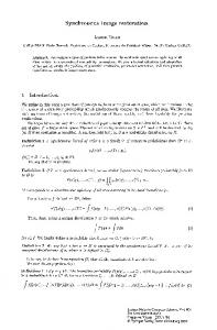
A total of 802 patients (481 men and 321 women, mean age 65.4 ± 10.5) with 802 colonic polyps >20 mm were included in the analysis. Large flat polyps were removed in 582 cases by complete colonoscopy, in 119 cases by incomplete colonoscopy and in 101 cases by sigmoidoscopy (Fig. 1). Mean size of all large polyps was 34.1 mm (range 20–150 mm, standard deviation 16.1 mm). For all removed large polyps, tissue histopathology was retrieved. The most frequent histological subtypes were adenoma with low-grade intraepithelial dysplasia (LGD) with n = 421 (52.5 %), followed by high-grade intraepithelial dysplasia (HGD) with n = 214 (26.7 %), adenocarcinoma (both invasive and carcinoma in situ) with n = 90 (11.2 %) and serrated polyps (SP) with n = 77 (9.6 %). The majority of serrated polyps were hyperplastic polyps or sessile serrated adenomas/polyps with no or low grade dysplasia. Nonserrated adenomas included 254 tubular, 372 tubulovillous and 9 villous adenoma. Large flat polyps were localized in 60.7 % (n = 487) in the proximal colon and in 39.3 % (n = 315) in the distal colon (see Table 1). Adenocarcinomas in large flat polyps were predominantly stage T1 cancer (Additional file 1: Table S1).
Page 3 of 8
Impact of endoscopic and demographic features on histology of large polyps
In order to identify factors that are associated with the occurrence of specific histological subtypes, we analyzed the following parameters: localization and size of flat polyp, patient age and sex. We found no difference in distribution of histological subtypes (p = 0.97) between different sex (Table 2). Large polyps containing SP were in average smaller than polyps with other histological subtypes (p < 0.0001). In addition, there was a trend towards HGD/adenocarcinoma with increase of polyp size in non-serrated adenomas (significant JT-test, see Table 2). The mean age of patients with SP was lower compared to patients with other histology (59.4 vs 66.1, p < 0.0001). We observed a distinct anatomic distribution for polyps with specific histological subtypes. Serrated polyps were predominantly found in the proximal colon (80.5 % of all SP) while adenocarcinomas were preferentially localized in the distal colon (72.2 % of all adenocarcinomas). For LGD or HGD, there was no preference for a specific location within the colon. However, villous adenomas were mainly detected in the distal colon (67 % of all villous adenomas) and tubular adenomas in the proximal colon (74 % of all tubular adenomas) (Additional file 2: Table S2). Localization of polyps also influenced mean size of polyps, with the highest average polyp size in the rectum compared to other sites (44.2 mm vs. 31.2 mm, p < 0.0001). General characteristics of synchronous polyps
Data from complete colonoscopies were available from 582 patients, allowing for a characterization of synchronous polyps. Synchronous polyps were detected in 391 patients undergoing complete colonoscopies (67.2 %). Of those, histological assessment was available in 355 and location described in 378 cases. A total of 1487 synchronous
Fig. 1 Flow chart showing basic distribution of patients and endoscopic procedures
Zhan et al. BMC Gastroenterology (2015) 15:82
Page 4 of 8
Table 1 Baseline characteristics of patients and large flat polyps Characteristic
Number of patients
Number of patients
802
Age, mean ± SD (range), y
65.4 ± 10.5 (31–92)
Female, no. (%)
321 (40)
Polyp size, mean ± SD (range), mm
34.1 ± 16.1 (20–150)
Polyp location, no. (% of total) Caecum
187 (23.3)
Ascending colon
150 (18.7)
Hepatic flexure
71 (8.9)
Transverse colon
79 (9.9)
Splenic flexure
13 (1.6)
Descending colon
40 (5.0)
Sigmoid colon
82 (10.2)
Rectum
180 (22.4)
Histology, no. (% of total) Tubular adenoma
254 (31.7)
Tubulovillous adenoma
372 (46.4)
Villous adenoma
9 (1.1)
Adenocarcinoma
90 (11.2)
LGD (without SP with LGD)
421 (52.5)
HGD (without SP with HGD)
214 (26.7)
Serrated polyps
77 (9.6)
Hyperplastic polyps
32 (4)
Sessile serrated adenoma/polyp (SSA/P) without dysplasia
32 (4)
Sessile serrated adenoma/polyp (SSA/P) with LGD
11 (1.37)
Sessile serrated adenoma/polyp (SSA/P) with HGD
2 (0.2)
polyps were removed. The mean number of polyps of patients with synchronous polyps was 3.8 (range 1–40). We observed a sex specific difference in the number of detected synchronous polyps. The mean number of polyps was slightly, but significantly higher for male than female (mean 2.0 vs. 2.9, p = 0.0029). The total proportion of male patients with at least one synchronous polyp was also higher (71 % vs 61 %, p = 0.01). The majority of patients
were found to have synchronous polyps at multiple sites within the colon (222 of 378 cases) and there was a general trend toward occurrence in the proximal colon (see Table 3). Interestingly, patients with any synchronous polyps in the rectosigmoid colon had a higher overall polyp burden compared to those with synchronous polyps elsewhere in the colon (mean 3.2 vs. 4.6, p < 0.0001). In the majority of cases (243 of 355 colonoscopies), removed synchronous polyps consisted of multiple histological subtypes. Adenocarcinoma was found in 6 %, HGD in 13.8 %, LGD in 85.4 % and SP in 28.2 % of all patients with histologically assessed synchronous polyps (Table 3). The histology of synchronous polyps was associated with overall polyp load, as patients with HGD in any synchronous polyp had a higher average number of polyps (SP: 2.5 vs. HGD: 6.8, p < 0.0001). Factors associated with occurrence of high-grade dysplasia and adenocarcinoma
Based on our data, we sought to identify factors that were associated with the occurrence of adenoma with HGD/adenocarcinoma, for both large flat polyps and synchronous polyps. By multivariate logistic regression analysis based on all patients with synchronous polyps, we found that increase in polyp size (OR 1.29, 95 % CI 1.09–1.55, per 10 mm increase, p = 0.0041), location of the large polyp in the rectosigmoid colon (OR 3.89, 95 % CI 2.26–6.79, p < 0.0001) and increase in age (OR 1.13, 95 % CI 1.00–1.29, per 5 year increase, p = 0.0471) were independently associated with presence of HGD/adenocarcinoma in large polyps. In contrast, patient sex, the location and the number of synchronous polyps had no significant effect on histology of the large polyp (Table 4). Biopsies of polyps prior to EMR did not detect the presence of adenocarcinoma in most cases (see Additional file 1: Table S1). We also analyzed parameters potentially associated with occurrence of HGD/adenocarcinoma in synchronous polyps (Table 5). Location of any synchronous polyp in the rectosigmoid colon (OR 2.65, 95 % CI 1.44–5.0, p = 0.002) and a high number of synchronous polyps
Table 2 Characteristics of large flat polyps for histological subtypes Histology (total = 802) p value
Variable
SP
LGD
HGD
adenocarcinoma
Patients, no. (% of total)
77 (9.6)
421 (52.5)
214 (26.7)
90 (11.2)
Female, no. (% of group with specific histology)
29 (37.7)
171 (40.6)
86 (40.2)
35 (38.9)
0.97
Age, mean ± SD (range), y
59.4 ± 11 (41–87)
65.6 ± 10.3 (31–92)
67 ± 9.6 (38–92)
65.7 ± 11.6 (41–91)