May 12, 1987 - In contrast to most inbred strains, P mice fail to develop significant resistance to Schistosoma mansoni infection as a result of vaccination with ...
Vol. 55, No. 8
INFECTION AND IMMUNITY, Aug. 1987, p. 1884-1889
0019-9567/87/081884-06$02.00/0 Copyright © 1987, American Society for Microbiology
Genetic Association of Defects in Macrophage Larvicidal Activity and Vaccine-Induced Resistance to Schistosoma mansoni in P Strain Mice STEPHANIE L.
JAMES,l'2* RODRIGO CORREA-OLIVEIRA,3t
ALAN SHER,3 LISA A. MEDVITZ,1 R. DALE McCALL4 Departments of Medicine' and Microbiology,2 George Washington University Medical Center, Washington, D.C. 20037; Laboratory of Parasitic Diseases, National Institute of Allergy and Infectious Diseases, Bethesda, Maryland 208923; and Institute of Marine Biomedical Research, University of North Carolina, Wilmington, North Carolina 284034 AND
Received 20 February 1987/Accepted 12 May 1987
In contrast to most inbred strains, P mice fail to develop significant resistance to Schistosoma mansoni infection as a result of vaccination with irradiated cercariae. Vaccinated P mice also exhibit a defect in macrophage activation for killing of larval schistosomes upon specific-antigen challenge in vivo. To examine the genetic basis of these defects in vaccine-induced immunity, inheritance of the two traits was examined in (C57BL/6 x P)F1, F2, and reciprocal backcross generations. The defect in macrophage function which characterizes the P strain parent was found to be inherited in a fully recessive manner and to be controlled by only one or two major genetic loci. Moreover, a highly significant correlation (P < 0.0025) was observed between the level of macrophage larvicidal activity and the level of resistance to challenge infection in segregating generations. Such an association is consistent with a cause-and-effect relationship, providing strong in vivo evidence implicating activated macrophages as immune effector cells of resistance to S. mansoni in the mouse-irradiated-vaccine model.
well as other cell-mediated immune responses (including lymphocyte proliferation, cytotoxic T-cell function, interleukin production, and macrophage antigen presentation), were quantitatively indistinguishable from those of the high responder B6 strain (4, 9). Identification of a mouse strain that does not respond to vaccination against S. mansoni has provided the opportunity for genetic analysis of the relationship between various antischistosome immune responses and resistance in vivo. Examination of the segregation of resistance and IgM antibody level in B6 x P crosses has shown that the P-associated defect in each of these responses is inherited in a recessive manner and is predominantly controlled at a single major genetic locus (2). However, the loci controlling IgM antischistosomulum antibody production and vaccine-induced resistance are unlinked (2), indicating that the humoral immune defect is not responsible for the lack of vaccine-induced resistance in P mice. This conclusion was substantiated by the observation that xid mice, which fail to produce IgM antilarval antibodies, are significantly protected by vaccination (3). In the present study, we analyzed inheritance of the P-associated defect in macrophage activation in B6 x P crosses and tested for association with protective immunity in vaccinated progeny to assess the possible role of activated macrophages as effector cells of resistance to S. mansoni in
It has been reasoned that definition of immune mechanisms participating in resistance to the helminth parasite Schistosoma mansoni in animal models of protective immunity will simplify development of a defined vaccine useful against human schistosomiasis. Although a number of immunologic effector mechanisms have been shown to damage larval schistosomes (schistosomula) in vitro (reviewed in reference 19), the participation of any of these mechanisms in acquired immunity in vivo has been difficult to assess. Indeed, in some cases it has been shown that induction of elements capable of killing schistosomula in vitro (e.g., lethal antibody and complement) is not sufficient to protect animals against S. mansoni infection in vivo (18). For this reason, we have attempted to define protective antischistosomal immune responses in vivo in a well-characterized model using mice vaccinated with radiation-attenuated parasites. Most inbred mouse strains develop moderate to high levels of resistance to challenge infection as a result of such vaccination. However, P/N-J (P) mice fail to display significant protection even after multiple immunizations (4, 11). When compared with C57BL/6 (B6) mice, a strain that develops high levels of vaccine-induced resistance (11), vaccinated P mice were found to be defective in schistosome-specific, cell-mediated immune responses, including delayed hypersensitivity, production of macrophageactivating lymphokine, and macrophage activation for direct cytotoxicity toward schistosomula or tumor cell targets (8). In addition, P mice were also defective in their immunoglobulin M (IgM) antibody responses to schistosomulum surface antigens (4). These defects were relatively specific since levels of P mouse antibodies belonging to other isotypes, as *
vivo.
MATERIALS AND METHODS Parasites and antigens. Cercariae of S. mansoni (NMRI strain) were obtained from infected Biomphalaria glabrata snails purchased from the Biomedical Research Institute (Rockville, Md.). Schistosomula were prepared from cercariae by mechanical transformation and gradient purification as previously described (8). A soluble adult worm
Corresponding author.
t Present address: Centro de Pesquisas Rene Rachou, 30,000 Belo
Horizonte, Minas Gerais, Brazil. 1884
VOL. 55, 1987
RESISTANCE TO S. MANSONI LINKED TO MACROPHAGE ACTIVITY
antigenic preparation (SWAP) was made with the salinesoluble constituents of homogenized adult parasites (8). Mice and genetic crosses. Inbred P and B6 mice were obtained from the Animal Genetics and Production Branch, National Cancer Institute, Frederick, Md. All experiments were initiated when animals were 6 to 9 weeks old. Genetic crosses were performed by using females and males at a ratio of 4:1 as previously described (2). Vaccination. Mice were vaccinated by percutaneous exposure of tail skin to approximately 500 cercariae that were attenuated by prior exposure to 50 kR of gamma radiation from a 'Co source (2, 4, 8, 9, 11). Assay of macrophage-activating lymphokine. Lymphokinecontaining supernatant fluids were prepared by in vitro stimulation of splenocytes (107/ml) from vaccinated or control mice with SWAP (500 ,ug/ml) for 48 h (8, 9). Macrophage-activating factor in these fluids was measured as previously described (8, 9) by examining the ability of three-fold dilutions of lymphokine to stimulate in vitro schistosomulum killing by sodium caseinate-elicited peritoneal macrophages from normal C3H/HeN mice in suspension cultures. Quantitaion of macrophage activation in vivo. Peritoneal cells were collected from vaccinated mice or unimmunized controls that were injected intraperitoneally (i.p.) 16 to 20 h previously with 1 ml of phosphate-buffered saline (PBS) containing 250 ,ug of SWAP or with PBS alone. Cells were harvested by peritoneal lavage and counted as previously described (8). Plastic adherent monolayers were prepared by adding 8 x 10⁵ macrophages to 16-mm (diameter) culture wells (Cluster24; Costar, Cambridge, Mass.) in Dulbecco modified Eagle medium (GIBCO Laboratories, Grand Island, N.Y.) containing 10% heat-inactivated fetal bovine sera (Hyclone, Logan, Utah), 2 mM 1-glutamine, and 50 jig of gentamicin per ml. Nonadherent cells were removed by washing after 2 h of incubation. Activated macrophages can kill schistosomula directly; however, antibody opsonization enhances larval killing by activated but not inactive cells (8). Therefore, to maximize the difference in larvicidal activity of cells from high- and low-responder mouse strains in these experiments, the larvae were opsonized by prior exposure to antibody-containing sera from infected mice as previously described (8). Schistosomula were added at 80 per well. After 48 h of incubation at 37°C, macrophage-mediated larval killing was evaluated microscopically by the criteria of internal granularity, loss of internal structure, and motility (8). Cytotoxicity was expressed as the mean percentage of dead larvae in duplicate cultures. In this and previous studies (8), SWAP was inactive in normal animals and PBS was inactive in immunized mice, indicating that elicitation of activated macrophages by SWAP in vivo is an immune response to specific-antigen recognition. In experiments in which the animals were kept alive during macrophage collection for future measurement of resistance to challenge infection, mice were lightly anesthetized with Metofane (2,2-dichloro-1,1-difluoroethyl methyl ether; Pitman-Moore Inc., Washington Crossing, N.J.) before peritoneal lavage with 5 to 10 ml of Dulbecco modified Eagle medium-2% fetal bovine serum-5 U of heparin per ml (The Upjohn Co., Kalamazoo, Mich.). Quantitation of resistance. Animals were allowed 2 days to recover from macrophage collection before challenge infection. Shaved abdominal skin was exposed to about 150 cercariae for 20 min. Recovery of challenge parasites was assayed 6 weeks later by perfusion of the hepatic portal system (2, 20). Resistance was measured as the percent
1885
TABLE 1. Comparison of macrophage larvicidal activities in vaccinated B6, P, and (B6 x P)F1 mice Micea
Larvicidal
activityb
B6 Control ....... Vaccinated .......
11 + 3 34 ± 6'
p
Control ....... Vaccinated ........
11 ± 6
Control ...... Vaccinated ......
11 ± 8 41 ± 12e
11
6d
F1 a Peritoneal macrophages were elicited by i.p. injection of SWAP in control mice or mice vaccinated with irradiated cercariae. b The ability of macrophages in monolayer culture to kill opsonized schistosomula in vitro was analyzed by microscopic evaluation of larval motility and internal structure after 48 h. Values represent the mean ± the standard deviation for four to seven experiments. Statistical significance was defined by Student's t test. C p < 0.001. d p > 0.05 (no significant difference). P < 0.002.
reduction in worm burden calculated according to the equation [1 - (number of worms recovered from vaccinated mice/number recovered from control mice)] x 100. Statistical analyses. Comparisons of differences in macrophage larvicidal activity or resistance between control and immunized groups were performed with Student's two-tailed t test. Because macrophage larvicidal activity was
continuously
distributed in segregating generations, the pattern of inheritance was assessed by a powerful and general maximumlikelihood procedure specifically designed for data of this type (5). The version of computer-driven statistical subroutine MAXLIK used (5) tests the fit of the data to 11 different models, including models of inheritance of one or two simple Mendelian elements and influence of one or two major loci, and polygenic models (2). Association of the two quantitative variables macrophage larvicidal activity and worm recovery was tested by Spearman's rank correlation, a nonparametric analysis. RESULTS Recessive inheritance of the P mouse defect in macrophage larvicidal activity. Analysis of cytotoxic function in (B6 x P)F1 hybrid mice showed that the defective macrophage response of P mice is inherited as a recessive trait (Table 1). As previously reported (8), SWAP-elicited macrophages from vaccinated responder strain animals showed statistically significant larvicidal activity (approximately 300% of background). Peritoneal macrophages from vaccinated F1 mice reacted as did those from the B6 parent, whereas antigen-elicited cells from vaccinated P mice were inactive under all conditions tested. All nonimmunized control mice were unresponsive to SWAP stimulation. Since previous experiments had shown that the Passociated defect in macrophage activation was at least in part due to low production of macrophage-activating lymphokine (1, 8), the abilities of supernatant fluids from coculture of parental or F1 lymphocytes with SWAP to stimulate control macrophages to kill schistosomula were
1886
INFECT. IMMUN.
JAMES ET AL. EXP.1
EXP.3
EXP.2
7c
sa
0
-J
401 -((00%) (41 %)
-C
30j
* (92%) (44%)
*(75%) . (60%)
-~~~~~~~~
z
20
w
*
*(40%)
.l(59%)
a.
.! (56%) *
10
* I
B6
FlxS6 F1xP
Fl
P
B6
Fl
F1xB6 F1xP
P
B6
FlxB6 FlxP
Fl
P
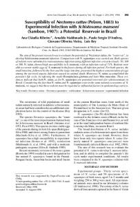
FIG. 1. Scatter diagram of macrophage larvicidal activity in vaccinated B6, (B6 x P)F1, and P parental mice and reciprocal backcross progeny. The line dividing the two parental distributions was estimated by calculating the average of the means of activity in the high and low responder parental groups. Only one mouse in a parental group fell outside this range (Exp. 2).
compared. The titer of lymphokine produced by cells from immunized P mice was a 1:54 dilution (15 + 8% larval killing versus 11 ± 6% with supernatants from control cultures of normal splenocytes with SWAP). In contrast, lymphokines produced by immune B6 and F1 cells remained strongly reactive at the equivalent titer (99 + 1% and 98 ± 1% killing, respectively) and approached background levels only at ninefold higher dilutions (1:486). Similar results were observed when these lymphokines were tested in a tumoricidal assay (data not shown). These observations substantiate the immune basis of in vivo macrophage activation by SWAP. Male and female F1 hybrid mice produced by reciprocal matings of male and female B6 and P mice were indistinguishable in terms of antigen-specific lymphokine production as well as macrophage activation. These results confirm that the P-associated cellular defects are fully recessive and under autosomal control. Genetic control of macrophage activation defects in P mice. To estimate the number of genes controlling the P mouse EXP. 2
EXP.1
EXP~3
defect in macrophage larvicidal function, reciprocal backcross and F2 progeny were bred, vaccinated, and assayed for SWAP-elicited macrophage activation (Fig. 1 and 2). Whereas the macrophage activities of the parental strains fell into two sepairate groups (high larval killing by cells from B6 and F1 animals and low activity by cells from P mice), the reactivity of cells from the backcross and F2 generations was continuously distributed over the entire range of the parental response. By constructing approximate dividing lines based on parental distribution, it was possible to designate each of the progeny provisionally into high- and low-responder groups. The resulting distributions were in close agreement with those predicted for control of macrophage function by a single locus (Table 2). In total, 28 of 31 F1 x B6 mice tested (90%), 30 of 62 F1 x P mice tested (48%), and 45 of 60 F2 mice tested (75%) responded similarly to the B6 or F1 high-responder parent within the respective experiments. Because the trait was continuously distributed in backcross and F2 individuals, designation of progeny iulto either parental type based on approximation of the parental range introduced a potential source of error. Therefore, the backcross data were also analyzed by the maximum-likelihood procedure, which is specifically designed for data of this type (5, 14). The macrophage activation patterns in the F1 x P
¢ 50