Annotating PDFs for eReturn version 1.0; August 28, 2007; full release
1. Introduction eProof files are self-contained PDF documents for viewing on-screen and for printing. They contain all appropriate formatting and fonts to ensure correct rendering on-screen and when printing hardcopy. DJS sends eProofs that can be viewed, annotated, and printed using the free version of Acrobat Reader 7 (or greater). These eProofs are “enabled” with commenting rights, therefore they can be modified by using special markup tools in Acrobat Reader that are not normally available unless using the Standard or Professional version. The screen images in this document were captured on a PC running Adobe Acrobat Reader version 8.1.0. Though some of the images may differ in appearance from your platform/version, basic functionality remains similar. At the time of this writing, Acrobat Reader v8.1.0 is freely available and can be downloaded from: http://www.adobe.com/products/acrobat/readstep2.html
2. Comment & Markup toolbar functionality
A
B
C
D
E
G
F
H
A. Sticky Note tool; B. Text Edits tool; C. Stamp tool; D. Highlight Text tool; E. Callout tool; F. Text Box tool; G. Various Object tools; H. Pencil tool
A. Show the Comment & Markup toolbar The Comment & Markup toolbar doesn’t appear by default. Do one of the following: • Select View > Toolbars > Comment & Markup. • Select Tools > Comment & Markup > Show Comment & Markup Toolbar. • Click the Review & Comment button in the Task toolbar, and choose Show Comment & Markup Toolbar. To add or remove tools for this toolbar, right-click the toolbar and select the tool. Or, select Tools > Customize Toolbars.
B. Select a commenting or markup tool Do one of the following: • Select a tool from the Comment & Markup toolbar. • Select Tools > Comment & Markup > [tool]. Note: After an initial comment is made, the tool changes back to the Select tool so that the comment can be moved, resized, or edited. (The Pencil, Highlight Text, and Line tools stay selected.)
C. Keep a commenting tool selected Multiple comments can be added without reselecting the tool. Select the tool to use (but don’t use it yet). Choose Tools > Customize Toolbars to remove unnecessary items from the toolbar (see Section 7 for suggested toolbar layout)
• Select View > Toolbars > Properties Bar. • Select Keep Tool Selected.
1
3. The Properties bar The Properties bar can be used to format text and select options for individual tools. To view the Properties bar, do one of the following: • Choose View > Toolbars > Properties Bar. • Right-click the toolbar area; choose Properties Bar. • Select [Ctrl-E]
4. Using the comment and markup tools To insert, delete, or replace text, use the Text Edits tool. Select the Text Edits tool, then select the text with the cursor (or simply position it) and begin typing. A pop-up note will appear based upon the modification (e.g., inserted text, replacement text, etc.). Use the Properties bar to format text in pop-up notes. A pop-up note can be minimized by selecting the button inside it.
A
B
C
D
E
A. Attached file; B. Highlighted text; C. Crossed-out (strike-through) text; D. Inserted text; E. Replaced text 2
5. Inserting symbols or special characters An ‘insert symbol’ feature is not available for annotations, and copying/pasting symbols or non-keyboard characters from Microsoft Word does not always work. Use angle brackets < > to indicate these special characters (e.g., , ).
6. Editing near watermarks and hyperlinked text eProof documents often contain watermarks and/or hyperlinked text. Selecting characters near these items can be difficult using the mouse alone. To edit an eProof which contains text in these areas, do the following: • Without selecting the watermark or hyperlink, place the cursor near the area for editing. • Use the arrow keys to move the cursor beside the text to be edited. • Hold down the shift key while simultaneously using arrow keys to select the block of text, if necessary. • Insert, replace, or delete text, as needed.
7. Summary of main functions Insert text - Use Text Edits tool (position cursor and begin typing) Replace text - Use Text Edits tool (select text and begin typing) Delete text - Use Text Edits tool (select text and press delete key) Highlight text - Use Highlight Text tool (select text) Attach a file - Use the Attach a File with Comment tool (select tool, position cursor and click mouse, select file)
Suggested toolbar layout
8. Reviewing changes To review all changes, do the following: • Select the Show button on the Comment & Markup toolbar. • Select Show Comments List. Note: Selecting a correction in the list will highlight the corresponding item in the document, and vice versa.
Use the Comments list to review all changes
9. The eReturn process A. An email is received that contains a link to the eProof of the article: http://eproofing.dartmouthjournals.com/pdfproofing/journal1234.pdf B. Click on the link to open the proof with the internet browser. Select “Save As” from the browser’s ‘File’ menu to save a copy of the PDF to the desktop or other folder. C. Close the browser and open the saved PDF file with Acrobat. D. Make corrections using Acrobat’s Comment & Markup tools. E. Save the PDF file, now with annotations, and return according to the instructions provided by the DJS journal manager. 3
NUMBER
OF
AUTHOR QUERIES DATE 8/27/2011 JOB NAME TAS ARTICLE 1100207 QUERIES FOR AUTHORS
Brief Reports
THIS QUERY FORM MUST BE RETURNED WITH ALL PROOFS; HOWEVER, PLEASE MARK YOUR CORRECTIONS DIRECTLY ONTO THE PROOFS, NOT ONTO THIS SHEET.
AU1: Are the changes to this sentence ok? ‘This represents a ‘‘Pandora’s Box’’ for the enormous typology of possible symptoms for which etiology is not ever easily found.’’ AU2: Are the changes to this sentence ok? ‘‘This particular site of metastasis is probably favored by the complex arterial, venous, and lymphatic drainage of the region that consists of many anastomotic branches with abdominal vessels, deriving from fetal circulation.’’
Brief Reports Brief Reports should be submitted online to www.editorialmanager.com/ amsurg. (See details online under ‘‘Instructions for Authors’’.) They should be no more than 4 double-spaced pages with no Abstract or sub-headings, with a maximum of four (4) references. If figures are included, they should be limited to two (2). The cost of printing color figures is the responsibility of the author. In general, authors of case reports should use the Brief Report format.
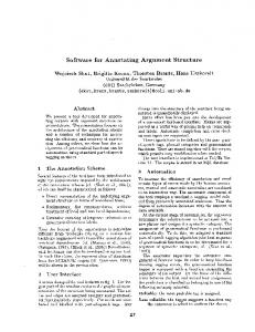
Umbilical Node as First Clinical Appearance in a Patient with a High Tumor Burden
AU1
F1
disappearance of abdominal pain and reduction of umbilical metastasis. The lymphatic metastasis of the umbilical region is also named Sister Mary Joseph’s nodule. It is the first appearance of cancer in 40 per cent of cases and the only manifestation of disease in 60 per cent of these. Generally, the primary tumors originate from the stomach (30% in men and 9% in women), rectum, colon, and small bowel (25% in man and 12% in women), or from pancreatic carcinoma (10%). In women, the first site for the primary tumor is the ovary (34%), whereas umbilical metastases have an unknown origin in 11 per cent of cases.3 This particular site of metastasis is probably favored by the complex arterial, venous, and lymphatic drainage of the region that consists of many anastomotic branches with abdominal vessels, deriving from fetal circulation. Umbilical metastasis usually may be part of an advanced metastatic disease, and it is related to a poor prognosis, with an average survival of 11 months. Fine-needle aspirate with cytological examination is recommended for the diagnosis of malignancy, because of its good sensitivity and positive-predictive value (92.8% and 100%, respectively).4 However, fine-needle aspirate is not always able to diagnose the primary tumor and a careful radiological examination after the clinical appearance of umbilical lesion could help to detect the primary site and to orient both the appropriate diagnostic procedures and therapy.
The term ‘‘abdominal pain’’ is generally used to describe pain originating from organs within the abdominal cavity. This represents a ‘‘Pandora’s Box’’ for the enormous typology of possible symptoms for which etiology is not ever easily found. Moreover, a nodule originating from the umbilical region may represent a sign of various diseases such as benign dermatologic conditions, including melanocytic nevi, epithelial inclusion cysts, epidermoid cysts, omphaliths, keloids, pyogenic granulomas, and pilonidal sinuses.1 Primary neoplasms of the umbilicus account for 38 per cent of nodules; endometriosis accounts for 32 per cent; and metastatic disease from a distant site accounts for 30 per cent of umbilical nodules.2 A 58-year-old woman reported mild abdominal pain, which started 4 months before presentation, and the appearance of a red not-ulcerating cutaneous nodule in the umbilical region (Fig. 1). Patient did not refer any history of abdominal or cutaneous diseases. The abdomen ultrasonography revealed the presence of multiple liver lesions suspected for metastasis. A subsequent computed tomographic scan showed a large lesion originating from the transverse colon with multiple peritoneal metastases and a mass through the umbilicus (Fig. 1). Multiple pulmonary and mediastinal metastases were also described. Colonoscopy confirmed the presence of a large lesion, which was biopsied and the histological examination was diagnostic for high-grade adenocarcinoma. Considering the type and the stage of disease, patient started a first line poly-chemotherapy with 5-fluorouracil, oxaliplatin, and bevacizumab. To date, she is presently under chemotherapy 7 months from diagnosis and has obtained a significant clinical benefit characterized by Address correspondence and reprint requests to Roberto Iacovelli, M.D., Sapienza University of Rome, Department of Radiology, Oncology, and Human Pathology, Viale regina Elena 324, 00161 Rome, Italy. E-mail:
[email protected].
FIG. 1. Clinical and radiological signs of the Sister Mary Joseph’s nodule.
1
AU2
2
THE AMERICAN SURGEON
Roberto Iacovelli, M.D. Patrizia Trenta, M.D. Alessandro Tuzi, M.D. Antonella Palazzo, M.D. Enrico Cortesi, M.D., Prof Sapienza University of Rome Department of Radiology, Oncology, and Human Pathology Rome, Italy
November 2011
Vol. 77
REFERENCES
1. Cohen DC. A man with an umbilical ulcer. Medscape J Med 2008;10:11. 2. Barrow MV. Metastatic tumors of the umbilicus. J Chronic Dis 1966;19:1113–7. 3. Dubreuil A, Dompmartin A, Barjot P, et al. Umbilical metastasis or Sister Mary Joseph’s nodule. Int J Dermatol 1998;37: 7–13. 4. Edoute Y, Malberger E, Kuten A. Umbilical metastasis diagnosed by fine needle aspiration. J Surg Oncol 1990;45: 56–8.