acidic polymer (Rang, 2011) which is highly polar. Positively charged ionic ... ological structure of Gram negative bacterium (Rang et al, 2011). Ophthalmic ..... Pharmacology an applied approach for ... (2011) Rang and Dales. Pharmacology ...
Clinical
Antibiotics in ophthalmology Abstract Antibiotics, in any practice, are a signiicant asset to clinically effective treatments. This paper presents is a clinical overview of antibiotic agents which are commonly used in ophthalmology. It seeks to advise on how particular agents are identiied, grouped, and how they are chosen so that they may affect speciic organisms. The aim of this article, is to refresh the reader’s knowledge with regards to common pathogens, how they are identiied and how their patients may be appropriately treated in practice.
T
his paper presents an overview of the types of antibiotics commonly utilized on certain clinical presentations in the ophthalmic setting. It identiies some common pathogens found in the eye and how they might be identiied in cellular physiological sense, so that nurses gain an awareness of the underlying physiological processes which lead to effective treatments. It also highlights clinical considerations when treating patients with antibiotic agents and concludes with the requirement to enhance patient concordance with treatment.
Antibiotics overview
Amanda Sherratt RGN, RM, RNP. MSc NP, BhSC (Hons) PGDip ANP, Dip Ed. Cert ANP. Lecturer, Hull University Department of Health Professional Studies. Member of Expert Advisory Group MHRA Pharmacovigilance, External Examiner University of Dundee Advanced Practice Programs
204
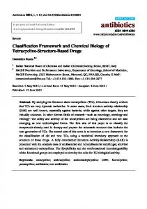
The term ‘antibiotic’ is often utilized to encompass descriptors for antimicrobial, antibacterial and antifungal agents. They are used to affect or prevent live microbes, such as bacteria, fungi or protozoa from proliferation. They can work in two different ways, bacteriostatic or bactericidal. Bacteriostatic agents inhibit growth of a bacterium thus producing a systemic effect whereby the individual humeral disease mechanism can work to complete the process of destroying the bacterium. Bactericidal antibiotics work to destroy susceptible bacteria. They break down peptidoglycan in the cell walls so that the cell becomes fragile, cellular apoptosis occurs through subsequent ‘bursting’ of the bacterial cells. All bacteriostatic and bactericidal agents work in a number of ways which are identiied in Figure 1.
Gram staining On laboratory examination, bacteria are subjected to a ‘Gram stain’. This reveals the bacterial cellular wall typology. Gram positive bacterium are able to retain the Gram stain, as they have a thick layer of peptidoglycan. Peptidoglycan allows certain antibiotics access to penetrate and destroy the cell wall. The remaining
40–50% of a Gram positive bacteria consists of an acidic polymer (Rang, 2011) which is highly polar. Positively charged ionic antibiotics, such as streptomycin, are therefore very effective in cellular penetration of Gram positive bacterium (Joint Formulary Committee, 2011). Gram negative bacterium have a more complex cell wall structure. An outer layer of lipopolysaccharides, with a thin inner layer of peptidoglycan, differ with each bacterium (Rang et al, 2011). The complex layers mean certain antibiotics are extremely ineffective for Gram negative bacterial strains. Benzylpenicilin, vancomycin and methicillin are a few of the identiied agents which have reduced effects due to the physiological structure of Gram negative bacterium (Rang et al, 2011).
Ophthalmic considerations The physiology and anatomy of the eye provides a number of protective barriers to infective agents (Zhang et al, 2005). A variety of ophthalmic infections occur across the structures, but the corneal epithelium acts as both a barrier and inlammatory responder to potential pathogens (Zhang et al, 2008). The host’s usual physiological cellular response with the release of pro-inlammatory cytokines, acts to contain the infection. However, subsequent inlammation can also predispose further pain and potential damage to the fragile surrounding structures (Wilson et al, 2001; Narayanan et al, 2005). Often, neoadjuvant anti-inlammatory agents (such as steroids) are used to treat ocular
Inhibit cell wall synthesis • β lactam antibiotics (Penicillins) Cephallosporins Inhibit protein synthesis • Fluoroquinolines • Chloramphenicol • Erythromycin Inhibit nucleic acid synthesis • Aminoglycosides • Quinolines Inhibit metabolites which are essential to growth • Trimethoprim Folate synthesis • Sulfonamides Figure 1. Effects of antibiotics.
Vol 3 No 6 • December/January 2012/2013 • International Journal of Ophthalmic Practice
Clinical infections (Zhang et al, 2008). Some broad spectrum antibiotics, which are no longer used for systemic treatments due to their toxic effects, can be used in the eye. The eye provides a local site, which greatly reduces risks of systemic toxicity. In addition to this, the localized nature of instillation also reduces the potential for multi-resistant bacterial strains developing resultant from over prescribing of certain antibacterials.
Pathogens and treatments Common pathogens which affect the eye include Gram positive bacteria such as staphlycoccus, streptococcus and pneumococcus. Staphylococcal and streptococcal bacteria are responsive to the ß lactam antibiotic penicillin (Rang et al, 2011). The bacterial cell wall is destroyed through transpeptidation enzyme which attaches to the peptidoglycan of the cell walls, causing breakdown of the walls and subsequent lysis of the bacterium (Tarabishy, 2008). This action means that it is often beneicial to administer ß lactams in timed intervals (i.e. 4–6 hourly doses), so that each dose is able to act on the bacterium as the bacterium proliferate (Joint Formulary Committee, 2011). Following oral ingestion, elimination is through the renal tract and is very rapid. 90% of the drug is excreted through tubular secretion (Rang et al, 2011). ß lactam antibiotics are lipid insoluble, so do not enter human cells, but can distribute across all bodily luids. This means they have very limited systemic effects when administered via ocular route, unless there is a predisposing allergy to penicillin (Galbraith et al, 2004). Streptococcus aureus, Haemophyllus inluenzae, Neisseria gonorrhoea, Proteus and Klebsiella normally cause an inlammatory response which is self limiting, and therefore, antibiotic therapies are not always indicated in these patients (Joint Formulary Committee, 2011; Sherratt and Needham, 2012). In some cases, gonococcal conjunctivitis may be treated with chloramphenicol, or erythromycin (Sherratt and Needham, 2012), with adjuvant systemic therapies (Tarabishy, 2008). Chloramphenicol works at genetic level. It works on the 50s ribosome of DNA to prevent synthesis of bacterial proteins. It is a wide spectrum antibiotic (Joint Formulary Committee, 2011), which penetrates structures of the conjunctiva and cornea easily. There are side effects of local irritation, itching burning and hypersensitivity (Joint Formulary Committee, 2011). With systemic chloramphenicol, there is a risk of severe idiosyncratic depression of the bone marrow, pancytopenia and resultant anaemia (Joint Formulary Committee, 2011; Rang et al, 2011). As there is a very small risk of systemic absorption, chloramphenicol is not recommended for use in children 206
(Joint Formulary Committee, 2011). As an alternative, fusidic acid is identiied, probably because it is only required as a twice daily dosage regimen, rather than three times daily (Lee, 2006). However, in children, most children presenting with bacterial conjunctivitis require no treatment (Rose et al, 2005). Gram negative bacteria are also responsible for a number of ocular infections. Pseudomonas aeruginosa is treated with aminoglycosides (gentamycin). Aminoglycosides inhibit nucleic acid synthesis but are dependent on oxygen to penetrate the cell wall. For this reason, the aminoglycosides have limited effect on anaerobic bacterium. They will not cross the vitreous humour of the eye. Elimination is renal, with 50–60% of the drug excreted unchanged within 24 hours in systemic treatment (Rang et al, 2011). In studies relating to systemic effects of aminoglycosides, they have been shown to be ototoxic and nephrotoxic. They are mainly excreted renally. Gentamycin and neomycin are commonly used aminoglycosides used in ophthalmology. They inhibit bacterial protein synthesis, but penetration depends on oxygendependent active transport across the cells. They are therefore only effective against aerobic bacterium. Ciproloxacin and oloxacin are luoroquinolines which are used to treat corneal ulceration and supericial bacterial infections. Topical quinolines are more cost effective than aminoglycosides, with comparable eficacy on trial (Robert and Adenis, 2001; Sherratt and Needham, 2012). However, it must be noted that the extremely regular application of eye drops within the initial stage of treatment, may predispose reduction in patient concordance. If patients to not conform to the required dosage regimens, then treatments will not be effective and outcomes will be impaired through non compliance. Blepharitis is often caused by staphylococci. Fusidic acid is a narrow spectrum antibiotic which again inhibits protein synthesis and is effective against Gram positive staphylococcal infections (Rang et al, 2011). It penetrates purulent sites and aqueous humour and is recommended for topical staphylococcal conjunctivitis (National Institute for Health and Clinical Excellence, 2011). Bacterial blepharitis may require tetracyclines for a long duration, with ointments applied to the conjunctival sac and lid margins (Joint Formulary Committee, 2011). The treatment will be adjunct to a course of systemic antibiotics. Acute bacterial conjunctivitis is usually self limiting, but gonococcal conjunctivitis is usually treated with topical and systemic antibacterials. Keratitis, and endophthalamus may be bacterial, viral or fungal (Henry et al, 2012). Corneal ulceration requires more specialist treatment and more
Vol 3 No 6 • December/January 2012/2013 • International Journal of Ophthalmic Practice
Clinical intensive therapy which will be speciic to the causative agent. Endophthalmitis is an emergency, often requiring intravitreal administration of antimicrobials as well as concomitant corticosteroids. In some cases, a vitrectomy may be required (Field and Tillotson, 2008).
Corneal infections The most common pathogen associated with corneal infections, is the rapidly aggressive Gram negative organism, P. aeruginosa (Ozkiris et al, 2005). The antibiotics which effect pseudomonal infections in ophthalmology are aminoglycosides gentamycin, tobramycin and the quinoloines (Joint National Formulary, 2011). Acanthomeabic keratitis is an increasing issue which is a threat to sight. It is a painful condition associated with contact lens wearers following cross contamination with acanthomoeba. Acanthamoeba is an amoeba which is found in stagnant water and the rise has been linked with multipurpose contact lens solutions (Jasim et al, 2012). Propamidine isotionate is a broad spectrum antimicrobial which is unlicensed, but available over the counter. It has been identiied as being useful in the reduction of microbes (Joint Formulary Committee, 2011). For chlamydial keratoconjunctivitis, oral sulphonamides or tetracyclines may be used. Bacteria require folate to synthesize DNA and RNA. Sulfonamides compete with bacterial enzymes to inhibit further growth and proliferation. They are bacteriostatic, rather than bactericidal. For children, oral erythromycin or azithromycin may instead be used as sulphonamides are contraindicated. Topical eye dropped sulphonamides may also be used as adjunct to the systemic antibiotics. It must be noted, however, that presence of pus, or products of tissue breakdown will artiicially feed the bacteria folates, so rendering sulphonamide activity ineffective. Although this article is related to antibiotics in ophthalmology, there are also a number of viral agents which are responsible for ocular infections.Viral herpes simplex can be treated with acyclovir, which interferes with viral DNA synthesis. Ganciclovir is used for Cytomegalovirus (CMV), a virus commonly found in patients who are already immunocompromised. Ganciclovir is implanted as slow release intraocular device to treat the issue. Ganciclovir is an analogue of guancystine. It competes with guanosine triphosphate for incorporation into the viral DNA, suppressing replication. There are a number of serious side effects, so this drug is usually only administered for sight threatening CMV infections when there is a degree of immunocompromisation. Foscarnet is the
second line choice for CMV eye infection. Subconjunctival injections are often utilized to instil anti-infectives where topical therapies have been trialled, but found to be ineffective. The water soluble drug diffuses through the cornea and sclera to anterior and posterior chambers and vitreous humour to take effect.
Application of antibiotics Eye drops are aqueous solutions which disperse across the eye rapidly. They are sterile in order to prevent microbial contamination, and come in packaging suited to each individual drug. Often these medicines may be affected by sunlight, oxidation, heat or hydrolysis, so correct packaging and storage is an essential component of prescribing and enabling concordance of an effective drug regimen (Joint National Formulary, 2011). All unused medications of this type must not be used beyond 4 weeks, (Joint National Formulary, 2011), but this depends on the drug in use. All medications are affected by breakdown of active components, and prolonged periods with an open bottle, may preclude the possibility that sterility of the solution will be compromised. Therefore, the potential for proliferation of pathogens within the bottle will arise. With eye drops, instillation advice is to gently pull down the lower eyelid and keep the eye closed for as long as possible after application. More than one drop should be avoided due to risk of increased systemic effects (Joint Formulary Committee, 2011). Systemic effects are caused through a number of factors. Absorption through conjunctival vessels may occur (Sherratt and Needham, 2012). Or when excess amounts of the preparation have drained down the tear ducts to nasal mucosa, especially with eye drops, systemic effects may be caused. Light pressure on the lacrimal punctum for at least a minute has been identiied as a procedure to prevent this occurrence (Joint Formulary Committee, 2011; Rang et al, 2011). Eye ointments on the other hand, are instilled in the same way, but blinking will assist in the transmission of the ointment across the eye. Ointments are useful as they are oil based, therefore, maintain contact with the lower fornix for a longer duration. The oil also makes the drug retain effectiveness for a longer duration. It is important to remember any instillation of medication to the eye may cause visual disturbances, such as blurring or stinging. However, this depends on which medication is used. All medications should be clearly understood, and patients should be informed of potential effects of any medical treatment. All ocular instillations should be
International Journal of Ophthalmic Practice • Vol 3 No 6 • December/January 2012/2013
207
Clinical eye, as the area is likely to become re-infected.
Key points
Conclusion
• Antibiotic is a term used to describe a number of agents which includes antimicrobial, antibacterial and antifungal agents. • It is essential to be aware of types of bacterium causing infection, so that speciic agents will effectively treat the clinical issue. • An understanding of bacteriostatic and bactericidal agents and how they work provides an insight into what we use in everyday clinical treatments within ophthalmology.
Antibiotics • Antimicrobials • Occular infection
preceded with a warning about driving or operating machinery during the period following instillation (Karpecki et al, 2010). Usually, disturbances in sight are short lived, and so the patient may undertake usual activities when they feel able to do so. In any ocular infection, an essential consideration is to always cleanse the eye carefully, prevent cross contamination through application of good hygiene techniques. It is advisable to mention to the patient that they should never cover, or pad their infected
This paper identiied some of the more common infections in ophthalmology. It did not address surgical considerations, or prophylaxis in ophthalmology. When considering any form of antibiotic treatment for any patient, it is essential to consider concordance, to highlight all treatment rationales, and ensure application will be continued for the duration of treatment. The main consideration for any prescriber or administrator of the medication, is that the treatment must be palatable and accessible to their patients. A sound understanding of how these agents work, underpins an evidence-based IJOP approach to practice. Conlict of interest statement: none.
References Field D, Tillotson J (2008) Eye Emergencies The Practitioners Guide. M&K Update, Keswick
British Medical Association and Royal Pharmaceutical Society of Great Britain, London
Galbraith A, Bullock S, Manias E, Hunt B, Richards A (2004) Fundamentals of Pharmacology an applied approach for nursing and health (2nd ed.) Pearson, Essex
Jasim H, Knox-Cartwright S, Cook D, Tole D (2012) Increase in acanthamoeba keratitis may be associated with use of multipurpose contact lens solution. Br Med J 344(e1246)
Henry CR, Flynn HW, Miller D, Forster R, Alfonso E (2012) Infectious Keratitis Progressing to Endophthalmitis : A 15-Year-Study of Microbiology, Associated Factors, and Clinical Outcomes Ophthalmology 2012. Accessed via http:// www.sciencedirect. com/science/article/pii/ S0161642012005611 04/10/2012
Karpecki P, Paterno M, Comstock T (2010) Limitations of Current Antibiotics for the Treatment of Bacterial Conjunctivitis. Optom Vis Sci 87(11): 908–19
Joint Formulary Committee (2011) British National Formulary. 62nd Edition.
208
Lee A (2006) The conjunctiva: In: Marsden J, ed. Ophthalmic Care. Chichester, Wiley Narayanan S, Glasser A, Hu YS, McDermott AM (2005) The effect of interleukin 1 on cytokine gene expression by human corneal epithelial cells. Exp Eye
Res 80{2}: 175–83 National Institute for Health and Clinical Excellence (2011) Evidence on topical antibiotics for blepharitis http://www.cks.nhs.uk/ blepharitis/evidence/ supporting_evidence/ topical_antibiotics (accessed 15 October, 2012) Ozkiris A, Evereklioglu C, Esel D, Akgün H, Göktas S, Erkiliç K. (2005) The eficacy of piperacillin/ tazobactam in experimental Pseudomonas aeruginosa endophthalmitis: a histopathological and microbiological evaluation. Curr Eye Res 30(1): 13–9 Rang H, Dale M, Ritter J, Flower R, Henderson G. (2011) Rang and Dales Pharmacology (7th ed.) Churchill Livingstone, Edinburgh Robert PY, Adenis JP (2001)
Comparative review of topical ophthalmic antibacterial preparations. Drugs 61(2) : 175–85 Rose PW, Harnden A, Brueggemann AB et al (2005) Chloramphenicol treatment for acute infective conjunctivitis in children in primary care: a randomised doubleblind placebo-controlled trial. Lancet 366(9479): 37–43 Sherratt A, Needham Y (2012) Clinical Update – Pharmacological issues in ophthalmology. International Journal of Ophthalmic Practice 3(1): 43–7
healing response: Cytokine mediated interaction of the epithelium, stroma and inlammatory cells. Progress in Retinal Eye Research 20(5): 625–37 Zhang J, Wu XY, Yu FS (2005) Inlammatory response of corneal epithelial cells to pseudomonas aeruginosa infection. Curr Eye Res 30(7): 527–34 Zhang JZ, Cavet M, Ward K (2008) Anti inlammatory effects of besiloxacin, a novel luoroquinolone in primary human corneal epithelial cells. Curr Eye Res 33(3): 923–32
Tarabishy K (2008) Bacterial conjunctivitis – a review for internists. Cleveland clinic Journal of Medicine 75(7): 507–12 Wilson SE, Mohan RR, Ambrosio R, Hong J, Lee J (2001) The corneal wound
Vol 3 No 6 • December/January 2012/2013 • International Journal of Ophthalmic Practice