Am. J. Trop. Med. Hyg., 93(2), 2015, pp. 241–243 doi:10.4269/ajtmh.15-0180 Copyright © 2015 by The American Society of Tropical Medicine and Hygiene
Antimicrobial Disk Susceptibility Testing of Leptospira spp. Using Leptospira Vanaporn Wuthiekanun (LVW) Agar Vanaporn Wuthiekanun,* Premjit Amornchai, Sayan Langla, Nicholas J. White, Nicholas P. J. Day, Direk Limmathurotsakul,* and Sharon J. Peacock Mahidol-Oxford Tropical Medicine Research Unit, Faculty of Tropical Medicine, Mahidol University, Bangkok, Thailand; Nuffield Department of Medicine, Centre for Tropical Medicine and Global Health, University of Oxford, Headington, Oxford, United Kingdom; Department of Tropical Hygiene, Faculty of Tropical Medicine, Mahidol University, Bangkok, Thailand; Department of Medicine, University of Cambridge, Addenbrooke’s Hospital, Cambridge, United Kingdom
Abstract. Leptospira Vanaporn Wuthiekanun (LVW) agar was used to develop a disk diffusion assay for Leptospira spp. Ten pathogenic Leptospira isolates were tested, all of which were susceptible to 17 antimicrobial agents (amoxicillin/ clavulanic acid, amoxicillin, azithromycin, cefoxitin, ceftazidime, ceftriaxone, chloramphenicol, ciprofloxacin, clindamycin, doripenem, doxycycline, gentamicin, linezolid, nitrofurantoin, penicillin, piperacillin/tazobactam, and tetracycline). All 10 isolates had no zone of growth inhibition for four antimicrobials (fosfomycin, nalidixic acid, rifampicin, and trimethoprim/ sulfamethoxazole). Of the ten Leptospira, seven had a growth inhibition zone of £ 21 mm for aztreonam, the zone diameter susceptibility break point for Enterobacteriaceae. This assay could find utility as a simple screening method during the epidemiological surveillance of antimicrobial resistance in Leptospira spp.
vate (Merck), 2.3 g/L Leptospira Medium Base EMJH (Difco), 100 mL/L Leptospira Enrichment EMJH (Difco), and 10% rabbit serum (Gibco). Twenty-five mL of LVW agar was poured into a 90-mm diameter petri dish to a depth of 4 mm. The antimicrobial agents selected for testing (N = 22) represent the spectrum of drugs used in tropical settings for the treatment of suspected bacterial sepsis. Disk susceptibility testing was performed by spread plating 300 mL of each isolate (108 CFU/mL) across the surface of a LVW agar plate. These were preincubated at 30 °C in 5% CO2 for 2 days (the established optimal incubation conditions for LVW agar), after which a standard disk was applied in the center of a
Leptospirosis is a worldwide zoonotic infection caused by pathogenic members of the genus Leptospira. Clinical manifestations range from a mild influenza-like illness to multiorgan failure and death, although the most common clinical presentation is an undifferentiated febrile illness.1,2 Antibiotics should be commenced as soon as leptospirosis is suspected, using high-dose intravenous penicillin for patients with severe leptospirosis and oral agents such as doxycycline or amoxicillin for milder cases.3 Third generation cephalosporins such as ceftriaxone and cefotaxime, and fluoroquinolone antibiotics may also be effective.3 There is currently no accepted standard method for assessing the in vitro activity of antimicrobial agents against Leptospira species. Routine laboratories do not culture leptospires because of their very slow growth rate and need for specialist expertise, but the recent development of a solid culture medium (Leptospira Vanaporn Wuthiekanun [LVW] agar) led to the description of susceptibility testing of Leptospira spp. using the Etest method.4 Here, we report the results of a pilot study in which we used LVW agar to determine the feasibility of the disk diffusion method for pathogenic Leptospira. Ten Leptospira isolates representing four species were tested: seven Leptospira interrogans (three serovar Autumnalis, and one each of serovars Bataviae, Canicola, Medanensis, and Pyrogenes), and one L. borgpetersenii (Javanica), L. kirschneri (Grippotyphosa), and L. weilii (Mengdeng). All organisms were maintained in LVW agar tubes at room temperature, as described previously.5 One milliliter EMJH broth with 3% rabbit serum was added into the tube, left in air at 30°C for 1 week, and the surface fluid then transferred into 12 mL EMJH broth and incubated at 30°C to reach a final concentration at 108 CFU/mL (assessed by dilution colony counts on solid agar). LVW agar was prepared as previously described,4 and contained 1% Noble agar base (Becton Dickinson), 10 mg/L sodium pyru-
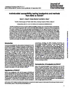
Figure 1. Zone of inhibition (50 mm) for penicillin G disk diffusion method on Leptospira Vanaporn Wuthiekanun (LVW) agar for Leptospira interrogans serovar Autumnalis strain NR-20161. The plate was prepared by spreading 300 mL of 108 CFU/mL and preincubating at 30 °C in 5% CO2 for 2 days followed by application of the disk and further incubation at 30 °C in air for a total of 7 days.
*Address correspondence to Vanaporn Wuthiekanun or Direk Limmathurotsakul, Mahidol-Oxford Tropical Medicine Research Unit, Faculty of Tropical Medicine, Mahidol University, 420/6 Rajvithi Road, Bangkok 10400, Thailand. E-mails: lek@tropmedres .ac or
[email protected]
241
242
WUTHIEKANUN AND OTHERS
single plate for the following antimicrobials (disk content): amoxicillin/clavulanic acid (20/10 mg), amoxicillin (10 mg), azithromycin (15 mg), aztreonam (30 mg), cefoxitin (30 mg), ceftazidime (30 mg), ceftriaxone (30 mg), chloramphenicol (30 mg), ciprofloxacin (5 mg), clindamycin (2 mg), doripenem (10 mg), doxycycline (30 mg), fosfomycin (50 mg), gentamicin (10 mg), linezolid (30 mg), nalidixic acid (30 mg), nitrofurantoin (300 mg), penicillin (10 units), piperacillin/tazobactam (100/10 mg), rifampicin (5 mg), tetracycline (30 mg), and trimethoprim/sulfamethoxazole (1.25/23.75 mg) (Oxoid Ltd, Basingstoke, United Kingdom). An additional plate (without discs) was used as a growth control. Plates were then incubated at 30 °C in air and observed every day for 7 days. The growth inhibition zone sizes were measured at the point at which a bacterial lawn was clearly discernible by the naked eye (usually at day 7) (Figure 1). As disk diffusion testing has not been performed previously for Leptospira, we used Clinical and Laboratory Standards Institute (CLSI) performance standards (M100-S25) for threshold zone sizes primarily for Enterobacteriaceae, extending to Pseudomonas aeruginosa or Staphylococcus spp. where zone sizes were not available for the drug being tested (Supplemental Table 1). The results for four antimicrobials (penicillin, doxycycline, ceftriaxone, and chloramphenicol) were also compared with susceptibility testing using a published minimum inhibitory concentration
(MIC) method (Etest),4 which was performed in parallel with disk testing. All 10 Leptospira isolates were susceptible to 17 antimicrobials (amoxicillin/clavulanic acid, amoxicillin, azithromycin, cefoxitin, ceftazidime, ceftriaxone, chloramphenicol, ciprofloxacin, clindamycin, doripenem, doxycycline, gentamicin, linezolid, nitrofurantoin, penicillin, piperacillin/tazobactam, and tetracycline) (Table 1). All 10 isolates had no zone of growth inhibition for four antimicrobials (fosfomycin, nalidixic acid, rifampicin, and trimethoprim/sulfamethoxazole) (Table 1). Of the 10 Leptospira, seven had a growth inhibition zone of £ 21 mm for aztreonam, the zone diameter susceptibility break point of Enterobacteriaceae. Comparison between disk and Etest results for penicillin, doxycycline, ceftriaxone, and chloramphenicol showed concordance between the two methods (all susceptible). Since LVW agar was developed, it has found use for the isolation of Leptospira from the environment,6 for longterm maintenance of the organism in agar tubes (> 1 year) without frequent media transfer,5 and for susceptibility testing using the Etest method.4 In this preliminary evaluation, the disk diffusion method was performed with an individual single antimicrobial disk per LVW agar plate, since preliminary testing demonstrated very large zones of inhibition. Break points have not been established for
Table 1 Zone diameter (millimeters) of the 10 Leptospira isolates tested Species
Serovars
Strains
Amoxicillin/ clavulanic acid†
Amoxicillin†
Aztreonam†
Cefoxitin†
Ceftazidime†
Ceftriaxone†
Leptospira interrogans L. interrogans L. interrogans L. interrogans L. interrogans L. interrogans L. interrogans L. borgpetersenii L. kirschneri L. weilii
Autumnalis Autumnalis Autumnalis Bataviae Canicola Medanensis Pyrogenes Javanica Grippotyphosa Mengdeng
L0013 L0752 NR-20161* UT0229 NR-20170* NR-20178* NR-20157* NR-20151* NR-20327* NR-20181*
85 85 77 85 70 78 80 80 76 85
85 85 73 85 74 80 44 80 76 85
57 30 16 40 20 13 10 20 19 18
85 85 70 85 64 80 80 76 76 85
85 85 67 85 70 72 80 76 82 85
77 70 42 72 42 64 44 62 64 72
Species
Chloramphenicol†
Ciprofloxacin†
Doxycycline†
Gentamicin†
Nitrofurantoin†
Piperacillin/ tazobactam†
Tetracycline†
Doripenem‡
Leptospira interrogans L. interrogans L. interrogans L. interrogans L. interrogans L. interrogans L. interrogans L. borgpetersenii L. kirschneri L. weilii
49 64 44 75 32 50 28 30 42 42
76 85 42 74 52 62 36 67 38 52
68 66 22 64 34 24 38 38 35 50
34 33 20 32 25 30 25 30 37 26
74 85 40 38 28 34 42 32 76 62
85 85 69 72 50 74 80 80 80 70
73 74 43 66 32 38 42 28 55 61
85 85 73 85 60 78 30 86 80 70
Species
Azithromycin§
Clindamycin§
Linezolid§
Penicillin§
Fosfomycin†
Nalidixic acid†
Trimethoprim sulfamethoxazole†
Rifampicin§
Leptospira interrogans L. interrogans L. interrogans L. interrogans L. interrogans L. interrogans L. interrogans L. borgpetersenii L. kirschneri L. weilii
85 85 72 85 62 76 70 76 70 85
64 60 24 51 26 35 40 30 34 50
75 85 50 72 26 78 72 40 74 60
76 67 50 65 40 70 54 76 59 61
0 0 0 0 0 0 0 0 0 0
0 0 0 0 0 0 0 0 0 0
0 0 0 0 0 0 0 0 0 0
0 0 0 0 0 0 0 0 0 0
*NR represents strains deposited with Biodefense and Emerging Infections Research Resources Repository (N = 7). Clinical and Laboratory Standards Institute (CLSI) threshold zone sizes for †Enterobacteriaceae, ‡Pseudomonas aeruginosa, and §Staphylococcus spp.
ANTIMICROBIAL DISK SUSCEPTIBILITY TESTING OF LEPTOSPIRA SPP.
Leptospira, but four antimicrobial agents were apparently inactive and gave no inhibition zones. These drugs may prove useful as inhibitors of contamination in clinical and environmental samples, and could be incorporated in selective Leptospira culture media. The findings of our study are consistent with prior reports (using broth MIC) of Leptospira susceptibility to amoxicillin, azithromycin, cefoxitin, ceftriaxone, chloramphenicol, ciprofloxacin, doxycycline, erythromycin, and tetracycline7 and resistance to fosfomycin, trimethoprim, and sulfamethoxazole.8 The disk diffusion method is easy to perform and could become a useful, initial screening test for the epidemiological surveillance of antimicrobial resistance. Received March 5, 2015. Accepted for publication April 1, 2015. Published online June 8, 2015. Note: Supplemental table appears at www.ajtmh.org. Acknowledgments: We thank Paul Newton, LOMWRU, Mahosot hospital, Vientiane, Lao PDR, who provided Leptospira isolates. We also thank Prapass Wannapinij for technical assistance. Financial support: This work was supported by the Wellcome Trust (089275/Z/09/Z) and the Faculty of Tropical Medicine, Mahidol University, Bangkok, Thailand. Authors’ addresses: Vanaporn Wuthiekanun, Premjit Amornchai, Sayan Langla, Nicholas J. White, and Nicholas P. J. Day, MahidolOxford Tropical Medicine Research Unit, Faculty of Tropical Medicine, Mahidol University, Bangkok, Thailand, E-mails: lek@ tropmedres.ac,
[email protected],
[email protected], nick@ tropmedres.ac, and
[email protected]. Direk Limmathurotsakul, Faculty of Tropical Medicine, Mahidol University, Bangkok, Thailand, E-mail:
[email protected]. Sharon J. Peacock, Department of Medicine, University of Cambridge, Addenbrooke’s Hospital, Cambridge, United Kingdom, E-mail:
[email protected].
243
This is an open-access article distributed under the terms of the Creative Commons Attribution License, which permits unrestricted use, distribution, and reproduction in any medium, provided the original author and source are credited.
REFERENCES 1. Levett PN, 2001. Leptospirosis. Clin Microbiol Rev 14: 296–326. 2. Bharti AR, Nally JE, Ricaldi JN, Matthias MA, Diaz MM, Lovett MA, Levett PN, Gilman RH, Willig MR, Gotuzzo E, Vinetz JM, 2003. Leptospirosis: a zoonotic disease of global importance. Lancet Infect Dis 3: 757–771. 3. World Health Organization, 2003. Human Leptospirosis: Guidance for Diagnosis, Surveillance and Control. Available at: http://www.who.int/zoonoses/resources/Leptospirosis/en/. 4. Wuthiekanun V, Amornchai P, Paris DH, Langla S, Thaipadunpanit J, Chierakul W, Smythe LD, White NJ, Day NP, Limmathurotsakul D, Peacock SJ, 2012. Rapid isolation and susceptibility testing of Leptospira spp. using a new solid medium (LVW agar). Antimicrob Agents Chemother 57: 297–302. 5. Wuthiekanun V, Amornchai P, Langla S, Oyuchua M, Day NP, Limmathurotsakul D, 2014. Maintenance of Leptospira species in Leptospira Vanaporn Wuthiekanun agar. J Clin Microbiol 52: 4350–4352. 6. Thaipadungpanit J, Wuthiekanun V, Chantratita N, Yimsamran S, Amornchai P, Boonsilp S, Maneeboonyang W, Tharnpoophasiam P, Saiprom N, Mahakunkijcharoen Y, Day NP, Singhasivanon P, Peacock SJ, Limmathurotsakul D, 2013. Leptospira species in floodwater during the 2011 floods in the Bangkok Metropolitan Region, Thailand. Am J Trop Med Hyg 89: 794–796. 7. Murray CK, Hospenthal DR, 2004. Determination of susceptibilities of 26 Leptospira sp. serovars to 24 antimicrobial agents by a broth microdilution technique. Antimicrob Agents Chemother 48: 4002–4005. 8. Chakraborty A, Miyahara S, Villanueva SY, Gloriani NG, Yoshida S, 2010. In vitro sensitivity and resistance of 46 Leptospira strains isolated from rats in the Philippines to 14 antimicrobial agents. Antimicrob Agents Chemother 54: 5403–5405.