Application of a classification system and description of a combined manual therapy intervention: a case with low back related leg pain Shannon M. Petersen1,2, Daphne R. Scott3 1
Physical Therapy Program, Des Moines University, USA, 2Regis University Manual Therapy Fellowship Program, USA, 3AthletiCo, USA
Low back pain and leg pain commonly occur together. Multiple factors can cause low back related leg pain; therefore, identification of the source of symptoms is required in order to develop an appropriate intervention program. The patient in this case presented with low back and leg pain. A patho-mechanism based classification is described in combination with the patient’s subjective and objective examination findings to guide treatment. The patient’s symptoms improved marginally with intervention addressing primarily the musculoskeletal impairments and with intervention addressing primarily the neurodynamic impairments. Full functional improvements were attained with a manual therapy intervention directed at both mechanisms simultaneously. The approach described in this case address a mixed pathology utilizing passive accessory and passive physiological lumbar mobilizations in combination with lower extremity neurodynamic mobilization. The patient reported complete resolution of symptoms after a total of seven visits over a period of 6 weeks. While specific guidelines do not yet exist for treatment based on the classification approach utilized, this case report provides an example of manual therapy to address low back related leg pain of mixed pathology. Keywords: Classification, Combined intervention, Manual therapy, Neurodynamics, Neuropathic
Posterior thigh pain occurs commonly in conjunction with low back pain (LBP).1 Low back related leg pain (LBRLP) is of clinical interest because it is a predictor of high rates of sick leave from work and is associated with development of chronic LBP and disability.1–3 There are many potential sources of leg pain in individuals with lumbar dysfunction including musculoskeletal and neuromuscular mechanisms.4,5 Scha¨fer and colleagues have proposed a system for classification of patients with LBRLP to guide intervention and improve outcomes.5 Inter-rater reliability of this classification system has been shown to be good.6 They5 recommend classification of patients into one of four subgroups including: (1) central sensitization; (2) denervation; (3) peripheral nerve sensitization; or (4) musculoskeletal referred pain. Central sensitization involves altered transmission of information about pain by the nervous system resulting in amplified responses to pain signals.7,8 This hypersensitivity may result in shooting or Correspondence to: Shannon Petersen, Physical Therapy Program, Des Moines University, IA, USA. Email:
[email protected]
ß W. S. Maney & Son Ltd 2010 DOI 10.1179/106698110X12640740712572
burning pain associated with allodynia and mechanical hyperalgesia;5,9 non-noxious stimuli may produce responses that normally occur with noxious stimuli.8,10 Sensory and motor function may or may not be impaired.9,10 Denervation involves impaired nerve function secondary to Wallerian degeneration or demyelination.5,11,12 Altered nerve conduction can occur following injury or with disease processes. Denervation results in pain, sensory or motor deficits, and changes in reflexes.5,9,13 Peripheral nerve sensitization is caused by nerve root or nerve trunk irritation. It is a condition in which nerves exhibit an adverse response to mechanical stimulation14–16 and can be examined through nerve palpation or neurodynamic tests.16 In cases of peripheral nerve sensitization, neural mechanosensitivity may be accompanied by subtle changes in sensory nerve function; however, no significant neruological dysfunction is present upon examination.5,14,16 Musculoskeletal pain can be referred into the lower extremity (LE) from various sources.4,5,17–19 Facet
Journal of Manual and Manipulative Therapy
2010
VOL .
18
NO .
2
89
Petersen and Scott
Description of a combined manual therapy intervention
in intensity to 3/10 following sprinting and to 7/10 after 20 minutes of sitting. He reported that his job required driving up to 2 hours at a time. The patient stated that once aggravated, his symptoms would dissipate within a few minutes of standing. He denied LE weakness, paresthesias or bowel and bladder changes. His Oswestry low back pain disability questionnaire (OSW) score was 18/100, suggesting minimal self report of disability.
Previous tests and interventions The interpretation of plain film radiographs and magnetic resonance imaging studies of the lumbar spine were normal. Past interventions included two lumbar epidural injections and a corticosteroid injection to the right sacroiliac joint; each provided 1 day of relief. Previous physical therapy intervention consisted of lumbar stabilization and stretching exercises which did not relieve the patient’s symptoms. He denied use of medication and denied any additional health problems or significant past medical history. Based on the subjective examination alone, there was not enough information to determine the pathologic mechanisms involved with the patient’s complaints. Because he presented with posterior thigh pain in conjunction with LBP, he was an appropriate candidate for application of the classification system for LBRLP proposed by Scha¨fer and colleagues.5 Although there were no subjective complaints of sensory or motor changes, an objective examination was required to determine if neurological or musculoskeletal involvement was present. The Leeds assessment of neuropathic symptoms and signs (LANSS) is a component of the LBRLP classification system.5 The LANSS is a two-part examination tool used to score sensory function and has been shown reliable and valid in identification of neuropathic pain.9 A score of 12/24 or greater is associated with central sensitization.5 This patient’s score for section A of the LANSS, the pain questionnaire component, was 0/16.9 The score for section B of the LANSS, the sensory component, was 0/8 yielding a composite LANSS score of 0/24.9
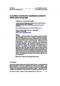
Figure 1 Body diagram depicting areas of symptoms as described by the patient.
referral patterns into the LE have been determined through diagnostic facet blocks in addition to radiofrequency denervation of the medial branches supplying the facet.17,20 Lumbar intervertebral discs have also been shown to refer pain;4,21 noxious stimuli applied to the disc itself can produce pain in the low back, thigh, and lower leg.21 The hamstring muscle can produce pain in the posterior thigh following trauma, strain, or overuse.18,19 In cases of musculoskeletal causes of LE symptoms, physical examination stressing the musculoskeletal structures involved should reproduce the patients’ symptoms.5 Although this classification has been found to be reliable, its validity has not been established.6 There is currently no literature describing application of this classification system followed by intervention directed toward the patho-mechanisms indicated. The purpose of this case report was to demonstrate application of the classification system proposed by Scha¨fer and colleagues5 to a patient with LBRLP and to describe the intervention provided.
Patient characteristics Chief complaint/onset A 35-year-old male presented to physical therapy with chronic low back and posterior thigh pain. He attributed his symptoms to a weightlifting incident 2 years earlier while lifting a barbell from a squatted position. He experienced no symptoms immediately but pain began in the right low back and posterior thigh 1 week later. He denied any other trauma or change in activity preceding symptom onset. This patient had no prior history of LBP or LE pain. Despite current symptoms, he reported regular participation in baseball, weight training, and cardiovascular conditioning.
Physical examination Findings from the physical examination are summarized in Table 1.3,9,16,22–30 Although lumbar range of motion (ROM) was found to be normal with singleplane movements, combined motion testing for flexion and left lateral flexion reproduced LBP and buttock symptoms. Repeated lumbar flexion and extension in standing did not produce symptoms,3,22 but buttock pain was produced with repeated lumbar flexion in supine. These symptoms were only present at end-range and did not extend into the thigh. There were no residual symptoms and no subsequent loss of extension ROM. Passive physiological intervertebral motion (PPIVM) testing revealed hypomobility at the L4/L5 segment
Aggravating/relieving factors He described his LBP as a mild ache that was constantly present and increased in intensity to 3/10 with driving and following exercise. The patient’s primary complaint was his LE symptoms, which he described as a deep ache in the proximal third of the right posterior thigh (Fig. 1). LE symptoms increased
90
Journal of Manual and Manipulative Therapy
2010
VOL .
18
NO .
2
Petersen and Scott
on the right with both flexion and left lateral flexion.23 This reproduced the patient’s LBP and right buttock symptoms. Posterior to anterior (PA) intervertebral joint examination revealed hypomobility at L4/L5 without symptoms.23,24 Pain was present upon palpation to the proximal hamstring although tissue texture appeared symmetrical and there was no muscle guarding. The sciatic nerve was palpated just lateral to the ischial tuberosity26,27 and was painful on the right. Neurodynamics were examined using the straight leg raise (SLR) and slump tests.16,30 The SLR was measured at the onset of resistance as described by Maitland,23 then continued through range until firm resistance was encountered. Sensitizing maneuvers of cervical flexion and ankle dorsiflexion were added to the SLR test and did not elicit symptoms.16,29 The slump test produced right posterior thigh pain with the knee fully extended and the cervical spine flexed. When overpressure was applied through the trunk and the patient’s ankle was dorsiflexed, posterior thigh pain was produced. In this provocative position, the patient was asked to extend the cervical spine which resulted in a decrease in pain intensity.
Clinical impression Based on the low LANSS score, central sensitization was ruled out as the source of this patient’s LBRLP. This conclusion was consistent with the patient’s presentation since his signs and symptoms did not include paresthesia, hyperesthesia, or hyperalgesia. There was no adverse response to stimulating the skin overlying the hamstring muscle or in the region where the sciatic nerve was found to be tender to palpation, but the proximal portion of the hamstring muscle was
Description of a combined manual therapy intervention
painful with palpation. Since the hamstring was not painful with resisted testing or length testing, it is possible that the pain with palpation to this muscle was due to allodynia.5,9 The patient did not demonstrate signs or symptoms associated with denervation. He had no signs of hypoesthesia or diminished sensation, his reflexes were normal and he showed no signs of motor weakness. Based on the lack of neurological deficit, it was determined that the patient did not fit into the denervation classification.5 Classification into the peripheral nerve sensitization category requires neural mechanosensitivity including sensitivity to mechanical pressure.5,16,27 This patient had pain with palpation to the sciatic nerve on the involved side but presented with inconsistent findings upon neurodynamic testing. The SLR test did not elicit symptoms despite addition of sensitizing maneuvers. The patient demonstrated less motion on the involved compared to uninvolved extremity, but had excessive mobility with the SLR test on both lower extremities.30,31 The slump test however did reproduce the patient’s symptoms. This finding was consistent with his functional limitation because the slump test position includes spinal and LE positions similar to those involved with driving. Because the patient’s symptoms were reproduced with mechanical pressure to the nerve and the slump test, his presentation of LBRLP fit into the peripheral nerve sensitization classification. The musculoskeletal classification was also appropriate for this patient. Right sided symptom reproduction with combined movements of lumbar flexion and left lateral flexion is consistent with a regular opening pattern for posterior structures on the right but this position also increases the distance the neural tissue
Table 1 Summary of objective examination findings at initial visit Examination procedure Posture
Gait Lumbar ROM Hip ROM Lumbar combined ROM Repeated motion testing3,22 Lumbar PPIVM23 Lumbar PA23,24 accessory motion SIJ provocative tests Hip joint mobility Flexibility 90/90 hamstring test25 Palpation26,27 Strength28 Neurological exam9 Neurodynamic tests16,23,29,30
Observations/measurements No lumbar or LE deviations observed Symmetrical weight-bearing No lateral shift Equal stance time and step length No antalgia Full Full Flexion with left LF*{ Lumbar flexion and extension in standing and lying{ Hypomobile at L4/L5 on the right with flexion and left LF*{ Hypomobile at L4/L5 Compression; distraction; sacral PA Accessory motion testing normal Full knee extension Proximal hamstring{ Sciatic nerve{ Gross hip and knee strength normal Specific hamstring strength normal LE myotomes, deep tendon reflexes, and dermatomes intact SLR right 110u; left 120u Slump test{
Note: LE, lower extremity; ROM, range of motion; LF, lateral flexion; PPIVM, passive physiological intervertebral motion; PA, posterior to anterior; SIJ, sacroiliac joint; SLR, straight leg raise. *Indicates reproduced low back pain. {Indicates reproduced LE pain.
Journal of Manual and Manipulative Therapy
2010
VOL .
18
NO .
2
91
Petersen and Scott
Description of a combined manual therapy intervention
Table 2 Summary of patient visits including examination findings, interventions provided, and outcome Treatment session Visit 1
Examination findings
Intervention
N Pain sprinting 3/10
N Postural education
N Pain driving 20 minutes 7/10
N PA L4 Grade IV sustained 30 seconds, 5 repetitions N Lumbar PPIVM combined flexion and left LF, Grade III sustained 30 seconds, 5 repetitions N HEP supine stretch of lumbar flexion with LF
N OSW 18/100
N SLR 110u
Visit 2 (2 days later)
LBP and LE pain: N Combined lumbar flexion and left LF ROM N PPIVM L4/L5 LE pain: N Repeated flexion in lying N Slump test pain 5/10 N Decreased LBP
N Postural education for driving
N No change LE pain N SLR 110u
Visit 3 (1 week later)
LBP and LE pain: N Combined lumbar flexion and left LF ROM with overpressure LE pain: N Slump test pain 4/10 N Decreased LBP
N Pain driving 30 minutes 5/10
Visit 4 (1 week later)
N SLR 110u LE pain: N Combined lumbar flexion and left LF ROM with overpressure N Combined PPIVM L4/L5 N Slump test positive after sustained 20 seconds N Pain driving 30 minutes 3/10
N SLR 115u
LE pain: N Combined lumbar flexion and left LF ROM with overpressure N Combined PPIVM L4/L5 N PA at L4 in prone SLR N Slump test positive after sustained 30 seconds N Pain driving 60 minutes 3/10
N SLR 120u
92
Journal of Manual and Manipulative Therapy
2010
N Lumbar PPIVM combined flexion and left LF, Grade III sustained 30 seconds, 10 repetitions N HEP of seated slump with ankle dorsiflexion sustained 30 seconds; 10 repetitions; 3 times per day
N Decreased pain with combined lumbar flexion and left LF ROM N Repeated flexion in lying pain 1/10 N Slump test pain 4/10
N Slump test positive after sustained 15 seconds N SLR 115u
N Pain-free combined lumbar flexion and left LF ROM with overpressure N Slump test positive after sustained 30 seconds
N SLR 115u
N OSW 5/100
Visits 5 and 6 (1 week apart)
N PA L4 Grade IV sustained 30 seconds, 5 repetitions N Lumbar PPIVM combined flexion and left LF, Grade III sustained 30 seconds, 10 repetitions N Continuation of HEP
Outcome
VOL .
N Lumbar PA in prone SLR position with ankle dorsiflexion, Grade IV sustained 10 seconds, 10 repetitions N Continued HEP; added cervical flexion to slump stretch and decreased hold time to 10 seconds due to c/o calf paresthesias N Prone SLR with active ankle dorsiflexion, 15 second holds, 10 repetitions
N Pain-free combined lumbar flexion and left LF ROM with overpressure
N Lumbar PA in prone SLR position with ankle dorsiflexion, Grade IV sustained 30 seconds, 10 repetitions N Lumbar PPIVM combined flexion and left LF in SLR position
N Slump test negative sustained for 60 seconds
18
NO .
2
N Slump test negative sustained for 30 seconds
N SLR 115u
N SLR to 120u
Petersen and Scott
Description of a combined manual therapy intervention
Table 2 Continued. Treatment session
Examination findings LE pain:
Visit 7 (1 week later)
Intervention
Outcome
N Prone SLR with active ankle dorsiflexion, 20 second holds, 10 repetitions, 3 times per day
N PA at L4 in prone SLR N Slump test positive after sustained 30 seconds N No pain driving up to 4 hours the past 4 days N OSW 0/100 N SLR 120u N Slump test negative sustained for 60 seconds N Prone SLR with PA and ankle dorsiflexion 20 seconds pain-free
N Patient discharged
Note: LF, lateral flexion; ROM, range of motion; PA, posterior to anterior; HEP, home exercise program; OSW, Oswestry low back pain disability questionnaire; PPIVM, passive physiological intervertebral motion; SLR, straight leg raise; LBP, low back pain; LE, lower extremity; c/o, complaint of.
travels when considering the right sciatic nerve and the spinal dura.16,32 Reproduction of comparable sign with these combined movements could have been due to peripheral nerve sensitization or musculoskeletal structures. Repeated lumbar flexion in lying produced right buttock pain at end-range and symptoms did not peripheralize; this result fit the McKenzie classification of tissue dysfunction but did not fit for neural tissue involvement or disc derangement.22 Lumbar joint mobility testing reproduced low back and LE symptoms, therefore the intervertebral joints could have been contributing to this patient’s pain. Since he presented with a mixed pathology, both peripheral nerve sensitization and muscluloskeletal impairments were addressed during intervention.5
Intervention/outcomes The patient was seen in physical therapy 2 times the first week, then one time per week for a total of seven visits over a period of 6 weeks. Examination findings, intervention, and outcomes are summarized in Table 2. Impairments associated with the musculoskeletal classification were addressed during the first two visits. Lumbar combined ROM and PPIVMs reproduced the patient’s low back symptoms during examination, therefore, were the focus of manual interventions and the home exercise program. Although these interventions resulted in a decrease in LBP and some improvement while driving, minimal change had occurred in LE symptoms. Impairments associated with the peripheral nerve sensitization classification were addressed at visit 3 through the addition of a neurodynamic intervention to the patient’s home program. This consisted of sitting with the back and shoulders relaxed into a flexed position while the patient extended his knee and dorsiflexed his ankle. Cervical flexion was not added to the home program due to production of paresthesias in the LE with maintaining the position. Following
this intervention he continued to improve and tolerated driving longer with less intense symptoms. By the fourth visit, the patient’s Oswestry Disability Index score had improved to 5/100. At this time, lumbar PA was examined in the prone SLR position. This combination of musculoskeletal and neurodynamic testing reproduced the patient’s LE symptoms. The need to combine tests in order to elicit symptoms was consistent with the patient’s mixed pathology classification; therefore, both pathomechanisms were addressed through a combined intervention. Mobilization was executed at visits 4–6 by performing lumbar manual interventions of PPIVM and PA in the SLR position to incorporate neurodynamics (Fig. 2). For the combined PA intervention, the patient was positioned in prone with his right LE over the edge of the plinth and his foot resting on the floor with his knee slightly flexed (Fig. 2A). Lumbar PA mobilization was administered while the patient dorsiflexed the ankle. He reported reproduction of posterior thigh symptoms with PA which was immediately alleviated once mobilization ceased. The patient returned to a neutral ankle position, and lumbar PAs were performed at the L4 level with a Grade III pressure sustained for 15 seconds and repeated 10 times. The patient was instructed to repeat active dorsiflexion in this prone SLR position as part of his home program. The lumbar flexion and left lateral flexion PPIVM was also progressed to incorporate neurodynamics. The PPIVM was performed with the patient in left side lying as previously; however, this time the addition of an SLR was included (Fig. 2B). The L4/ L5 segment was localized through hip flexion until motion was palpated at L5. Left lateral flexion of the lumbar spine was introduced from above by drawing the left upper extremity caudally until motion was palpated at L4. This Grade IV mobilization was sustained as the patient’s right hip was passively
Journal of Manual and Manipulative Therapy
2010
VOL .
18
NO .
2
93
Petersen and Scott
Description of a combined manual therapy intervention
A
B
Figure 2 Combined interventions of lumbar joint mobilization with neurodynamic SLR. (A) lumbar PPIVM with SLR; (B) lumbar PA with SLR.
Joint mobilization can influence facets but can also impact neurodynamics because of the facets’ close anatomical proximity to nerve roots as they exit the lateral foramen.16,36 Creating motion at the mechanical interface between the facet joint and the neural tissue is also thought to affect fascia and circulation in the region.16,36 The patient responded to the lumbar mobilization with a decrease in LBP but demonstrated minimal improvement in LE symptoms. The peripheral sensitization classification was addressed next. The patient’s neural tissue was not irritable based on the fact that his SLR was negative for symptom reproduction and he was able to attain full knee extension in the slump test position before symptoms were produced.16,36 Both the SLR and slump tests can be indicative of neural mechanosensitivity. He demonstrated excessive mobility with the SLR test although limitations were noted on the involved side compared to the non-involved side. In a study investigating agreement of SLR and slump tests in subjects with LBRLP, it was found that only 44% of study participants demonstrated both a positive SLR test and a positive slump test.37 Forty-nine percent of the subjects demonstrated a positive slump test but a negative SLR. Both the slump test and SLR test have been shown to have good specificity (0.83; 0.89) when used to examine patients with lumbar disc herniation, but the slump test is more sensitive (0.84) than the SLR (0.52) in this population.4 These statistics are not available for patients with peripheral nerve sensitization. In addition to the positive slump test, the patient in this case also had an adverse response to palpation of the sciatic nerve which is consistent with neural mechanosensitivity.37,38 Criteria have been described to determine which patients are likely to benefit from slump stretching.39 Slump stretching has been shown to result in decreased pain, improved function, and centralization of symptoms in a subgroup of patients who met these criteria.40 The patient in this case met all three criteria including LBP with LE symptoms; no improvement or worsening of LE symptoms with
flexed, knee was extended and ankle dorsiflexed. Low back and LE symptoms with driving and sports had resolved by visit 7 and the Oswestry Disability Index score was 0/100. The patient’s goals were met and he was discharged from physical therapy.
Discussion This case demonstrates application of a classification system for a patient with LBRLP to assist in identification of the pathological mechanisms involved. Patients who undergo classification and are treated according to the classification matrix have been shown to demonstrate improved outcomes.33,34 Although no intervention guidelines have been established in association with the classification system proposed by Scha¨fer and colleagues,5 the current case provides an example of its application and describes the intervention provided. The classification system utilized suggests four potential mechanisms of pathology; in cases of mixed pathologies there may be overlap among classification categories.5 It is suggested that the physical examination protocol be used to determine the predominant mechanism of pathology.5,35 The patient in this case presented with signs and symptoms consistent with two different categories. Intervention is described for both the musculoskeletal and peripheral nerve sensitization components of his presentation. Although his symptoms partially resolved with intervention directed toward the musculoskeletal impairments, they did not completely resolve until interventions were combined to address both systems. Interventions directed toward the patient’s limitations associated with the musculoskeletal pain classification were addressed first. Maitland recommended initiating manual therapy intervention with the procedure that most closely reproduces the patient’s comparable sign with the least amount of force.23 Since the patient’s LBP was reproduced with combined ROM testing and with intervertebral joint mobility examination, combined ROM and joint mobilization were chosen as the first interventions.
94
Journal of Manual and Manipulative Therapy
2010
VOL .
18
NO .
2
Petersen and Scott
repeated motion testing; and a positive slump test with no sensory, motor, or reflexive changes. The patient in this case continued to show subjective and objective improvement with intervention, but it was not until lumbar mobilization was combined with neurodynamic intervention that his symptoms completely resolved. This intervention addressed the patient’s mixed pathology classification. Spinal mobilization has been described in conjunction with neurodynamic intervention for patients with neurogenic cervicobrachial pain, but no similar description has been presented for the LBRLP.41 Cervical lateral glide mobilization has been shown to result in decreased pain and improved neurodynamics in patients with lateral epicondylalgia and arm pain of cervical origin.41,42 Because this patient’s symptoms were reproduced with a combination of lumbar intervertebral mobility examination and the LE neurodynamic position, the two interventions were combined by performing the lumbar mobilization in a position that increased the distance the neural tissue traveled in the LE while creating proximal mobility at the spine.16 The patient in this case presented with a mixed pathology according to the patho-mechanism-based approach utilized. Since he displayed examination findings consistent with both peripheral nerve sensitization and musculoskeletal impairment, both were addressed through intervention. Combined manual interventions addressing joint mobility and neurodynamic limitations resulted in resolution of the patient’s symptoms.
Conclusion Classification of patients with LBRLP may be useful in examination and intervention planning for patients with mixed pathologies. Future investigations should examine the presence of mixed pathologies in a larger patient population in addition to offering guidelines for classification-based intervention. Further research should focus on the effects of combined lumbar mobilization with LE neurodynamic intervention in addition to identification of patients likely to benefit from this combined intervention. References 1 Selim AJ, Ren XS, Fincke G, Deyo RA, Rogers W, Miller D, et al. The importance of radiating leg pain in assessing health outcomes among patients with low back pain. Results from the Veterans Health Study. Spine. 1998;23:470–4. 2 Puolakka K, Ylinen J, Neva MH, Kautiainen H, Ha¨kkinen A. Risk factors for back pain-related loss of working time after surgery for lumbar disc herniation: a 5-year follow-up study. Eur Spine J. 2008;17:386–92. 3 Werneke M, Hart DL. Centralization phenomenon as a prognostic factor for chronic low back pain and disability. Spine. 2001;26:758–64. ¨ nalan H, Toprak S. The sensitivity and 4 Majlesi J, Togay H, U specificity of the slump and the straight leg raising tests in patients with lumbar disc herniation. J Clin Rheumatol. 2008;14:87–91. 5 Scha¨fer A, Hall T, Briffa N. Classification of low back-related leg pain: a proposed patho-mechanism-based approach. Man Ther. 2009;14:222–30.
Description of a combined manual therapy intervention
6 Scha¨fer A, Hall T, Ludtke K, Mallwitz J, Briffa N. Interrater reliability of a new classification system for patients with neural low back-related leg pain. J Man Manip Ther. 2009;17:109–17. 7 Carlton SM, Du J, Tan HY, Nesic O, Hargett GL, Bopp AC, et al. Peripheral and central sensitization in remote spinal cord regions contribute to central neuropathic pain after spinal cord injury. Pain. 2009;147:265–76. 8 Winkelstein BA, DeLeo JA. Mechanical thresholds for initiation and persistence of pain following nerve root injury: mechanical and chemical contributions at injury. J Biomech Eng. 2004;126:258–63. 9 Bennett M. The LANSS pain scale: the Leeds Assessment of Neuropathic Symptoms and Signs. Pain. 2001;92:147–57. 10 Latremoliere A, Woolf CJ. Central sensitization: a generator of pain hypersensitivity by central neural plasticity. J Pain. 2009;10:895–926. 11 Kropf N, Krishnan K, Chao M, Schweitzer M, Rosenberg Z, Russell SM. Sciatic nerve injury model in the axolotl: functional, electrophysiological, and radiographic outcomes. J Neurosurg. 2009;112:880–9. 12 Ronchia G, Nicolinoa S, Raimondob S, Tos P, Battiston B, Papalia I, et al. Functional and morphological assessment of a standardized crush injury of the rat median nerve. J Neurosci Methods. 2009;179:51–7. 13 Shim S, Ming G. Roles of channels and receptors in the growth cone during PNS axonal regeneration. Exp Neurol. 2010;223:38–44. 14 Elvey RL, Hall T. Neural tissue evaluation and treatment. In: Donatelli RA, editor. Physical therapy of the shoulder. New York: Churchill Livingstone; 1997. 15 Butler D. The sensitive nervous system. Adelaide: NOI Group Publications; 2000. 16 Shacklock M. Clinical neurodynamics. Philadelphia (PA): Elsevier Limited, 2005. 17 Boswell M, Colson J, Sehgal N, Dunbar E, Epter R. A systematic review of therapeutic facet joint interventions in chronic spinal pain. Pain Physician. 2007;10:229–53. 18 Schache A, Wrigley T, Baker R, Pandy M. Biomechanical response to hamstring muscle strain injury. Gait Posture. 2009;29:332–8. 19 McGregor C, Gohosh S, Young D, Maffulli N. Traumatic and overuse injuries of the ischial origin of the hamstrings. Disabil Rehabil. 2008;30:1597–1601. 20 Windsor RE, King FJ, Roman SJ, Tata NS, Cone-Sullivan LA, Thampi S, et al. Electrical stimulation induced lumbar medial branch referral patterns. Pain Physician. 2002;5:347–53. 21 O’Neill CW, Kurgansky ME, Derby R, Ryan DP. Disc stimulation and patterns of referred pain. Spine. 2002;27:2776–81. 22 McKenzie RA. The lumbar spine: mechanical diagnosis and therapy. Waikanae: Spinal Publications; 1981. 23 Maitland GD, Hengeveld E, Banks K, English K, editors. Maitland’s vertebral manipulation. 7th ed. New York: Butterworth-Heinemann; 2005. 24 Phillips DR, Twomey LT. A comparison of manual diagnosis with diagnosis established by a uni-level lumbar spinal block procedure. Man Ther. 1996;1:82–7. 25 Davis D, Quinn R, Whiteman C, Williams J, Young C. Concurrent validity of four clinical tests used to measure hamstring flexibility. J Strength Cond Res. 2008;2:583–8. 26 Walsh J, Hall T. Reliability, validity and diagnostic accuracy of palpation of the sciatic, tibial and common peroneal nerves in the examination of low back related leg pain. Man Ther. 2009;14:623–9. 27 Hall TM, Elvey RL. Management of mechanosensitivity of the nervous system in spinal pain syndromes. In: Boyling JD, Jull G, editors. Grieves modern manual therapy. 3rd ed. Edinburgh: Churchill Livingstone; 2004. 28 Kendall F, McCreary E, Provance P. Muscles: testing and function with posture and pain. 5th ed. Baltimore (MD): Williams & Wilkins; 2005. 29 Deville W, van der Windt D, Dzaferagic A, Bezemer P, Boutler L. The test of Lase´gue. Systematic review of the accuracy in diagnosing herniated discs. Spine. 2000;25:1140–7. 30 Herrington L, Bendix K, Cornwell C, Fielden N, Hankey K. What is the normal response to structural differentiation within the slump and straight leg raise tests? Man Ther. 2008;13:289–94. 31 Boyd B, Wanek L, Gray A, Topp K. Mechanosensitivity of the lower extremity nervous system during straight-leg raise neurodynamic testing in healthy individuals. J Orthop Sports Phys Ther. 2009;39:780–90. 32 Edwards B. Manual of combined movements. 2nd ed. Woburn (MA): Butterworth-Heinemann; 1999.
Journal of Manual and Manipulative Therapy
2010
VOL .
18
NO .
2
95
Petersen and Scott
Description of a combined manual therapy intervention
33 Brennan G, Fritz J, Hunter S, Thackeray A, Delitto A, Erhard R. Identifying subgroups of patients with acute/subacute ‘nonspecific’ low back pain: results of a randomized clinical trial. Spine. 2006;31:623–31. 34 Fritz JM, Delitto A, Erhard RE. Comparison of classificationbased on clinical practice guidelines for patients with acute low back pain: a randomized clinical trial. Spine. 2003;28:1363– 71. 35 Finnerup N, Otto M, McQuay H, Jensen T, Sindrup S. Algorithm for neuropathic pain treatment: an evidence based proposal. Pain. 2005;118:289–05. 36 Butler D. Mobilisation of the nervous system. Melbourne: Churchill Livingstone, 1991. 37 Walsh J, Hall T. Agreement and correlation between the straight leg raise and slump tests in subjects with leg pain. J Manipulative Physiol Ther. 2009;32:184–92.
96
Journal of Manual and Manipulative Therapy
2010
VOL .
18
38 Hall T, Elvey R. Nerve trunk pain: physical diagnosis and treatment. Man Ther. 1999;4:63–73. 39 George S. Characteristics of patients with lower extremity symptoms treated with slump stretching: a case series. J Orthop Sports Phys Ther. 2002;32:391–8. 40 Cleland J, Childs J, Palmer J, Eberhart S. Slump stretching in the management of non-radicular low back pain: a pilot clinical trial. Man Ther. 2006;11:279–86. 41 Coppieters M, Stappaerts K, Wouters L, Janssens K. The immediate effects of a cervical lateral glide treatment technique in patients with neurogenic cervicobrachial pain. J Orthop Sports Phys Ther. 2003;33:369–78. 42 Vicenzino B, Collins D, Wright A. The initial effects of a cervical spine manipulative physiotherapy treatment on the pain and dysfunction of lateral epicondylalgia. Pain. 1996;68:69–74.
NO .
2