University of Connecticut
DigitalCommons@UConn Doctoral Dissertations
University of Connecticut Graduate School
3-31-2016
Application of a Portable Hyperspectral Imaging System to Field Studies in Animal Camouflage and Coral Reef Symbiosis Brandon J. Russell University of Connecticut - Storrs,
[email protected]
Follow this and additional works at: http://digitalcommons.uconn.edu/dissertations Recommended Citation Russell, Brandon J., "Application of a Portable Hyperspectral Imaging System to Field Studies in Animal Camouflage and Coral Reef Symbiosis" (2016). Doctoral Dissertations. 1093. http://digitalcommons.uconn.edu/dissertations/1093
This Open Access is brought to you for free and open access by the University of Connecticut Graduate School at DigitalCommons@UConn. It has been accepted for inclusion in Doctoral Dissertations by an authorized administrator of DigitalCommons@UConn. For more information, please contact
[email protected].
Application of a Portable Hyperspectral Imaging System to Field Studies in Animal Camouflage and Coral Reef Symbiosis
Brandon James Russell University of Connecticut 2016 Abstract Hyperspectral imaging (HSI) represents a powerful tool for measuring both the spatial and spectral components of a target. It has a wide variety of uses in marine science, but has previously been restricted to either large spatial scales or laboratory studies. Here, a portable imager is used in field research on biological camouflage and coral/dinoflagellate symbiosis. Mats of the pelagic macroalgae Sargassum represent a complex environment for the study of marine camouflage at the air-sea interface, where endemic organisms have convergently evolved similar colors and patterns. Using HSI, spectral camouflage of two crab species (Portunus sayi and Planes minutus) was assessed. Crabs matched Sargassum reflectance across blue and green wavelengths (400-550 nm) and diverged at longer wavelengths, with maximum discrepancy in the far-red (i.e., 675 nm) due to Chlorophyll a absorption in Sargassum. Predator visual modeling showed that both species have effective color matching against blue/green sensitive dichromat fish, but are discernible to red sensitive, tetrachromat birds. The two species showed opposing trends in background matching with relation to body size. Held in a naturalistic light regime, P. sayi displayed a distinct diel cycle of dark/pale color change not observed under constant illumination. Individuals changed color in response to monochromatic black, grey, and white backgrounds, as integrated reflectance (ΣR) of crabs generally followed background
Brandon James Russell – University of Connecticut, 2016 albedo. Dynamic color change in this species may play a photoprotective role, with possible use in enhancing cryptic color matching. The imaging technology and methodology utilized in studying camouflage in the Sargassum environment was then applied to a different area of optical marine science. For stony corals, reflectance is driven by the pigments of photosynthetic endosymbionts. The warm inshore bays and cooler offshore reefs of Palau share a variety of coral species with differing symbiotic dinoflagellates (genus: Symbiodinium). Hyperspectral imagery revealed that coral integrated reflectance (ΣR 400 – 700 nm) had an inverse correlation to symbiont cell density. As hypothesized, coral colonies from offshore (Clade C symbionts) showed greater bleaching response to experimental heating than inshore counterparts with thermally resistant S. trenchii. Although no unique reflectance features were found to distinguish symbiont species, differences related to symbiont density could prove useful in field and remote sensing studies. This dissertation demonstrates the suitability of portable hyperspectral imaging for a variety of field studies in marine science. This includes a unique at-sea use of HSI to study animal camouflage.
Application of a Portable Hyperspectral Imaging System to Field Studies in Animal Camouflage and Coral Reef Symbiosis
Brandon James Russell
B.S. Boston College 2007
A Dissertation Submitted in Partial Fulfillment of the Requirements for the Degree of Doctor of Philosophy at the University of Connecticut 2016
i
Copyright by Brandon James Russell
2016 ii
APPROVAL PAGE
Doctor of Philosophy Dissertation
Application of a Portable Hyperspectral Imaging System to Field Studies in Animal Camouflage and Coral Reef Symbiosis
Presented by Brandon James Russell, B.S. Major Advisor:_________________________________________________________________ Heidi Dierssen Associate Advisor:______________________________________________________________ Peter Auster Associate Advisor:______________________________________________________________ Molly Cummings
University of Connecticut 2016 iii
Acknowledgements First and foremost, I would like to express my deepest gratitude to my advisor and mentor, Dr. Heidi Dierssen. In the past six years she has shown unflagging support of this work and my independent development of the questions, methods, and hypotheses which became this dissertation. Heidi made certain that I was not only producing quality science, but that I became an active and known member of the ocean optics community. She has an amazing ability to generate personal connections and recognize potential collaborations. For me personally, this has resulted in several important opportunities during my time in her lab, one of which became a chapter of this dissertation. When doing research, Heidi has always encouraged me to keep an open, observing mind and to be ready to ask new questions beyond what I have originally planned. The first two chapters of this dissertation were the result of just such flexibility. I am also grateful to the other members of my advisory committee, Dr. Peter Auster and Dr. Molly Cummings of UT Austin. Both provided important insight, as did former committee member Dr. Robert Whitlatch. The faculty of the Department of Marine Sciences provided me with an excellent oceanographic education. For his assistance in the field I would like to thank Eric Heupel, as well as the crew and researchers of the SSV Corwith Cramer, SV Sea Dragon, and the staff of Keys Marine Lab who helped sort through massive quantities of algae chasing small crabs on multiple occasions. I would also like to acknowledge the support and administrative staff of UConn Marine Sciences, who made it possible to conduct the research presented. During the past 6 years, my fellow lab members Kelley Bostrom and Kate Randolph have provided technical assistance and moral support. This work was supported primarily through a grant to my advisor from the Office of Naval Research. I also received direct funding from the UConn Department of Marine Sciences, and educational support from NASA and UNIS. Valuable ship-time was donated by the Sea Education Alliance, as well as the Bermuda Alliance for the Sargasso Sea and Bermuda Aquarium, Museum, and Zoo. Personally, I am deeply grateful for the love and support of my parents and family. My mother, herself trained in biology, provided unending support and a sounding board for my research. After spending many years working with optical electronics, my father has helped me to understand the technology of my instruments and to develop my own research equipment.
iv
Table of Contents 1. Introduction ..................................................................................................................................1 2. Use of Hyperspectral Imagery to Assess Cryptic Color Matching in Sargassum Associated Crabs ..............................................................................................................................................13 2.1 Introduction ..................................................................................................................14 2.2 Methods........................................................................................................................16 2.2.1 Collection of Crabs and Sargassum ..............................................................16 2.2.2 Hyperspectral Imaging and Analysis ............................................................16 2.2.3 Discrimination by Predators .........................................................................17 2.3 Results ..........................................................................................................................19 2.3.1 Environment and Organism Description ......................................................19 2.3.2 Reflectance of Organism and Background ...................................................23 2.3.3 Discrimination of Crab and Background by Predators .................................26 2.3.4 Discrimination of Crab and Background—Model Variation........................27 2.4 Discussion ....................................................................................................................30 2.1 Conclusions ..................................................................................................................35 3. Color Change in the Sargassum Crab, Portunus sayi: Response to Diel Illumination Cycle and Background Albedo .......................................................................................................................40 3.1 Introduction ..................................................................................................................42 3.2 Methods........................................................................................................................44 3.2.1 Collection of Crabs and Algae ......................................................................44 3.2.2 Diel Color Change ........................................................................................45 3.2.3 Response to Background Albedo ..................................................................45 3.2.4 Spectral Reflectance......................................................................................46 3.3 Results ..........................................................................................................................47 3.3.1 Diel Color Change ........................................................................................49 3.3.2 Response to Background Albedo ..................................................................53 3.4 Discussion ....................................................................................................................57 v
3.4.1 Color Change in Response to Environmental Conditions ............................57 3.4.2 Camouflage Through Morphological Color Change ....................................60 3.5 Conclusions ..................................................................................................................60 4. Spectral Reflectance of Palauan Reef-Building Coral with Different Symbionts in Response to Elevated Temperature ....................................................................................................................67 4.1 Introduction ..................................................................................................................68 4.2 Methods........................................................................................................................70 4.2.1 Study Sites ....................................................................................................70 4.2.2 Coral Collection and Maintenance................................................................71 4.2.3 Experimental Setup .......................................................................................71 4.2.4 Symbiont Species and Density......................................................................72 4.2.5 Image and Reflectance Processing................................................................72 4.2.6 Reflectance and Symbiont Density Comparison ..........................................74 4.2.7 Reflectance Analysis .....................................................................................74 4.3 Results ..........................................................................................................................74 4.3.1 Symbiont Species and Density......................................................................74 4.3.2 Spectral Reflectance......................................................................................75 4.3.3 Reflectance and Symbiont Density Comparison ..........................................77 4.3.4 Response to Heating .....................................................................................77 4.4 Discussion ....................................................................................................................80 4.4.1 Reflectance of Different Coral/Symbiont Systems .......................................80 4.4.2 Spectral Variability with Symbiont Concentration .......................................80 4.4.3 Spectral Response to Heating .......................................................................81 4.5 Conclusions and Outlook for Remote Sensing ............................................................81 Appendix: Supplementary Material Chapter 2 ..............................................................................87
vi
1. Introduction 1.1. Reflectance in Marine Science In marine sciences, remote sensing may be used to study an immense range of biological, geological, and physical subjects at scales ranging from individual phytoplankton to the global ocean. This involves measuring the light, or electromagnetic radiation, from a target without being in contact with the object or surface in question in order to gain information [1]. One of the basic quantities in optical remote sensing is spectral reflectance, a powerful tool that can be utilized in a diverse array of marine studies including benthic mapping [2–4], estimation and modeling of water column properties [5–8], primary productivity estimation [9,10], identification of plants, macroalgae, and phytoplankton [2,3,11,12], coral reef composition and health [13–16], and visual modeling [17–19]. In its simplest form, reflectance is the ratio of photons scattered backwards from a target to those incident on it, at every wavelength measured [6,17]. The resultant spectrum can be used to infer properties of the target based on the manner in which photons are absorbed or scattered. Reflectance is therefore an intrinsic property of the target, largely independent of the local light field. 1.2. Measurement Techniques For close-range, small spatial-scale studies, the primary method of measuring reflectance has historically been fiber optic spectrometry [17,20]. Photons reflected by the target are collected in a fiber optic probe, diffracted, and channeled to a detector where the intensity and spectral distribution of the incoming light can be measured. This signal is then compared to the incoming light, either by using the same fiber optic to measure the signal from a standard of known reflectance, or by comparison to an intercalibrated reference sensor. This technique has
1
been successfully applied to a wide variety of subjects, including measuring benthic reflectance for sediments, coral reefs and seagrasses [2,5,21,22], animal coloration [19,23], and validation for airborne and satellite sensors [3,11]. The technique is relatively simple with a wide variety of affordable and customizable units commercially available, and can be employed in the lab or in the field. Fiber optic spectrometers are capable of providing high-resolution spectral data (e.g. 2 nm) over wide ranges of wavelengths (e.g. 350 – 2500 nm), depending on manufacturer configuration. While spectrally robust, fiber optic techniques are spatially poor, producing a single spectrum that is the result of light integrated from the probe’s field of view, which may be 25° for a typical flat-faced probe. For small or spatially complex targets such as individual coral polyps or animals with complex color patterns, adequately characterizing a sample with a fiber optic probe can be extremely difficult, requiring equipment modification and rigorous, time consuming techniques [19,24]. Spectra derived from fiber optics may not be statistically representative of the entire subject. Further, signal contamination by glare or saturation, often present for smooth or curved surfaces, can occur when using a fiber probe [25] (see chapters 2 and 3 of this dissertation). By contrast, photography provides data that is spatially robust but spectrally poor. A typical color photograph captures data in only 3 wide spectral bands. Modification of the lenses and sensor, as well as calibration against known light sources [26–29] can be utilized to expand the spectral range and utility of photographic cameras, but all data is necessarily of low spectral resolution. Hyperspectral imagery (HSI) represents a combination of these traditional techniques, and is a powerful technology in marine science that is being used in an increasing number of applications. An imager provides a data cube, a virtually synoptic image with full spectral or “hyperspectral” information for every pixel. The cube is a 3-dimensional image, with spatial 2
information in the X and Y axes, and spectral information stored in the Z axis [17,30]. Both imagery and spectral analysis techniques can be applied to HSI data cubes, including manually or automatically selecting data by object identity or reflectance properties as described in this dissertation. The imager used here is a spatial-scanning style system. Light from a viewing area enters through a focusing and collection lens and is passed through a narrow slit such that only a small fraction of the visible scene is sampled at a given time. This light is dispersed by a diffraction grating onto a detector, which reads the relative amount of light at each wavelength [31,32]. In this way, the light from a single line of the image, one pixel wide, is spread across the detector at a given moment and spectrally measured. This data is saved to a computer, and a scanning mirror pans through the entire visual scene, building the data cube line by line. An entire cube is collected in a matter of seconds using this system. The user can set the spatial area and resolution through the selection of lens and distance to target. Hyper or multi- spectral imaging technology has been utilized extensively for remote sensing of large spatial areas for over 30 years [1,11,33], and has more recently been used in laboratory-based studies of smaller subjects [13,14,30,34]. Developments in highly portable imagers allow the use of hyperspectral imagery in the field and onboard small vessels, as presented here. 1.3. Dissertation Organization and Objectives While HSI has historically been utilized for regional or even global remote sensing, there has been relatively limited application to spatial scales at the level of individual marine animals. The overall goal of the dissertation was to use portable hyperspectral imaging to investigate phenomena in marine biota and showcase the utility of this tool in different field-based
3
applications. Two areas of study are presented: animal camouflage and coral/dinoflagellate symbiosis. The first component of the animal camouflage investigation (Chapter 2) was published in PLoS ONE on September 9, 2015. The Appendix appeared as supplementary material at publication. The second component, Chapter 3, is prepared for submission to PLoS ONE. An investigation on coral symbiosis (Chapter 4) was published in the journal Remote Sensing on February 23, 2016 to a special issue: “Remote Sensing for Coral Reef Monitoring.” The chapters are presented as intact, independent research articles written in the style of their respective journals. A brief introduction and summary of each chapter is provided below. 1.3.2. Animal Camouflage One of the most common camouflage strategies in the marine environment is background matching, in which an animal’s appearance generally matches the color, shade, or pattern of one or more background types [35]. The degree to which this matching is effective depends largely on the visual system of the predator or prey that the cryptic animal is hiding from. Additionally, the ambient light field and optical properties of the water will play a role in underwater color discrimination [17,30]. The main body of this dissertation is a study of spectral camouflage in fauna associated with floating mats of the pelagic algae Sargassum, a unique and complex environment at the airsea interface. Due to patchy habitat distribution and lack of physical cover, Sargassum fauna must rely on camouflage to remain hidden from both water- and air-borne predators that may view them from many directions simultaneously. Endemic organisms have convergently evolved similar colors and patterns, but quantitative assessments of camouflage strategies are lacking.
4
The first objective of this dissertation was to evaluate the ability of Sargassum endemics to match the color of the algae in the view of the multiple predator types present in this habitat. In Chapter 2, a field study detailing the ability of two crab species, Portunus sayi and Planes minutus, to match the spectral reflectance of the floating macroalgae on which they live and hide is presented. In a unique at-sea study, I quantified the degree of general color matching for both species in the view of two known predator types: fish, which have two cone cell types sensitive to blue and green light, and seabirds, which possess four cone types and are sensitive to wavelengths in the far red. Further, I used optical modeling to investigate the impact of water column properties, distance, and the light field on camouflage effectiveness. Chapter 3 presents an experiment building on these results, by examining the ability of P. sayi to change its coloration. Rapid color change can serve a variety of ecological functions [36]. For Sargassum fauna, which lack hard cover from predation and must rely on crypsis, dramatic color shifts can potentially have important consequences for camouflage. I describe the presence of a diel color cycle, in which crabs are dark during daylight hours and have a paler body color at night. I demonstrate that this cycle is not an endogenous circadian rhythm, but is at least largely regulated by ambient light, as crabs exposed to constant illumination displayed no such cycle. Crabs placed on backgrounds representing a range in albedo (0.04 – 0.73) were imaged at multiple time points to investigate the ability of P. sayi to change color in order to improve background matching. 1.3.3. Coral Symbiosis In Chapter 4, I apply the measurement and processing techniques developed in the previous chapters to approach a very different subject matter, which may also be studied using
5
marine optics: coral reefs. The objective of this study was to use hyperspectral imagery to investigate potential reflectance differences in specific lineages of the photosynthetic endosymbionts that are critical in maintaining coral reef health. Reef building corals represent a symbiosis between colonial cnidarians and endosymbiotic dinoflagellates (genus: Symbiodinium). Globally, coral reefs are threatened by frequent episodes of coral bleaching and mortality [37,38]. Coral bleaching or whitening occurs when symbiotic algae are expelled from the host in response to environmental stressors like warming sea surface temperature. The diversity of endosymbiotic dinoflagellates is tremendous, with significant differences in assemblages between ocean basins [39,40]. The same coral species from habitats less than several kilometers may even host different symbionts [39–41]. Thermal tolerance varies among Symbiodinium lineages, and corals with heat-tolerant symbionts may resist thermally induced bleaching [42–44]. In particular, Symbiodinium trenchii is known for being a generalist inhabiting corals in marginal conditions, and coral colonies with this symbiont can often tolerate higher temperatures than conspecifics with other endosymbionts [40,42,45]. This species is native to the western Pacific but has successfully invaded reef communities in the Caribbean, where it may offer corals increased resistance to thermal bleaching but at reduced calcification rates [43,46]. The interaction of white calcium carbonate skeleton, photoprotective and fluorescent host pigments, and the photosynthetic pigments of the endosymbiotic algae determine the spectral reflectance of the coral colony [16,47]. Reflectance has been used to differentiate between coral and other benthic objects [21] and may allow discrimination between different coral/dinoflagellate systems [48,49]. We are aware of no published research on whether communities of corals hosting different symbionts can be distinguished from colony-level 6
reflectance. Therefore, I investigated the reflectance of corals with different symbiont lineages from Palau, Micronesia. As part of a larger collaborative study, multiple colonies of two coral species were collected from sites with distinct temperature regimes that hosted different lineages of endosymbiotic algae. Here, fragments from each colony were imaged in an attempt to identify reflectance features or markers of the thermally resistant S. trenchii not found in corals with other symbiont species. The density of endosymbiotic dinoflagellates within the host tissue, a physiological metric of coral health [50,51], is routinely measured through destructive sampling [52,53]. I present an initial method for estimating symbiont cell density from reflectance, which can be measured in situ or in the laboratory without damaging the coral. In a manipulative experiment, colony replicate fragments were exposed to control and elevated temperatures. I measured the change in symbiont density and spectral reflectance in response to heating and compared the response of the same coral species containing S. trenchii and symbionts from a different lineage (Clade C types).
7
References 1. Martin, S. An Introduction to Ocean Remote Sensing; Cambridge University Press: New York, 2004. 2. Hedley, J.; Russell, B.; Randolph, K.; Dierssen, H. A physics-based method for the remote sensing of seagrasses. Remote Sens. Environ. 2016, 174, 134–147. 3. Dierssen, H. M. Overview of hyperspectral remote sensing for mapping marine benthic habitats from airborne and underwater sensors. In; Mouroulis, P.; Pagano, T. S., Eds.; 2013; p. 88700L. 4. Johnsen, G.; Volent, Z.; Dierssen, H.; Pettersen, R.; Ardelan, M. V.; Søreide, F.; Fearns, P.; Ludvigsen, M.; Moline, M. Underwater hyperspectral imagery to create biogeochemical maps of seafloor properties. In Subsea Optics and Imaging; Elsevier, 2013; pp. 508– 540e. 5. Gilerson, A. A.; Stepinski, J.; Ibrahim, A. I.; You, Y.; Sullivan, J. M.; Twardowski, M. S.; Dierssen, H. M.; Russell, B.; Cummings, M. E.; Brady, P.; Ahmed, S. A.; Kattawar, G. W. Benthic effects on the polarization of light in shallow waters. Appl. Opt. 2013, 52, 8685. 6. Mobley, C. D. Light and Water: Radiative Transfer in Natural Waters; Academic Press: San Diego, 1994. 7. Roesler, C. S.; Perry, M. J. In situ phytoplankton absorption, fluorescence emission, and particulate backscattering spectra determined from reflectance. J. Geophys. Res. 1995, 100, 13,279 – 13,294. 8. Garaba, S.; Voß, D.; Zielinski, O. Physical, Bio-Optical State and Correlations in North– Western European Shelf Seas. Remote Sens. 2014, 6, 5042–5066. 9. Hill, V. J.; Zimmerman, R. C.; Bissett, W. P.; Dierssen, H.; Kohler, D. D. R. Evaluating Light Availability, Seagrass Biomass, and Productivity Using Hyperspectral Airborne Remote Sensing in Saint Joseph’s Bay, Florida. Estuaries Coasts 2014, 37, 1467–1489. 10. Chennu, A.; Färber, P.; Volkenborn, N.; Al-Najjar, M. A. A.; Janssen, F.; de Beer, D.; Polerecky, L. Hyperspectral imaging of the microscale distribution and dynamics of microphytobenthos in intertidal sediments: Hyperspectral imaging of MPB biofilms. Limnol. Oceanogr. Methods 2013, 11, 511–528. 11. Dierssen, H. M.; Chlus, A.; Russell, B. Hyperspectral discrimination of floating mats of seagrass wrack and the macroalgae Sargassum in coastal waters of Greater Florida Bay using airborne remote sensing. Remote Sens. Environ. 2015, 167, 247–258. 8
12. Dierssen, H.; McManus, G. B.; Chlus, A.; Qiu, D.; Gao, B.-C.; Lin, S. Space station image captures a red tide ciliate bloom at high spectral and spatial resolution. Proc. Natl. Acad. Sci. 2015, 112, 14783–14787. 13. Caras, T.; Karnieli, A. Ground-level spectroscopy analyses and classification of coral reefs using a hyperspectral camera. Coral Reefs 2013, 32, 825–834. 14. Mehrubeoglu, M.; Smith, D. K.; Smith, S. W.; Strychar, K. B.; McLauchlan, L. Investigating coral hyperspectral properties across coral species and coral state using hyperspectral imaging. In; Mouroulis, P.; Pagano, T. S., Eds.; 2013; p. 88700M. 15. Anderson, D. A.; Armstrong, R. A.; Weil, E. Hyperspectral Sensing of Disease Stress in the Caribbean Reef-Building Coral, Orbicella faveolata - Perspectives for the Field of Coral Disease Monitoring. PLoS ONE 2013, 8, e81478. 16. Hedley, J. D.; Mumby, P. J. Biological and remote sensing perspectives of pigmentation in coral reef organisms. Adv. Mar. Biol. 2002, 43, 277–317. 17. Russell, B. J.; Dierssen, H. M. Use of Hyperspectral Imagery to Assess Cryptic Color Matching in Sargassum Associated Crabs. PloS One 2015, 10, e0136260. 18. Vorobyev, M.; Osorio, D. Receptor noise as a determinant of colour thresholds. Proc. R. Soc. Lond. B Biol. Sci. 1998, 265, 351–358. 19. Cummings, M. E.; Jordão, J. M.; Cronin, T. W.; Oliveira, R. F. Visual ecology of the fiddler crab, Uca tangeri: effects of sex, viewer and background on conspicuousness. Anim. Behav. 2008, 75, 175–188. 20. Johnsen, S. The Optics of Life: A Biologist’s Guide to Light in Nature; Princeton U. Press: Princeton, 2012. 21. Hochberg, E. J.; Atkinson, M. J. Spectral discrimination of coral reef benthic communities. Coral Reefs 2000, 19, 164–171. 22. Mazel, C. H.; Fuchs, E. Contribution of fluorescence to the spectral signature and perceived color of corals. Limnol. Oceanogr. 2003, 48, 390–401. 23. Akkaynak, D. Use of spectroscopy for assessment of color discrimination in animal vision. J. Opt. Soc. Am. A 2014, 31, A27. 24. Wangpraseurt, D.; Larkum, A. W. D.; Ralph, P. J.; Kühl, M. Light gradients and optical microniches in coral tissues. Front. Microbiol. 2012, 3. 25. Baldwin, J.; Johnsen, S. The male blue crab, Callinectes sapidus, uses both chromatic and achromatic cues during mate choice. J. Exp. Biol. 2012, 215, 1184–1191. 9
26. Troscianko, J.; Stevens, M. Image calibration and analysis toolbox - a free software suite for objectively measuring reflectance, colour and pattern. Methods Ecol. Evol. 2015, 6, 1320–1331. 27. Detto, T.; Hemmi, J. M.; Backwell, P. R. Y. Colouration and Colour Changes of the Fiddler Crab, Uca capricornis: A Descriptive Study. PLoS ONE 2008, 3, e1629. 28. Garcia, J. E.; Greentree, A. D.; Shrestha, M.; Dorin, A.; Dyer, A. G. Flower Colours through the Lens: Quantitative Measurement with Visible and Ultraviolet Digital Photography. PLoS ONE 2014, 9, e96646. 29. Leeuw, T. Crowdsourcing Water Quality Data Using the iPhone Camera. Master’s, University of Maine, 2014. 30. Chiao, C.-C.; Wickiser, J. K.; Allen, J. J.; Genter, B.; Hanlon, R. T. Hyperspectral imaging of cuttlefish camouflage indicates good color match in the eyes of fish predators. Proc. Natl. Acad. Sci. 2011, 108, 9148–9153. 31. Lerner, J. M. Imaging spectrometer fundamentals for researchers in the biosciences—A tutorial. Cytometry A 2006, 69A, 712–734. 32. Dell’Endice, F. Improving the performance of hyperspectral pushbroom imaging spectrometers for specific science applications. Int. Arch. Photogramm. Remote Sens. Spat. Inf. Sci. 2008, 37, B7. 33. Gordon, H. R.; Morel, A. Y. Remote Assessment of Ocean Color for Interpretation of Satellite Visible Imagery; Lecture Notes on Coastal and Estuarine Studies; SpringerVerlag: New York, 1983; Vol. 4. 34. Caras, T.; Karnieli, A. Ground-Level Classification of a Coral Reef Using a Hyperspectral Camera. Remote Sens. 2015, 7, 7521–7544. 35. Animal camouflage: mechanisms and function; Stevens, M.; Merilaita, S., Eds.; Cambridge University Press: Cambridge, UK ; New York, 2011. 36. Stuart-Fox, D.; Moussalli, A. Camouflage, communication and thermoregulation: lessons from colour changing organisms. Philos. Trans. R. Soc. B Biol. Sci. 2009, 364, 463–470. 37. Hoegh-Guldberg, O.; Mumby, P. J.; Hooten, A. J.; Steneck, R. S.; Greenfield, P.; Gomez, E.; Harvell, C. D.; Sale, P. F.; Edwards, A. J.; Caldeira, K.; others Coral reefs under rapid climate change and ocean acidification. science 2007, 318, 1737–1742. 38. Baker, A. C.; Glynn, P. W.; Riegl, B. Climate change and coral reef bleaching: An ecological assessment of long-term impacts, recovery trends and future outlook. Estuar. Coast. Shelf Sci. 2008, 80, 435–471. 10
39. LaJeunesse, T. C. Diversity and community structure of symbiotic dinoflagellates from Caribbean coral reefs. Mar. Biol. 2002, 141, 387–400. 40. LaJeunesse, T. C.; Pettay, D. T.; Sampayo, E. M.; Phongsuwan, N.; Brown, B.; Obura, D. O.; Hoegh-Guldberg, O.; Fitt, W. K. Long-standing environmental conditions, geographic isolation and host–symbiont specificity influence the relative ecological dominance and genetic diversification of coral endosymbionts in the genus Symbiodinium. J. Biogeogr. 2010, 37, 785–800. 41. Tonk, L.; Sampayo, E. M.; LaJeunesse, T. C.; Schrameyer, V.; Hoegh-Guldberg, O. Symbiodinium (Dinophyceae) diversity in reef-invertebrates along an offshore to inshore reef gradient near Lizard Island, Great Barrier Reef. J. Phycol. 2014, 50, 552–563. 42. Kemp, D. W.; Hernandez-Pech, X.; Iglesias-Prieto, R.; Fitt, W. K.; Schmidt, G. W. Community dynamics and physiology of Symbiodinium spp. before, during, and after a coral bleaching event. Limnol. Oceanogr. 2014, 59, 788–797. 43. LaJeunesse, T. C.; Smith, R. T.; Finney, J.; Oxenford, H. Outbreak and persistence of opportunistic symbiotic dinoflagellates during the 2005 Caribbean mass coral “bleaching”event. Proc. R. Soc. B Biol. Sci. 2009, RSPB20091405. 44. LaJeunesse, T. C.; Wham, D. C.; Pettay, D. T.; Parkinson, J. E.; Keshavmurthy, S.; Chen, C. A. Ecologically differentiated stress-tolerant endosymbionts in the dinoflagellate genus Symbiodinium (Dinophyceae) Clade D are different species. Phycologia 2014, 53, 305– 319. 45. Berkelmans, R.; van Oppen, M. J. . The role of zooxanthellae in the thermal tolerance of corals: a “nugget of hope” for coral reefs in an era of climate change. Proc. R. Soc. B Biol. Sci. 2006, 273, 2305–2312. 46. Pettay, D. T.; Wham, D. C.; Smith, R. T.; Iglesias-Prieto, R.; LaJeunesse, T. C. Microbial invasion of the Caribbean by an Indo-Pacific coral zooxanthella. Proc. Natl. Acad. Sci. 2015, 112, 7513–7518. 47. Hochberg, E. Spectral reflectance of coral reef bottom-types worldwide and implications for coral reef remote sensing. Remote Sens. Environ. 2003, 85, 159–173. 48. Torres-Pérez, J.; Guild, L.; Armstrong, R. Hyperspectral Distinction of Two Caribbean Shallow-Water Corals Based on Their Pigments and Corresponding Reflectance. Remote Sens. 2012, 4, 3813–3832. 49. Torres-Pérez, J. L.; Guild, L. S.; Armstrong, R. A.; Corredor, J.; Zuluaga-Montero, A.; Polanco, R. Relative Pigment Composition and Remote Sensing Reflectance of Caribbean Shallow-Water Corals. PLOS ONE 2015, 10, e0143709. 11
50. Hoegh-Guldberg, O.; Smith, G. J. Influence of the population-density of zooxanthellae and supply of ammonium on the biomass and metabolic characteristics of the reef corals Seriatopora hystrix and Stylophora pistillata. Mar. Ecol. Prog. Ser. 1989, 57, 173–186. 51. Browne, N. K.; Tay, J. K. L.; Low, J.; Larson, O.; Todd, P. A. Fluctuations in coral health of four common inshore reef corals in response to seasonal and anthropogenic changes in water quality. Mar. Environ. Res. 2015, 105, 39–52. 52. Fitt, W. K.; McFarland, F. K.; Warner, M. E.; Chilcoat, G. C. Seasonal patterns of tissue biomass and densities of symbiotic dinoflagellates in reef corals and relation to coral bleaching. Limnol. Oceanogr. 2000, 45, 677–685. 53. Chlorophyll a Fluorescence in Aquatic Sciences: Methods and Applications; Suggett, D. J.; Prášil, O.; Borowitzka, M. A., Eds.; Springer Netherlands: Dordrecht, 2010.
12
2. Use of Hyperspectral Imagery to Assess Cryptic Color Matching in Sargassum Associated Crabs This chapter was published in PloS ONE on September 9, 2015 Russell, BJ; Dierssen, HM. (2015) Use of hyperspectral imagery to assess cryptic color matching in Sargassum associated crabs. PLOS ONE doi:10.1371/journal.pone.0136260
13
14
15
16
17
18
19
20
21
22
23
24
25
26
27
28
29
30
31
32
33
34
35
36
37
38
39
3. Color change in the Sargassum Crab, Portunus sayi: Response to diel illumination cycle and background albedo This chapter is prepared for submission to PloS ONE.
40
Color change in the Sargassum Crab, Portunus sayi: Response to diel illumination cycle and background albedo Russell, Brandon J; Dierssen, Heidi M University of Connecticut, Department of Marine Sciences 1080 Shennecossett Rd, Groton CT 06340
Abstract Physiological color change has been observed in many crab species, and allows relatively rapid change in overall coloration and patterning. Change can be both rhythmic and acute. This has relevance to thermal regulation, communication, and camouflage. The Sargassum crab Portunus sayi possesses a heterogeneous yellow and brown patterning which matches its algal background. Here, we show that this coloration can be altered within hours. P. sayi held in a naturalistic illumination and temperature regime displayed a distinct diel cycle of coloration, being pale at night and darker during the day. Individuals under constant illumination did not show this cycle, becoming progressively paler over time. Individuals held on monochromatic black, grey, and white containers showed an ability to change coloration in response to their backgrounds, as integrated reflectance (ΣR) of crabs generally followed background albedo. Dynamic color change in this species may play roles including photoprotection, with possible use in enhancing cryptic color matching.
41
1. INTRODUCTION Somatic color change in Crustacea has received a great deal of attention as a conspicuous and quantifiable phenomenon related to physiological and ecological factors. Both dramatic and subtle examples can be found which function in a variety of important biological responses, e.g. thermoregulation [1,2], background matching [3–6], and intra-specific communication [7,8]. Two general categories of reversible color change are present [9–11]. Morphological change involves the deposition or destruction of pigment in the dermal tissue or cuticle and production of new chromatophore cells, while physiological color change results from expansion and contraction of chromatophores and melanophores (reviewed in [9,11]). Physiological change occurs far more quickly than morphological (seconds to hours versus days to months), and can be utilized in timely response to acute demands. In crabs, color response to factors such as background, temperature and light levels are frequently superimposed on endogenous and exogenous diel, tidal, and lunar cycles [1,12–16]. Accordingly the control of somatic coloration in a given crab species is complex, depending upon specific adaptive pressures, and thus the full significance of observed pigmentation patterns often remains unclear [5,17]. Diel signals of pigment dispersion during the day and concentration at night are commonly observed, and some success has been made in linking these to behavior and ecological demands [2,5,9,14–16,18]. Pigmentation cycles and response to environmental stimulus has been extensively researched in a few species of the semi-terrestrial fiddler crabs Uca [1,2,4,7,8,13,16,17,19–32], while other groups remain comparatively unstudied (but see [5,6,14,33–38]).
42
Floating mats of pelagic Sargassum macroalgae form expansive yet variable habitat in the western North Atlantic, with a highly cryptic macrofaunal community [33,39–44]. These animals lack hard cover and must therefore rely largely on cryptic coloration to avoid detection by predators with distinct visual systems, making this a useful system for camouflage or animal color research in a unique and challenging habitat at the air/water interface [41,43–46]. Among the Sargassum macrofauna, crustaceans are perhaps the most abundant by biomass, number, and species [40], and have received the most attention in regards to coloration and crypsis [33,34,39,41,43,44,46]. Two species of crab are present in these mats: Portunus sayi and Planes minutus. Portunus sayi (Gibbes 1850) has a yellow/brown, mottled appearance which, to the human eye, blends into its algae habitat. Adults display varied pattern and shading on the dorsal surface, with a conspicuous central white spot that may mimic barnacles or calcareous tubes formed by worms. The smaller P. minutus is generally more uniform, ranging from light yellow to orange, with some individuals possessing irregular white patches [42,43,47]. In a previous study [43], we demonstrated that both species match their algal background well in the blue and green wavelengths (400 – 550 nm), but diverged at longer orange and red wavelengths. A visual model showed that while the crabs effectively matched the color of Sargassum against blue/green sensitive fish predators, birds were able to discriminate between crab and algae due to the presence of a far-red sensitive cone type [43]. Color change in response to background has previously been studied in P. minutus [33,34]. While this species appears to match the color of its background over long periods, i.e. morphological color change, the crabs failed to change appreciably when placed on colored backgrounds for short periods [33,43]. We are aware of no published studies on cryptic color 43
change in P. sayi. Preliminary investigations [45] revealed that P. sayi is able to rapidly change color, alternating between dark coloration during the day and paler shading at night. For a crab like P. sayi that has little protection from predators aside from camouflage, dynamic color change could have important ramifications for crypsis. We investigated physiological color change in P. sayi. We compared the color of individuals exposed to both natural and constant illumination over multiple diel cycles. Further, we exposed crabs to monochrome backgrounds to determine if dynamic color change in this species might be used to improve cryptic color matching. 2. METHODS P. sayi were captured from Sargassum in the field and transferred to the laboratory in order to study somatic coloration in response to background albedo and ambient light cycles. Coloration was assessed using spectral reflectance R(λ), the ratio of photons back scattered from a target relative to a standard at each wavelength [43,48]. This is an inherent property of the target and is independent of the light field. Patterns of color change in relation to time and background albedo were then examined. 2.1 Collection of Crabs and Algae Data collection took place during June 2012 at Keys Marine Lab in Layton, Florida, USA. Two days sampling effort yielded a total of 27 adult and large juvenile P. sayi (carapace width ≥ 8mm, size at which abdomen shape was discernible) from floating Sargassum mats in the vicinity of Long Key. Collection took place in accordance with permits (FL Fish and Wildlife Scientific Research SAL, NOAA National Marine Sanctuaries) and protocols established for a larger related study. No specific permits or protocols were required for collection of Sargassum 44
or associated invertebrates, and no protected or endangered species were collected. All care was taken to ensure humane treatment of animals, which were returned to the wild on clumps of live algae after experimentation. Mesh nets (hole size < 3 mm) were used to take samples of algae, which were then sorted by hand in buckets of seawater. Crabs were placed in a shaded, bubbled container for transport to the laboratory. Representative samples of both species of pelagic Sargassum (S. fluitans and S. natans) were also collected. Animals were maintained on live Sargassum in flow-through tanks prior to the start of experimentation. Males and females, both with and without egg masses, were included and randomly distributed among treatments. 2.2 Diel Color Change Seven individuals were held in transparent, shaded outdoor tanks with unfiltered flow-through sea water. Crabs were maintained on clumps of Sargassum which contained other associated organisms and allowed to move and feed freely. This set-up matched natural conditions as closely as possible while still allowing for regular retrieval and imaging of crabs. Individuals were tracked through the course of the experiment by sex and carapace width. Every 12 hours (starting at 12:00 EST), crabs were removed from the containers and imaged. Experimental duration was 48 hours, for a total of 5 measurements. These were compared to crabs held on live Sargassum under constant illumination. Following this, the group under constant illumination was returned to a natural light cycle for an additional 48 hours. 2.3 Response to Background Portunus sayi response to background albedo was assessed indoors under constant illumination (Philips Agro-Lite BR30 incandescent lamps). Twenty individuals were selected randomly from the collected crabs (acclimated in outdoor tanks on live Sargassum) and distributed among 4
45
groups. Individuals were placed on black, grey, and white backgrounds, with a control group remaining on a sample of algae in transparent containers. Colored plastic containers with nylon mesh were used as artificial substrate. The backgrounds were spectrally flat over the region considered, with reflectance values of approximately 0.04 (black), 0.16 (grey), and 0.73 (white). All crabs were placed in individual, aerated containers to avoid intra-specific interactions. Containers were kept in a flow-through seawater bath for temperature control. Crab color was assessed during transfer from the holding tanks to treatment backgrounds (initial measurements) and at 12 hour intervals for 48 hours, for a total of 5 measurements. 2.4 Spectral Reflectance Collection of Spectral Data Collection and processing of reflectance data for P. sayi and Sargassum followed a previously described method [43]. Crabs and algae were scanned with a 710 Hyperspectral Imager (Surface Optics Corp.). The instrument collects a 520 x 696 pixel image with radiance information for every pixel at 5 nm intervals between 380 – 1040 nm, known as a “data cube.” Imaging occurred under solar (day) or artificial (night) illumination with a diffusive, matte black cloth background. A 12% Spectralon (LabSphere) reflectance standard was included in every frame for calculation of spectral reflectance R(λ). Crabs were restrained using elastic bands, gently dried to minimize glare, and imaged in air. The spectral region initially considered is 400 – 700 nm. A comparison of crab images made under artificial light and then immediately under solar illumination showed good agreement of reflectance values at wavelengths longer than 450nm. At lower wavelengths, the illumination source produced insufficient photons for the imager, resulting in low signal-to-
46
noise ratio and reflectance artifacts. Therefore, spectra collected at night and reflectance-based metrics were restricted to 450 – 700 nm. Spectral Data Processing Data cubes were analyzed using ENVI (Exelis VIS) software. Cubes were converted from raw, dimensionless radiance data to R(λ) using the in-frame standard. Regions of Interest (ROIs) were generated for the carapace of each crab, from which mean R(λ) was extracted. Visibly glare contaminated pixels were avoided, and saturated pixels automatically masked. The reflectance of monochrome backgrounds was similarly determined from imagery of the containers. For the control group, Sargassum R(λ) was determined as the mean from all algal pixels in each of the control samples. A modified Normalized Difference Vegetation Index (NDVI) contrasting near infrared (700 nm) to red (660nm) wavelengths was used to automatically select Sargassum pixels. To quantify the overall shade of individuals, i.e. how light or dark it is, we calculated integrated reflectance [20,32,49] for each crab: 700
[2]
� 𝑅 = � 𝑅 (λ)𝑑𝑑 450
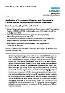
3. RESULTS Portunus sayi have a largely translucent cuticle, with their overall color patterns apparently determined by the distribution of chromatophores in the dermal tissue (Fig 1A, B). When molted, the cuticle shows a tint and may play a large role in the overall color of the animal (Fig 1C). The central dorsal white spot, as well as other smaller bright markings, is mirrored on the discarded 47
molt. The molted outer carapace is significantly more reflective across the region considered than the intact individual (Fig 1D).
Figure 1. Portunus sayi translucent cuticle. A: Magnified view of juvenile P. sayi. Chromatophores are evident beneath semi-translucent outer cuticle. B: RGB image of adult male, < 12 hrs before ecdysis. C: Psuedo RGB image of discarded cuticle. D: Mean carapace reflectance R(λ) for the pre-ecdysis individual (black line) is much lower than for the discarded cuticle (grey line), which displays a slight chlorophyll-like reflectance feature at 675 - 700 nm. Error bars are +/- 1 standard deviation. 48
3.1 Diel Color Change Five individuals survived for the duration of the experiment. Among these, a clear trend of dark, melanistic coloration during the day and paler shading at night was observed for individuals exposed to natural illumination (Fig 2). Median carapace reflectance spectra (Fig 2A) illustrate diurnal melanism (Fig 2B) with associated low reflectance, and pale nocturnal coloration (Fig 2C) with significantly higher R(λ). Spectrally, reflectance showed the greatest change in the red wavelengths, and lowest change in the blue.
49
Figure 2. Diurnal/nocturnal coloration of Portunus sayi. Median carapace reflectance of all individuals (A) was significantly higher at all wavelengths at night (dashed line) than during the day (solid line). Spectra are a median carapace R(λ) for all individuals, averaged over 2 observations. Error bars are +/- 1 pooled standard deviation. Subjectively, individuals (adult female shown for example) are considerably darker during daylight hours (B) than at night (C).
Crabs held on live Sargassum under constant illumination did not show the same diel cycle (Fig 3) as those in natural light. Integrated reflectance ΣR was low during daytime measurements (Fig
50
3A) and significantly higher at night for individuals under natural illumination. For those under constant illumination, ΣR increased over the experimental period regardless of time of measurement (Fig 3B). After being returned to a natural illumination cycle, crabs exposed to constant illumination darkened and returned to initial ΣR values by 96 hours.
51
Figure 3. Integrated reflectance ΣR of crabs exposed to natural illumination (A) and constant illumination (B). A clear cycle of low R during the day and higher R at night was observed for crabs exposed to natural lighting, which was absent from individuals under constant illumination. Double bars indicate return of crabs under constant illumination to natural light cycle. Outliers are marked by crosses. 52
3.2 Response to Background Aggregate reflectances do indicate group differences in coloration with background albedo after acclimation (Fig 4). Responses were highly variable on the individual level, and all individuals regardless of treatment group increased reflectance over time. At 36 hours however, crab reflectances were lower than at 24 hours, but increased by 48 hours.
53
Figure 4. Group mean reflectance R(λ) of P. sayi initially (A), and after exposure to Sargassum, white, grey, and black backgrounds for 24 (B) and 48 (C) hours. While individual response was
54
highly variable, group spectra do indicate a graded background albedo response. Reflectance of all individuals increased over time. Error bars are +/- 1 pooled standard deviation. For illustration, crabs acclimated to white backgrounds were significantly lighter than their initial state (inset photos).
Mean reflectance of all groups was equal initially (Fig 4A). After 24 hours (Fig 4B), black maintained crabs showed the lowest reflectance values, and acclimated reflectances were lower than the control group at all wavelengths (p < 0.05 400 – 440 nm, p < 0.1 400 – 700 nm). Grey crab reflectance was (non-significantly) lower than the control at wavelengths > 550 nm. Crabs maintained on white backgrounds showed (non-significantly) higher reflectance than the control at wavelengths > 550 nm by 24 hours (Fig 4B), and were noticeably lighter than their initial state (Fig 4 inset images). After 48 hours (Fig 4C), control, white, and grey treatment mean reflectance was virtually identical at all wavelengths. Black treatment reflectance was lower than the control at all wavelengths, significantly (p < 0.1) at < 500 nm. Integrated reflectance ΣR was not significantly different among treatments during initial measurements (Fig 5), and increased over time for all groups. At 12, 24, and 36 hours, ΣR of treatment groups followed background albedo. That is, median ΣR was greatest for white crabs and lowest for black treated crabs. Black treated crabs showed the lowest ΣR of all groups after acclimation at each measurement time, and was different from the control (Mann-Whitney U test) at 24 (p = 0.09), and 36 (p = 0.01) hours.
55
Figure 5. Integrated reflectance ΣR of crabs exposed to monochrome backgrounds. All crabs showed increased ΣR over time. Median ΣR of treatment groups followed background albedo at 12, 24, and 36 hours. Outliers are marked by crosses.
56
4. DISCUSSION 4.1 Color Change in Response to Environmental Conditions Data presented here show that P. sayi have the ability to drastically alter their overall carapace coloration within several hours. The absence of regular diel melanistic/pale cycles as seen in crabs exposed to constant illumination demonstrates that such cycles are partially controlled by ambient light and are at least largely non-circadian, unlike similar cycles in some other crabs [14,15,28]. The possibility of protection from ultra violet radiation has been posited for diel cycles involving a dark phase during daylight hours [9,12,22], particularly in semi-terrestrial Uca, in which direct melanistic response to exposure to wavelengths between 300 – 400 nm [23] and visible wavelengths [22,24] has been studied. For P. sayi, which are subject to high irradiance in lowlatitude surface waters, expanded chromatophores could serve a protective role by absorbing damaging UV photons [50]. The apparent cycle in coloration observed here may potentially be the result of energetic costs associated with pigmentation [51,52]. Maintaining expanded dark chromatophores, including the potential replacement of pigments damaged or destroyed by UV absorption, will presumably impose some metabolic cost. At night, the benefit for this cost (i.e. photoprotection) would be absent and paler nocturnal coloration would represent a simple decrease in metabolic expenditure. Further, visual predation is presumably reduced at night [53– 55] and trade-offs between body color, thermoregulation, photoprotection and crypsis could be largely irrelevant. Thermoregulation represents a possible function for light/dark pigmentation cycles. Uca fiddler crabs on exposed mudflats, for example, are subject to large temperature variation on short time
57
scales. U. pugilator changes from dark to light coloration in response to increasing temperature, with a demonstrated difference in body temperature between chromatic states [1,21]. A cycle of darker coloration during the day and lighter color at night has been observed in this species [12,13,21], similar to that seen here. Detailed temperature information was not collected during the present study, and is a possible factor influencing color change in P. sayi. An albedo response, where the individual changes shading in response to the reflectance of the background, has been observed to varying extents in crab species including multiple in the genus Uca [13,19,25], C. sapidus [37], Sesarma reticulatum [38,56], Carcinus maenas [57], Ocypode ceratophthalmus [5] and a variety of other crustaceans [9,11,39,58]. The trends in reflectance relative to certain backgrounds observed here indicates that physiological color change may have potential for dynamic camouflage in P. sayi, but further research is required. We are aware of a small number of previous studies on color change in Sargassum crustaceans. The other species of associated crab, Planes minutus, possesses three chromatophore classes: white, yellow, and black [34]. However, overall coloration of the animals was not adequately reported and no appreciable response to the reflectance of the background was observed. This was suggested to be due to “extra-chromatophoral” pigments in the dermal cells and faint coloration of the exoskeleton, observed upon molting. The shrimp Latreutes fucorum is extremely abundant in the pelagic Sargassum complex [40,42]. L. fucorum are found in a wide variety of colors and patterns, and responded to background albedo with chromatophore expansion on black backgrounds and contraction on white [39]. Chromatophore response in crabs can be divided into two mechanisms [11,59]. Primary chromatophore responses involve non-ocular pathways, such as the action of light directly on the chromatophore. Secondary responses in crustaceans are initiated by light perceived by the eye 58
and mediated by the sinus gland in the eyestalks [15,60,61]. This is the mechanism involved in albedo response [11,13,59,62]. As with other crustacean species, color change in P. sayi is likely controlled by a complex interaction of factors including background albedo, illumination, temperature, and possibly circadian or other rhythms [2,11,14,15,17,19,25]. At 36 hours, all crabs in the albedo treatment showed a decrease in reflectance from 24 hours, diverging from the otherwise linear increase observed. This could be the result of a factor outside of experimentation such as those previously discussed. Sex and reproductive status may also be relevant [1,2,35], and future work should examine color change in respect to such factors. The majority of individuals used in this study were female. It is unknown if this results from actual population dynamics or an unidentified bias in collection procedure. The impact of laboratory light levels seems to be a compounding variable when testing color response to background. The present observation of lighter coloration in crabs exposed to laboratory illumination was not expected, and may be due to physiological stress from experimental conditions. Interestingly, this response was opposite to the natural condition of paler shading in response to darkness and may indicate a response to lower light intensity in the lab compared to typical day-time light levels in the region. This and similar complications have been observed in other studies [13,37]. Those crabs exposed to constant illumination began to regain their diel color cycle after being returned to natural lighting. This would indicate that the progressive increase in ΣR was part of a response to experimental conditions, and not likely the result of any permanent degeneration or health issues induced by experimental stress.
59
4.2 Camouflage Through Morphological Color Change The slower acting mechanism of morphological color change, in which new pigments are created, has been observed to serve in background color matching for several crab species [63– 67]. The other Sargassum associated crab, Planes minutus, also appears to employ morphological color change. Crozier [33] noted that P. minutus was able to “harmonize” with the deep mahogany color of a drifting cedar log. Later, Hitchcock [34] documented their chromatophore response on different colored backgrounds, but found that extracellular pigmentation in hypodermis and exoskeleton prevented rapid color change. We have similarly found that P. minutus were able to achieve a deep red color when found on a red plastic bucket [43], though the time period required to match this background is unknown. The degree to which pigmentation in the cuticle impacts overall coloration in P. sayi has yet to be determined, but examination of the molts seen during this study indicates it is likely to play a role. 5. CONCLUSIONS Like many other marine crustaceans, Portunus sayi exhibits physiological color change in response to environmental conditions. Under natural illumination, the species displays a clear diel cycle of coloration through a physiological pathway. During daylight hours, adults have a highly contrasting pattern of light and dark, with brown, yellow, and white elements. This shifts to mostly yellow and white at night. Crabs exposed to continuous illumination did not show such a cycle. When placed on backgrounds of varying albedo, P. sayi appears to change coloration according to the background, being less reflective on black backgrounds and more reflective on brighter ones, though individual response was highly variable. Further research is needed to determine if sex or body size are important to camouflage or pigmentation cycles in P. sayi as in
60
other species, or if morphological change might function to improve background matching as in P. minutus.
61
REFERENCES 1. Munguia P, Levinton JS, Silbiger NJ. Latitudinal differences in thermoregulatory color change in Uca pugilator. J Exp Mar Biol Ecol. 2013 Feb;440:8–14. 2. Silbiger N, Munguia P. Carapace color change in Uca pugilator as a response to temperature. J Exp Mar Biol Ecol. 2008 Feb;355(1):41–6. 3. Fingerman M. Chromatophores. Physiol Rev. 1965;45:296–339. 4. Hemmi JM, Marshall J, Pix W, Vorobyev M, Zeil J. The variable colours of the fiddler crab Uca vomeris and their relation to background and predation. J Exp Biol. 2006 Oct 15;209(20):4140–53. 5. Stevens M, Rong CP, Todd PA. Colour change and camouflage in the horned ghost crab Ocypode ceratophthalmus. Biol J Linn Soc. 2013;109(2):257–70. 6. Stevens M, Lown AE, Wood LE. Camouflage and Individual Variation in Shore Crabs (Carcinus maenas) from Different Habitats. Eklöv P, editor. PLoS ONE. 2014 Dec 31;9(12):e115586. 7. Detto T, Backwell PR., Hemmi JM, Zeil J. Visually mediated species and neighbour recognition in fiddler crabs (Uca mjoebergi and Uca capricornis). Proc R Soc B Biol Sci. 2006 Jul 7;273(1594):1661–6. 8. Detto T. The fiddler crab Uca mjoebergi uses colour vision in mate choice. Proc R Soc B Biol Sci. 2007 Nov 22;274(1627):2785–90. 9. Umbers KDL, Fabricant SA, Gawryszewski FM, Seago AE, Herberstein ME. Reversible colour change in Arthropoda: Arthropod colour change. Biol Rev. 2014 Nov;89(4):820– 48. 10. Horst MN, Freeman JA. The Crustacean Integument: Morphology and Biochemistry. Boca Raton, FL: CRC Press; 1993. 240 p. 11. Fingerman M. The Control of Chromatophores. New York: Macmillan; 1963. 192 p. (International series of monographs on pure and applied biology. Zoology division). 12. Abramowitz AA. The chromatophorotropic hormone of the Crustacea: Standardization, properties and physiology of the eye-stalk glands. Biol Bull. 1937 Jun;72(3):344. 13. Brown FA, Sandeen MI. Responses of the chromatophores of the fiddler crab, Uca, to light and temperature. Physiol Zool. 1948;361–71. 14. Fingerman M. Persistent daily and tidal rhythms of color change in Callinectes sapidus. Biol Bull. 1955;109(2):255–64. 15. Thurman CL. Rhythmic physiological color change in crustacea: A review. Comp Biochem Physiol PART C Comp Pharmacol. 1988;91:171–85.
62
16. Fingerman M. Phase Difference in the Tidal Rhythms of Color Change of Two Species of Fiddler Crab. Biol Bull. 1956 Jun;110(3):274. 17. Detto T, Hemmi JM, Backwell PRY. Colouration and Colour Changes of the Fiddler Crab, Uca capricornis: A Descriptive Study. Rands S, editor. PLoS ONE. 2008 Feb 20;3(2):e1629. 18. Fingerman M, Lowe ME, Mobberly WC. Environmental Factors Involved in Setting the Phases of Tidal Rhythm of Color Change in the Fiddler Crabs Uca pugilator and Uca minax. Limnol Oceanogr. 1958;3(3):271–82. 19. Brown FA. Studies on the Physiology of Uca Red Chromatophores. Biol Bull. 1950 Jun;98(3):218. 20. Kronstadt SM, Darnell MZ, Munguia P. Background and temperature effects on Uca panacea color change. Mar Biol. 2013 Jun;160(6):1373–81. 21. Wilkens JL, Fingerman M. Heat tolerance and temperature relationships of the fiddler crab, Uca pugilator, with reference to body coloration. Biol Bull. 1965;128(1):133–41. 22. Coohill TP, Fingerman M. Relative effectiveness of ultraviolet and visible light in eliciting pigment dispersion in melanophores of the fiddler crab, Uca pugilator, through the secondary response. Physiol Zool. 1975;57–63. 23. Coohill TP, Bartell CK, Fingerman M. Relative effectiveness of ultraviolet and visible light in eliciting pigment dispersion directly in melanophores of the fiddler crab, Uca pugilator. Physiol Zool. 1970;43(3):232–9. 24. Coohill TP, Fingerman M. Comparison of the effects of illumination on the melanophores of intact and eyestalkless fiddler crabs,Uca pugilator, and inhibition of the primary response by cytochalasin B. Experientia. 1976;32(5):569–70. 25. Barnwell FH. Comparative Aspects of the chromatophoric responses to light and temperature in fiddler crabs of the genus Uca. Biol Bull. 1968 Apr;134(2):221. 26. Herreid CF, Mooney SM. Color change in exercising crabs: evidence for a hormone. J Comp Physiol B. 1984;154(2):207–12. 27. Thurman CL. Adaptive coloration in Texas fiddler crabs (Uca). In: Wickstein M, editor. Adaptive coloration in invertebrates. Texas: Texas A&M University Press; 1990. p. 109– 26. 28. Brown FA, Fingerman M, Sandeen MI, Webb HM. Persistent diurnal and tidal rhythms of color change in the fiddler crab, Uca pugnax. J Exp Zool. 1953;123(1):29–60. 29. Zeil J, Hofmann M. Signals from “crabworld”: cuticular reflections in a fiddler crab colony. J Exp Biol. 2001;204(14):2561–9. 30. Crane J. On the color changes of fiddler crabs in the field. Zoologica. 1944;29:161–8.
63
31. Shih H-T, Mok H-K, Chang H-W, Lee S-C. Morphology of Uca formosensis Rathbun, 1921 (Crustacea: Decapoda: Ocypodidae), an endemic fiddler crab from Taiwan, with notes on its ecology. Zool Stud-TAIPEI-. 1999;38:164–77. 32. Darnell MZ. Ecological physiology of the circadian pigmentation rhythm in the fiddler crab Uca panacea. J Exp Mar Biol Ecol. 2012 Sep;426-427:39–47. 33. Crozier WJ. Note on the coloration of Planes minutus. Am Nat. 1918 May;52(616/617):262– 3. 34. Hitchcock HB. The coloration and color changes of the gulf-weed crab, Planes minutus. Biol Bull. 1941;26–30. 35. Jensen GC, Egnotovich MS. A whiter shade of male: Color background matching as a function of size and sex in the yellow shore crab Hemigrapsus oregonensis (Dana, 1851). Curr Zool. 2015;61(4):729–38. 36. Granato FC, Tironi TS, Maciel FE, Rosa CE, Vargas MA, Nery LEM. Circadian rhythm of pigment migration induced by chromatrophorotropins in melanophores of the crab Chasmagnathus granulata. Comp Biochem Physiol A Mol Integr Physiol. 2004 Jul;138(3):313–9. 37. Fingerman M. Physiology of the black and red chromatophores of Callinectes sapidus. J Exp Zool. 1956;133(1):87–105. 38. Fingerman M, Nagabhushanam R, Philpott L. Physiology of the melanophores in the crab Sesarma reticulatum. Biol Bull. 1961;337–47. 39. Brown FA. The coloration and color changes of the gulf-weed shrimp, Latreutes fucorum. Am Nat. 1939;73:564–8. 40. Butler JN, Morris BF, Cadwallader J, Stoner AW. Studies of Sargassum and the Sargassum community. Bermuda Biological Station for Research, St Georges 22; 1983. 41. Hacker SD, Madin LP. Why habitat architecture and color are important to shrimps living in pelagic Sargassum: use of camouflage and plant-part mimicry. Mar Ecol Prog Ser. 1991;70:143–55. 42. Morris BF, Mogelberg DD. Identification manual to the pelagic Sargassum fauna. Bermuda Biological Station for Research, St Georges 22; 1973. 61 p. (Special Publication). 43. Russell BJ, Dierssen HM. Use of hyperspectral imagery to assess cryptic color matching in Sargassum associated crabs. PloS One. 2015;10(9):e0136260. 44. Brooks WR, Hutchinson KA, Tolbert MG. Pelagic Sargassum mediates predation among symbiotic fishes and shrimps. Gulf Mex Sci. 2007;2:144–52. 45. Russell BJ, Dierssen HM. Hyperspectral imaging as a tool for camouflage evaluation of the Sargassum crab Portunus sayi. In Glasgow, Scotland; 2012.
64
46. Jobe CF, Randy Brooks W. Habitat selection and host location by symbiotic shrimps associated with Sargassum communities: The role of chemical and visual cues. Symbiosis. 2009 Oct;49(2):77–85. 47. Chace FA. The oceanic crabs of the genera Planes and Pachygrapsus. Proc U S Natl Mus. 1951;101(3272). 48. Dierssen HM, Chlus A, Russell B. Hyperspectral discrimination of floating mats of seagrass wrack and the macroalgae Sargassum in coastal waters of Greater Florida Bay using airborne remote sensing. Remote Sens Environ. 2015 Sep;167:247–58. 49. Russell B, Dierssen H, LaJeunesse T, Hoadley K, Warner M, Kemp D, et al. Spectral reflectance of Palauan reef-building coral with different symbionts in response to elevated temperature. Remote Sens. 2016 Feb 23;8(3):164. 50. Gouveia GR, Lopes TM, Neves CA, Nery LEM, Trindade GS. Ultraviolet radiation induces dose-dependent pigment dispersion in Crustacean chromatophores. Pigment Cell Res. 2004;17(5):545–8. 51. Emery CJ. The ecological impact of near ultraviolet radiation on Daphnia pulex. [Master’s]. [Ontario]: University of Windsor; 1984. 52. Korínek V, Frey DG, editors. Biology of Cladocera: Proceedings of the Second International Symposium on Cladocera, Tatranska Lomnica, Czechoslovakia, 13–20, September 1989. Vol. 71. Springer Science & Business Media; 2013. 53. De Robertis A. Size-dependent visual predation risk and the timing of vertical migration: an optimization model. Limnol Oceanogr. 2002;47(4):925–33. 54. Ballance L, Pitman R. S34. 4: Foraging ecology of tropical seabirds. In: Proc 22 Int Ornithol Congr, Durban. Citeseer; 1999. p. 2057–71. 55. Oro D, Martínez-Abraín A. Ecology and behavior of seabirds. In: Duarte CM, Lota A, editors. Marine Ecology, Encyclopedia of Life Support Systems (EOLSS). Oxford: Eolss Publishers-UNESCO; 56. Fingerman M, Nagabhushanam R, Philpott L. Photomechanical responses of the proximal pigment in Palaemonetes and Orconectes. Biol Bull. 1962 Aug;123(1):121. 57. Stevens M, Lown AE, Wood LE. Color change and camouflage in juvenile shore crabs Carcinus maenas. Front Ecol Evol. 2014;2. 58. Hultgren KM, Mittelstaedt H. Color change in a marine isopod is adaptive in reducing predation. Curr Zool. 2015;6(4):739–48. 59. Fingerman M, Tinkle DW. Responses of the white chromatophores of two species of prawns (Palaemonetes) to light and temperature. Biol Bull. 1956;110(2):144–52. 60. Brown FA. The controlling mechanism of chromatophores in Palaemonetes. Proc Natl Acad Sci. 1933;19(3):327–9. 65
61. Welsh JH. Diurnal rhythms. Quart Rev Biol. 1938;13:123–39. 62. Powell BL. The responses of the chromatophores of Carcinus maenas (L., 1758) to light and temperature. Crustaceana. 1962;4(2):93–102. 63. Iampietro PJ. Distribution, diet, and pigmentation of the northern kelp crab, Pugettia producta (Randall) in central California kelp forests. [Master’s]. California State University, Stanislaus; 1999. 64. Hultgren KM, Stachowicz JJ. Alternative camouflage strategies mediate predation risk among closely related co-occurring kelp crabs. Oecologia. 2008 Mar;155(3):519–28. 65. Green JP. Morphological color change in the fiddler crab, Uca pugnax (S. I. Smith). Biol Bull. 1964 Oct;127(2):239. 66. Green JP. Morphological color change in the Hawaiian ghost crab, Ocypode ceratophthalma (Pallas). Biol Bull. 1964 Jun;126(3):407. 67. Kolwalkar DG, Rangnekar PV. Morphological color change in the marine crab Portunus pelagicus. J Bombay Nat Hist Soc. 1979;76(3):540–3.
ACKNOWLEDGEMENTS This work was funded by the Office of Naval Research MultiUniversity Research Initiative (N000140911054), and by a pre-doctoral award from the Unviersity of Connecticut Department of Marine Sciences. Statement concerning study animals: All live animals in this study were invertebrates and not subject to University of Connecticut IRB/IACUC requirements. All measures were taken to minimize suffering or stress of subjects. Author Contributions Conceived and designed the experiments: BJR. Performed the experiments: BJR. Analyzed the data: BJR, HMD. Contributed reagents/materials/analysis tools: BJR, HMD. Wrote the paper: BJR, HMD.
66
4. Spectral Reflectance of Palauan Reef-Building Coral with Different Symbionts in Response to Elevated Temperature
This chapter was published in Remote Sensing on February 23, 2016. It appeared in the special issue “Remote Sensing for Coral Reef Monitoring." Russell, BJ; Dierssen, HM; LaJeunesse, TC; Hoadley, KE; Warner, ME; Kemp, DW; Bateman, TG. Spectral Reflectance of Palauan Reef-Building Coral with Different Symbionts in Response to Elevated Temperature. Remote Sens. 2016, 8(3), 164; doi:10.3390/rs8030164
67
68
69
70
71
72
73
74
75
76
77
78
79
80
81
82
83
84
85
86
Appendix: Supplementary Material, Chapter 2 This material appeared with publication of Chapter 2 Russell, BJ; Dierssen, HM. (2015) Use of hyperspectral imagery to assess cryptic color matching in Sargassum associated crabs. PLOS ONE doi:10.1371/journal.pone.0136260
87
88
89
90
91