FTu2F.2.pdf
FiO/LS 2014 © OSA 2014
Application of computer vision technique with fluorescence imaging spectroscopy to differentiate citrus diseases 1
Caio B. Wetterich1, José Belasque Junior2, and Luis G. Marcassa1 Instituto de Física de São Carlos – Universidade de São Paulo, Cx. Postal 369, São Carlos, 13560-970, SP, Brazil. 2 Departamento de Fitopatologia e Nematologia, Universidade de São Paulo, Piracicaba, SP, 13418-900, Brazil
[email protected]
Abstract: We have used fluorescence imaging spectroscopy to investigate citrus diseases. Texture features were extracted and used as input into classifier. Results show that it is possible differentiate the diseases that have similar symptoms. OCIS codes: (000.1430) Biology and Medicine; (300.2530) Fluorescence, laser-induced 1. Introduction In recent years, there has been an increasing in the interest of early detection of plant stress in agricultural crops to prevent great economic losses due contamination of new plants. Fluorescence imaging spectroscopy (FIS) has been investigated as a tool in plant studies. The FIS has a potential to obtain spatial and spectral information than regular imaging techniques [1]. The most important aspect of fluorescence spectroscopy is that the technique is nondestructive and nonintrusive to the plant physiology [2]. The information contained in multispectral images allows the characterization, identification and classification of the diseased samples. The supervised learning method, support vector machines (SVMs), have been successfully used for the classification of multispectral remote sensing images [3,4]. In this work, we will present the development and evaluation of SVM technique for the detection of plant stress in citrus leaves using the fluorescence imaging spectroscopy. We applied the FIS system with the SVM technique for detection and classification of contaminated trees with citrus canker, citrus scab, HLB and zinc deficiency. 2. Material and Methods 2.1
Fluorescence imaging spectroscopy system
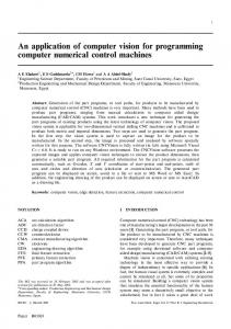
We developed two different systems to select the best emission region of the leaves and collect the images of the diseased samples. The main difference between the two systems is the way to select the wavelength. In the first system, we use a liquid crystal tunable filter (Liquid Crystal Tunable Filter, Meadowlark Optics, USA) that allows us selecting the wavelength of interest from 420 to 750 nm. In the second, we use a filter wheel with some bandpass filters. Initially, it was used the tunable filter to select the best excitations and wavelengths of interest, and then we use the bandpass filters to collect a large number of fluorescence images, because it is faster. Both systems are composed by a monochromatic charged couple device camera (CCD) (model mvBlueFOX120a, Matrix Vision, Germany), an objective lens, high power light emitting diodes (LEDs) at different wavelengths (365, 405, 470 and 530 nm), and a standard laptop computer. All the parts of the system, except the laptop are placed inside a closed box. The CCD, tunable filter and filter wheel were computer controlled. Figure 1 shows a schematic diagram of the portable FIS system. (a) (b)
Fig. 1. Schematics of the two FIS system developed. (1) CCD camera, (2) objective lens, (4) base for high power LEDs at different wavelengths (365, 405, 470 and 530 nm) for excitation. The difference between the two systems is the filter used. The component number 3 on (a) repsesrents the liquid crystal tunable filter and on (b) the filter wheel.
FTu2F.2.pdf
FiO/LS 2014 © OSA 2014
2.2 Leaf sample collection In the exploratory experiment was used the tunable filter system to collect fluorescence images from 10 samples for each of the following conditions: i) citrus canker, ii) citrus scab, iii) HLB and iv) zinc deficiency. So, with the filters selected, the fluorescence data were collected using the filter wheel system to 100 samples for each condition. The images were obtained for the excitations 405 and 470 nm, for the following filters: 530, 550, 560, 570, 580, 630, 690 and 740 nm. The leaf samples came from plants of Citrus reticulata (Blanco), Citrus sinensis (L. Osbeck), and Citrus limonia (L. Osbeck). Traditional diagnostics tests were performed on the samples after acquire de images. The results were used to compose statistical tests for experimental data analysis. 2.3 Segmentation, Feature Selection and Classification The data analysis procedures were written in MATLAB® (The MathWorks Inc., Natick, MA). There are several techniques for segmentation. Following segmentation, the leaf pixels were separated as rectangular areas. These rectangular regions were further processed for feature extraction and classification. Three metrics were computed from the image segment: uniformity, contrast, and homogeneity. The segmentation and feature selection was performed with the fluorescence images at 570, 630 and 690 nm for citrus canker and scab, and 530, 580 and 690 nm for HLB and zinc deficiency. These images were collected for the excitations 405 and 470 nm. The extracted features were used as an input in the classifier support vector machine (SVM). The classifier was applied on two groups of fluorescence images: i) Group 1, which comprises images of citrus canker and scab; ii) Group 2, with images of HLB and zinc deficiency. This separation is due only to the fact that we have problems to differentiate citrus canker from leaf miner, and HLB from zinc deficiency. 3.
Results and Discussion
The classification results are presented in Table 1. The best results were obtained when we considered the two excitations and all fluorescence measurements for the two sample groups. The overall accuracy and specificity of SVM for the Group 1 were 94% and 87%, and for the Group 2 were 94% and 89%, respectively. The classification accuracy was the same, but the specificity was a little lower for the Group 1.We noticed that some samples that were visually classified as HLB by the Fundecitrus technicians were negative for the disease. Furthermore, two samples of zinc deficiency were misclassified as HLB by the fluorescence method. Table 1. Classification results from support vector machine classifier. Diseases Sensitivity Specificity Accuracy Citrus Canker and Scab (Group 1) 100% 87% 93,5% HLB and Zinc Deficiency (Group 2) 100% 89,1% 94% 4.
Conclusions
In summary, we have proposed the use of a fluorescence imaging spectroscopy technique as a mean for fast identification of the main diseases that affect the citrus orchards. The fluorescence imaging system was developed and the diagnostic procedure was applied to citrus canker, citrus scab, HLB and zinc deficiency. The classification results indicated that this method presents a high accuracy when compared either samples with citrus canker and citrus scab (93,5%), or samples with HLB and zinc deficiency (94%). Furthermore, the specificity obtained for each group is also high. So, it is possible using this system to differentiate the diseases that have similar symptoms, like citrus canker with citrus scab, and HLB with zinc deficiency. 5.
Acknowledgements
This work was supported by Fapesp (Fundação de Amparo à Pesquisa do Estado de São Paulo). 6.
References
[1] F. M. V. Pereira, D. M. B. P. Miloria, E. R. Pereira-Filho, A. L. Venâncioa, M. T. Russo, M. C. Cardinali, P. K. Martins, J. Freitas-Astúa, “Laser - induced fluorescence imaging method to monitor citrus greening disease,” Computers and Electronics in Agriculture, v. 79, no 1, pp. 90-93, (2011). [2] C. B. Wetterich, R. Kumar, S. Sankaran, J. Belasque Junior, R. Ehsani, L. G. Marcassa, “A Comparative Study on Application of Computer Vision and Fluorescence Imaging Spectroscopy for Detection of Citrus Huanglongbing Disease in USA and Brazil,” Journal of Spectroscopy, v. 2013, p. 1-6, (2013). [3] S. Sankaran, A. Mishra, R. Ehsani, C. Davis, “A review of advanced techniques for detecting plant diseases,” Computers and Electronics in Agriculture, v.72, n.1, p. 1-13, (2010). [4] E. C. Lins, J. Belasque, Jr., and L. G. Marcassa, “Optical fiber laser induced fluorescence spectroscopy as a citrus canker diagnostic,” Applied Optics, v. 49, no 4, pp. 663-667, (2010).


![[PDF] OpenCV 3 Computer Vision Application Programming ...](https://m.moam.info/img/260x300/pdf-opencv-3-computer-vision-application-programmi_64786c04097c4737708cc9c6.jpg)
