DOI: 10.3171/2007.8.17566
Who is who: areas of the brain associated with recognizing and naming famous faces Clinical article Carlo Giussani, M.D.,1,2 Franck-Emmanuel Roux, M.D., Ph.D.,1,3 Lorenzo Bello, M.D., 2 Valérie Lauwers-Cances, M.D.,4 Costanza Papagno, M.D., Ph.D., 5 Sergio M. Gaini, M.D., 2 Michelle Puel, M.D.,1 and Jean-François Démonet, M.D., Ph.D.1 Institut National de la Santé et de la Recherche Médicale, Unité 825, and 3Federation of Neurosurgery, Hôpital Purpan; 4Service d’Epidemiologie, Centres Hospitalo-Universitaires, Toulouse, France; 2 Department of Neurological Sciences, Neurosurgery, University of Milan; and 5Neuropsychobiology of Language, Department of Psychology, University of Milano Bicocca, Italy 1
Object. It has been hypothesized that specific brain regions involved in face naming may exist in the brain. To spare these areas and to gain a better understanding of their organization, the authors studied patients who underwent surgery by using direct electrical stimulation mapping for brain tumors, and they compared an object-naming task to a famous face–naming task. Methods. Fifty-six patients with brain tumors (39 and 17 in the left and right hemispheres, respectively) and with no significant preoperative overall language deficit were prospectively studied over a 2-year period. Four patients who had a partially selective famous face anomia and 2 with prosopagnosia were not included in the final analysis. Results. Face-naming interferences were exclusively localized in small cortical areas (< 1 cm2). Among 35 patients whose dominant left hemisphere was studied, 26 face-naming specific areas (that is, sites of interference in face naming only and not in object naming) were found. These face naming–specific sites were significantly detected in 2 regions: in the left frontal areas of the superior, middle, and inferior frontal gyri (p < 0.001) and in the anterior part of the superior and middle temporal gyri (p < 0.01). Variable patterns of interference were observed (speech arrest, anomia, phonemic, or semantic paraphasia) probably related to the different stages in famous face processing. Only 4 famous face–naming interferences were found in the right hemisphere. Conclusions. Relative anatomical segregation of naming categories within language areas was detected. This study showed that famous face naming was preferentially processed in the left frontal and anterior temporal gyri. The authors think it is necessary to adapt naming tasks in neurosurgical patients to the brain region studied. (DOI: 10.3171/2007.8.17566)
Key Words • brain mapping • cortical stimulation • face naming
D
uring the Cubism Period in 1910 when Pablo Picasso (1881–1973) painted the “Portrait of Ambroise Vollard” (an art dealer), he perfectly captured the idea of prosopagnosia: in the painting the model’s face is split up in many facet-like shapes, confounding the features that would allow him to be recognized. Although accounts of prosopagnosia have been identified since antiquity in the writings of Thucydides, Abbreviations used in this paper: IFG = inferior frontal gyrus; ITG = inferior temporal gyrus; MFG = middle frontal gyrus; MTG = middle temporal gyrus; SFG = superior frontal gyrus; STG = superior temporal gyrus.
J. Neurosurg. / October 17, 2008
Pliny the elder, and Seneca, this term was coined only in 1947 by the German neurologist Joachim Bodamer from classical Greek “prosopon” meaning “face.” Authors of several brain lesion studies hypothesized that face recognition is composed of 4 sequential stages:1,3,8,11,13,14,18 1) facial feature encoding; 2) perceptual recognition; 3) semantic/biographic associations; and 4) face naming. Neurophysiological30,32,47 and neuroimaging2,23,24,35,36,40 studies have shown the crucial and specific roles of bilateral hippocampus/parahippocampus and particularly of the right ventral occipitotemporal areas (fusiform face area) in face perception and recognition. Face naming, the final end point of face recognition, 1
C. Giussani et al. could depend on the left temporal areas as suggested by studies focused on patients with temporal lobe epilepsy.17,38 A selective impairment in the production of people’s names without deficits in other naming categories (naming of handmade objects, fruits, and animals, for instance) has been described in patients harboring left temporal lobe lesions.25,28,33,39,41,46 Moreover some functional neuroimaging studies have shown the specific implication of the left premotor cortex and of the left temporal cortex in the face-naming process.15,16 These data suggest that the retrieval of proper names, which represent concrete entities belonging to distinct conceptual categories, could depend on partially segregated regions in higher-order cortices. Nevertheless, the existence of these areas and their localization is still a matter of debate.6,7,42 In our institutions, direct brain mapping is used routinely in patients with brain tumors to spare languagerelated cortical areas. In an attempt to spare hypothetical category-specific cortical areas at surgery and to better understand the anatomical basis of face specific–naming areas within language areas, we studied a prospective group of 56 patients with brain tumors. After neuropsychological and language assessments, 50 patients underwent craniotomies and brain mapping with a standard object-naming and category-specific task (famous face naming) using visually presented stimuli during direct cortical mapping. To the best of our knowledge, this is the first direct cortical stimulation study focused on famous face naming. The functional/anatomical organization of face-naming areas is analyzed in relation to the surgical data.
Preoperative and Postoperative Assessments
Methods
All patients in this series underwent pre- and postoperative language examination to rule out specific language deficits. The language evaluation of the Italian and French groups of patients was performed separately at each institution by neurologists and neuropsychologists. The 2 teams, specialized in language disorders, used standardized and equivalent tests. This testing included written and oral comprehension, language fluency, naming, computation, writing to dictation, repetition, and evaluation of apraxia. Possible category-specific naming deficits were evaluated by a subset of specific-naming categories. All patients underwent a famous face recognition and naming test. Patients were tested on their ability to match unfamiliar and familiar faces, and identify a familiar face among face triplets consisting of 1 famous face and 2 foils. They were also asked to identify facial expressions; finally, famous face naming was assessed by means of a data set of 30 familiar famous faces. The famous face–naming data sets were appropriately chosen according to the patient’s cultural degree, job, hobbies, and chronological adequacy between the patient’s age and the period of popularity of the famous person. Patients with language impairment (> 10% errors in naming tests) were not included in this study. Patients with famous–face naming deficits, even if able to retrieve biographic details about the stimuli, were excluded according to the objectives of this category-specific naming study. The degree of patients’ handedness was assessed using the Edinburgh Handedness Inventory test.31 One bilingual French/English patient was tested in both languages with famous faces of native English speakers. Postoperatively as well as preoperatively, the patients were asked to perform the same tests so that a language problem might be checked.
Between November 2004 and November 2006, 2 neurosurgical teams at the Universities of Toulouse and Milan used intraoperative direct cortical stimulation in a prospective group of 56 patients with brain tumors; 44 patients were native to France and 12 to Italy (mean age 47 years, range 22–74 years). The goal of these brain-mapping studies was to spare all language areas found during tumor removal. As suggested by other authors,44 it has been the policy of our 2 departments to perform, when feasible, the awake surgery technique with direct brain mapping in patients with brain tumors in whom functional areas are at risk during the resection. Although infrequently, the right hemisphere can sometimes be involved in some language or neurocognitive tasks,10,45 especially in face processing.11,12,17,38,41 Seventeen patients with brain tumors in the right hemisphere underwent surgery with the aid of a standard brain mapping technique because a mapping of the rolandic area was needed or a right language localization was suspected. Histopathological analysis of the lesions indicated the following tumor types: low-grade gliomas (World Health Organization Grades I and II, 29 patients), high-grade gliomas (World Health Organization Grades III and IV, 20 patients), cavernoma (4 patients), and metastasis (3 patients).
Our brain mapping procedure consists of an objectnaming task (animate and inanimate objects) because of its accepted reproducibility and efficacy in searching for standard anomia. Reading tasks are also used. Before concluding the direct brain mapping procedure, we routinely confirm essential naming sites by repeating an object-naming task. In this series, we used a famous face–naming task for the repeated naming procedure. Findings on different tasks may be affected or biased by the frequency or familiarity of the items presented during each task. Therefore, low-frequency and low-familiarity objects for which the names are more difficult to retrieve than those of common items could lead to a difference in task performance that may not reflect a category effect but rather a nonspecific one.5 The object-naming task and the famous face–naming task are not equivalent in terms of lexical frequency and complexity. This lack of equivalence could be explained by the differences existing between these 2 categories. In fact a face is a “complex object.” It is recognized as single exemplar of a category with specific geometric features and specific semantic and biographic features, whereas a single apple represents the apple category. For this reason, across-task differences in naming interferences can be accounted for by lexical-frequency effects.
Patient Population
2
Brain Mapping Tasks and Tasks Difficulty Comparison
J. Neurosurg. / October 17, 2008
Famous face naming Cortical Mapping Procedures
All patients gave their informed consent to this study of language areas. All surgical procedures at the 2 institutions were performed using the awake surgery technique.29 A neuronavigational system was used in all the patients (in Milan, Radionics; in Toulouse, StealthStation, Sofamor Danek). The brain was exposed in a standard fashion. Anatomical relationships among gyri were defined by using neuronavigation. Intraoperative cortical stimulation was used to localize areas of the functional cortex after the afterdischarge threshold was determined using electrocorticography. The cortex was directly stimulated by a bipolar electrode of a cortical stimulator with 1-mm electrodes separated by 5 mm (in Milan, Ojemann cortical stimulator, Lifesciences Corp.; in Toulouse, Nimbus Newmedic International s.a.s.). The current amplitude was progressively increased by 1-mA increments starting at 2 mA. We used a procedure of stimulation that included biphasic square wave pulses of 1 msec at 60 Hz, with a maximum train duration of 4 seconds. Cortical sites exposed by the craniotomy were randomly tested under direct stimulation during the different tasks. When a functional site was located, it was marked with a 0.25-cm2 sterile label and another area was tested. Our strategy was to spare the language areas found using these tasks by avoiding resection of tumor tissue located within 1 cm from the eloquent cortex (distance between the resection margin and the nearest functional site). Conditions of Validation of the Study
Strict conditions of language interference sites validation were applied and can be summarized in 5 points. 1) To ensure the conditions of uniformity and thus comparability of brain mappings between the 2 teams (Toulouse and Milan), the 2 neurosurgical teams applied prospectively the same criteria in the selection of candidates for awake surgery. We made uniform the intraoperative cortical stimulation technique and collected together the data from the successive brain mappings by using the same protocol and the same database. Finally, the data were analyzed and discussed collegially by the 2 teams. 2) To be accepted as a language area, the language sites that we localized were tested ≥ 3 times. The sites that did not present reproducible language interferences were not included in this study. 3) The cortical mapping procedures were recorded on video. Oral answers of each patient were recorded using a microphone placed near the patient’s mouth so that his or her responses could be further analyzed in team meetings. 4) Because they can be considered as nonspecific, the language interferences found in the pre- and postcentral gyrus (considered as language interferences due to blockade of articulatory mechanisms) were not included in the final analysis. 5) It must be emphasized that we qualified a site as “famous face–naming specific” when no interference in object naming was found at that site. However, we cannot completely exclude the possibility that other functions (such as other category-specific naming) not tested in this study could be revealed by stimulation of the famous face–specific site.
J. Neurosurg. / October 17, 2008
Statistical Analysis
During this study, all data regarding brain-mapping results were integrated into an Excel database (Microsoft Corp.). In presenting our data, we chose to define regions by using the gyral/sulcal anatomy. Fifteen regions were defined in the left hemisphere and 15 in the right hemisphere. For instance, the SFG, MFG, and IFG were considered as 3 distinct regions. Large gyri, such as the 3 temporal gyri, were arbitrarily divided into 3 segments by drawing an imaginary line extending to the pre- and postcentral sulci. The supramarginal and angular gyri, and the upper parietal lobe were also considered as 3 distinct regions. We analyzed the brain-mapping data by separating famous face–naming sites from object-naming sites. Secondarily, we analyzed the interdependence of famous face–naming sites to object-naming sites as a function of the location and the type of response obtained. For each stimulated zone, the percentage of responses was analyzed according to the number of stimulations performed. Statistical analysis was carried out with unbalanced repeated-measures analysis of variance after transformation of percentage values to obtain a normal distribution.50 The respective probability value was corrected for lack of sphericity by using the Box conservative epsilon. The differences were estimated as significant at a probability value < 0.05. The analysis was performed using Stata 7.0 Statistical Software (Stata Corp.). This statistical analysis was performed by an independent statistician (V.L.C.).
Results
A total of 51 cortical stimulation studies were performed in 50 patients (36 brain mappings in the left hemisphere [1 left-handed] and 15 in the right [2 left-handed]). One bilingual patient underwent 2 brain-mapping procedures in the left hemisphere. One hundred forty-two interference sites (excluding “motor” sites) were found: 138 in the left and 4 in the right hemisphere (all face-naming sites). Of the 138 interferences found in the left hemisphere, 70 were object-naming and 68 face-naming interferences. Some cortical regions were not studied in this series such as bilateral occipital lobes, interhemispheric regions, basal temporal lobes (lingual and fusiform gyri), and the nondominant inferior temporal gyrus. Other cortical regions were studied at least once. The maximal current that did not evoke afterdischarges ranged from 4.5 to 8 mA. All the object- or famous face–naming interference sites found were spared during surgery. Among patients tested preoperatively by our neuropsychological and physiotherapy team, 4 patients had a partially selective famous and relative’s face anomia. All of them had tumors in the anterior part of the temporal lobe (Fig. 1). Two patients with a right occipitotemporal lobe tumor had a typical prosopagnosia (Fig. 2). These patients could not undergo surgery and face-naming tasks. Patients’ data are summarized in Table 1. Famous Face Naming: Specific Findings
Concordance Between the Object Versus Face-Naming Tasks. Overall, in the left hemisphere, 138 interfer3
C. Giussani et al.
Fig. 1. Axial (A–left side of D) and coronal (right side of D) MR images obtained in each of the 4 patients with a selective deficit in famous face naming. Note that all these right-handed patients had tumors localized in the left anterior temporal lobe. These patients were not included in the study because of their initial famous face–naming difficulties. There is different enhancement between left and right images of each panel.
ence sites were found. There was no significant difference in the number of cortical sites found for each naming task in left hemisphere (70 for object naming and 68 for face naming), but only 42 sites were common to both tasks (in-
terferences detected for object and face naming). Furthermore, results obtained for both face and object naming were strictly identical in only 7 of the 36 brain mappings performed in left hemisphere.
Size of Face-Naming Interference Sites. As for object naming, face-naming interferences were localized in small parts of cortical areas. Among the 72 face-naming interference areas found in both hemispheres, 53 sites were single sites; stimulation of the closest areas located in the immediate vicinity of these areas did not show any facenaming interference. Thus, the areas of cortex involved in TABLE 1: Tumor location and initial preoperative selection in 56 patients Tumor Location* Variable
Lt Hemisphere Rt Hemisphere
no. of patients no. of ops† no. of brain mapping w/ face- & object-naming procedures
39 35 36‡
17 15 15
* Selected after overall language evalution.
† Four patients with left hemisphere tumors who had initial face-naming
Fig. 2. Sagittal (left) and axial (right) MR images obtained in the 2 patients with a high-grade glioma and prosopagnosia. These patients were not included in the study because of their prosopagnosia.
4
difficulties and 2 with right hemisphere tumors who had prosopagnosia (no face naming) did not undergo surgery. ‡ One bilingual patient underwent 2 brain mappings for each language.
J. Neurosurg. / October 17, 2008
Famous face naming
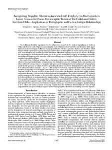
Fig. 3. Illustration of the findings of this study regarding object and famous face interferences. Twenty-six specific facenaming areas (that is, sites of interference in face naming only and not in object naming) were found. These face-naming specific sites were found in the left SFG, MFG, and IFG, and in the anterior part of the STG and MTG.
face naming were often extremely localized (< 1 cm2). The possibility of finding 2 neighboring sites involved in face naming was found 9 times. Three neighboring face-naming sites were only found 1 time.
Location of Face-Naming Interference Sites. Interferences in famous face naming were found in 3 regions: in the left frontal SFG, MFG, and IFG areas (p < 0.005); in the left temporal pole (p < 0.001); and in the left supramarginal gyrus (p < 0.001). Occasionally, some facenaming areas were found outside these areas. Regarding right hemispheric brain mappings, 4 interference sites were found in 3 patients. One patient exhibited a face-naming hesitation in the MFG. The other 2 patients exhibited 3 face-naming anomia sites in the right supramarginal gyrus. In 12 other patients (those who underwent surgery for right hemispheric lesions), no facenaming area was found.
Face Naming–Specific Areas. Among 35 patients whose dominant left hemisphere was studied (36 total brain mapping procedures), 26 face-naming specific areas (that is, sites of interference in face naming only and not in object naming) were found in 23 patients. These specific face-naming sites were found in 2 regions (Fig. 3): in the left SFG, MFG, and IFG (p < 0.001) (Fig. 4), and in the anterior part of the STG and MTG (p < 0.01) (Fig. 5). J. Neurosurg. / October 17, 2008
Among these face-naming specific sites, 1 was subcortical as illustrated in Fig. 6.
Types of Face-Naming Interferences. Different types of famous face interference occurred (Table 2). The different types of interference according to brain localization are summarized in Table 3. Most interferences (34 [50%]) were typical face anomia; patients acknowledged that they were able to recognize the face or to evoke related biographic information without being able to name the corresponding person. Semantic paraphasias (patients attributing repeatedly a wrong name to a famous face) were found mainly in frontal gyri and the STG. The patients were not always aware of being wrong. Phonemic paraphasias during famous face naming was only found in the supramarginal gyrus. Postoperative Testing
Difficulties in both naming tasks were detected postoperatively in 17 patients (34%), probably due to brain retraction or postoperative edema. These difficulties consisted in slow naming, naming hesitation or mistakes, and phonemic errors. No patient had a specific famous face– naming deficit. Among these 17 patients, 13 were retested a few weeks after surgery, and 5 of them had significant persistent object and face-naming difficulties. For vari5
C. Giussani et al.
Fig. 4. Intraoperative cortical brain mapping of famous face naming versus object naming in a right-handed patient revealed 2 different areas of dissociation between famous face and picture naming in the left frontal lobe. During tumor removal, both these areas were spared. F = famous face–naming area; L = object-naming area; N = negative area (no famous face interference).
ous reasons, the remaining 4 patients did not return to the hospital so they could not be followed up and retested. Among the 23 patients in whom a face-specific site was found, all had their face-specific areas spared. However, in 5 of them, the margins of tumor removal were very close (between 1 and 2 cm) to face-specific areas. These 5 patients had postoperative face-naming difficulties that disappeared, in 4 of them within 2 months. The remaining patient was lost to follow-up.
Discussion
The ability to recognize and to name known faces is crucial to create and maintain social relationships. The loss of these abilities represents a tragic event, frequently described in the literature.22,43,49 The face-naming process begins with face recognition. Face recognition is a specialized form of visual recognition and relies on mechanisms separate from object recognition.4,9,13,14,19 Positron emission tomography and functional MR imaging studies showed that face recognition specifically activated the inferior occipital gyrus and the lateral part of the middle fusiform gyrus mainly in the right hemisphere.23,35,36,40 This right occipital region has been called the fusiform face area.21,40 Nevertheless, other authors have underlined 6
that an evenly more distributed and bilateral neural system activity could be involved in face recognition.18 In this study, we found a relative anatomical segregation between famous face– and object-naming sites. The present study reiterates that brain mapping results are strongly influenced by the categories of items that have to be named.37 As object-naming sites, stimulation sites specific to famous face naming were exquisitely localized in small parts of cortical areas. Furthermore, 2 brain regions have been found to involve more frequently famous face– naming sites, the left frontal gyri and the anterior part of left temporal lobe. Although it has been demonstrated a fundamental role of the right hemisphere in learning new faces as well as in the face recognition and in the face emotion interpretation processes,1,11,12,17,38,41 we did not observe any trend to produce interferences of famous face naming in the areas of the right hemisphere that we explored. However, a specialization of the right hemisphere for face processing has been demonstrated for face perception processes, particularly in the posterior medial part of the right temporal lobe (fusiform gyrus).23,24,35,36,40 Stimulation-induced interferences did not concern perception of faces in the present study in which the inferior and medial parts of the temporal lobes were not explored. Instead we explored the presence of interference sites for J. Neurosurg. / October 17, 2008
Famous face naming
Fig. 5. Illustration of intraoperative subcortical brain mapping of famous face versus object naming of the left temporal lobe. Cortical intraoperative brain mapping of famous face versus object naming found different areas. One cortical area (F) in the anterior temporal lobe was involved in famous face naming. Interferences in object naming (L) were found elsewhere. To improve the understanding of the intraoperative photographs, cortical sites producing language impairment areas were not systematically labeled with a sterile ticket.
face naming, the final end point of the face recognition process and, similar to other authors, we showed that a lexical retrieval specific to famous faces depends mainly on the left hemisphere. Moreover this study showed that there are small areas in the SFG, MFG, IFG, and in the temporal cortex in which stimulation inhibited appropriate naming of famous faces without impairing object naming. In different cases of anomia for faces, the patient was able to recognize the corresponding person but could not retrieve his or her name. These results suggest that, even at its final stage involving access to a specific lexicon, naming of famous faces can be related to the activity of specific cortical territories, which can be anatomically distinguishable from the neighboring areas of the left lateral frontal and temporal cortex involved in object naming. This finding lends further support to earlier neuropsychological reports on naming deficits specific to famous faces.11,16,25 In few cases, semantic paraphasias for famous faces were observed in J. Neurosurg. / October 17, 2008
areas close to those eliciting anomia, suggesting again an anatomical specificity for appropriate association between semantic features retrieved from famous face perception and the lexical output specific to this category. Although face recognition preferentially relies on right hemispheric structures, most studies have shown that face naming is a function of the left hemisphere.11,12,17,38,41 Specific language sites for face naming have been identified in the left premotor and prefrontal cortex.15 In other studies, patients with lesions in the anterior region of the left temporal lobe were able to recognize faces and to give correct biographical information about the target, although they were unable to retrieve the proper name corresponding to that face.17,33,38,41 An involvement of the left temporal pole in face naming has been found by using functional neuroimaging, whereas the midposterior part of the left temporal lobe seems to be involved in retrieving other semantic categories’ nouns.6,7,26 In our study, all 4 patients 7
C. Giussani et al.
Fig. 6. Illustration showing the dissociation between famous face and picture naming in the left temporal lobe. Subcortical intraoperative brain mapping of famous face versus object naming found 2 different areas. One cortical area (12) was involved in object naming whereas a subcortical area was involved only in face naming (31).
with preoperative and selective famous face–naming difficulties had tumors in their anterior left temporal lobe. We observed the presence of specific famous face–naming sites in a nonclassical language area such as the anterior part of the left temporal lobe in the STG and MTG. The left temporal pole is usually considered by neurosurgeons not to contain eloquent areas. It is standard practice to remove the temporal pole without brain mapping. According to reports of patients with brain lesions17,33,38,41 or neuroimaging studies,16 as well as in this direct cortical stimulation study, this may not be completely true, at least as far as face naming is concerned. The finding of different neural regions correlated to semantic categories within the brain is an enlightening phenomenon. Findings of specific deficits20,27,48 in material acquired through education or special interest in individuals have favored the hypothesis that categorical knowledge could be, at least partially, organized in dedicated neuronal structures.2 Different evolutionary factors or a training ef8
fect in individuals for a specific topic could explain the development of category-specific units but are still a matter of debate.2,5
Conclusions
From a cognitive point of view, this prospective direct stimulation study showed that famous face naming is preferentially processed in the left frontal and anterior temporal gyri. These results match those of previous neuroimaging studies7,16 and studies in patients who have sustained brain damage.17,33,38,41 Relative anatomical segregation of lexical categories within language areas was detected. We think that it is necessary to adapt brain-mapping tasks in neurosurgical patients not only to the patient’s biography (that is, in bilingual individuals who need 2 language brain mappings) but also to the brain region involved. Indeed, the development of brain imaging tools over the past few years has J. Neurosurg. / October 17, 2008
Famous face naming TABLE 2: Classification used in this study of the different types of famous face–naming interferences found during direct brain mapping* Description speech arrest site
anomia site
paraphasia site
miscellaneous findings articulatory sites other findings single error ocular movements
Definition Patient stops speaking suddenly once stimulation is applied but no face/tongue contraction is visible. Once stimulation is stopped, patient can successfully name the famous face. Definition does not apply to stimulation in the pre- and postcentral gyri in which face & tongue contraction can be elicited. Often found in posterior temporal or Broca areas (especially in ramus operculare). Patient can speak (this is…yes, this face…I know her/him…I cannot find her/his name…) but is unable to find the name of famous face. Once stimulation is stopped, patient can successfully name the famous face. Despite fluent speech, patient’s responses to famous faces are incomprehensible or inappropriate. Type of paraphasias induced by electrostimulation can be either phonemic (substitution of pho nemes in the target name) or semantic (irrelevant name substituting for the correct name). Neolo gistic forms of paraphasia during famous face naming can also be observed. Repeated words, hesitations during naming. Patient stops speaking. Stimulation induces visible face/tongue contraction. Almost always found in pre- &/or postcentral gyri. In a given site, interference for number reading task used is found only once. Patient stops face naming because of ocular movements induced by stimulation.
* Articulatory sites comprise those considered to be nonspecific and related to motor mechanisms; single error sites, those with no reproducible reading interference; and ocular movements sites were not included in this study.
led to important breakthroughs in brain mapping. Brain regions involved in calculation, spatial neglect, as well as writing or reading have been more precisely defined. Some authors have shown that by using simple, acceptable, and not time-consuming tasks, direct brain mapping can replicate, at least partially, neuroimaging find-
ings.16,34,37 In the current study, we have shown that the use of different lexical categories may produce different brain-mapping results in left temporal and frontal lobes. We advocate the use of brain-mapping tasks more closely adapted to the functional properties of the brain region studied during surgery.
TABLE 3: Types of specific famous face interference in the left hemisphere (68 hemispheres) according to localization* No. of Hemispheres Brain Region Studied SFG MFG IFG anterior STG middle STG posterior STG anterior MTG middle MTG posterior MTG anterior ITG middle ITG posterior ITG supramarginal gyrus angular gyrus superior parietal area
Speech Arrest 1 1 3 1 4 4 1 1 0 0 0 0 0 0 1
Anomia
Phonemic Paraphasia
Semantic Paraphasia
Neologistic Paraphasia
Hesitations, Miscellaneous Findings
4 7 10 2 3 2 3 2 1 0 0 0 0 0 0
0 0 0 0 0 0 0 0 0 0 0 0 3 0 0
1 1 3 1 1 2 0 0 0 0 0 0 1 0 0
0 0 0 0 0 0 0 0 0 0 0 0 1 0 0
0 0 1 0 1 0 0 0 0 0 0 0 0 0 1
* The different types of interference could be related to the stages of famous face–name processing, supramarginal gyrus being involved in phonemic tasks and more anterior sites in naming or semantic tasks.
J. Neurosurg. / October 17, 2008
9
C. Giussani et al. Disclaimer The authors report no conflict of interest concerning the materials or methods used in this study or the findings specified in this paper. Acknowledgments We thank Denise Géblé for her help in supplying them with the references for this article, Leila Boukhatem and Alessandra Casarotti for all neuropsychological evaluations of the patients, and Catherine Raux and Antonio Ladislao for editorial assistance. References 1. Adolphs R, Damasio H, Tranel D: Cortical systems for the recognition of emotions in facial expressions. J Neurosci 16: 7678–7687, 1996 2. Allison T, Ginter H, McCarthy G, Nobre AC, Puce A, Luby M, et al: Face recognition in human extrastriate cortex. J Neurophysiol 71:821–825, 1994 3. Bruce V, Young A: Understanding face recognition. Br J Psychol 77:305–327, 1986 4. Buxbaum LJ, Glosser G, Coslett HB: Impaired face and word recognition without object agnosia. Neuropsychologia 37:41– 50, 1999 5. Caramazza A, Shelton JR: Domain-specific knowledge systems in the brain the animate-inanimate distinction. J Cogn Neurosci 10:1–34, 1998 6. Damasio H, Grabowski TJ, Tranel D, Hichwa RD, Damasio AR: A neural basis for lexical retrieval. Nature 380:499–505, 1996 7. Damasio H, Tranel D, Grabowski T, Adolphs R, Damasio A: Neural systems behind word and concept retrieval. Cognition 92:179–229, 2004 8. De Renzi E, Faglioni P, Grossi D: Apperceptive and associative forms of prosopagnosia. Cortex 27:213–221, 1991 9. Duchaine B, Nakayama K: Dissociation of face and object recognition in developmental prosopagnosia. J Cogn Neurosci 17:249–261, 2005 10. Duffau H: Lessons from brain mapping in surgery for lowgrade glioma: insights into associations between tumour and brain plasticity. Lancet Neurol 4:476–486, 2005 11. Ellis AW, Young AW, Critchley EM: Loss of memory for people following temporal lobe damage. Brain 112:1469–1483, 1989 12. Evans JJ, Heggs AJ, Antoun N: Progressive prosopagnosia associated with selective right temporal lobe atrophy. A new syndrome? Brain 118:1–13, 1995 13. Farah MJ, Levinson KL, Klein KL: Face perception and within-category discrimination in prosopagnosia. Neuropsychologia 33:661–674, 1995 14. Farah MJ, Wilson KD, Drain M: The inverted face inversion effect in prosopagnosia: evidence for mandatory, face-specific perceptual mechanisms. Vision Res 35:2089–2093, 1995 15. Grabowski TJ, Damasio H, Damasco AR: Premotor and prefrontal correlates of category-related lexical retrieval. Neuroimage 7:232–243, 1998 16. Grabowski TJ, Damasio H, Tranel D, Ponto LL, Hichwa RD, Damasio AR: A role for left temporal pole in the retrieval of words for unique entities. Hum Brain Mapp 13:199–212, 2001 17. Glosser G, Salvucci AE, Chiaravallotti ND: Naming and recognizing famous faces in temporal lobe epilepsy. Neurology 61:81–86, 2003 18. Haxby JV, Hoffman EA, Gobbini MI: The distributed human neural system for face perception. Trends Cogn Sci 4:223– 233, 2000 19. Henke K, Schweinberger SR, Grigo A: Specificity of face recognition: recognition of exemplars of non-face objects in prosopagnosia. Cortex 34:289–296, 1998
10
20. Hillis AE, Caramazza A: Category-specific naming and comprehension impairment: a double dissociation. Brain 114: 2081–2094, 1991 21. Hoffman EA, Haxby JV: Distinct representation of eye gaze and identity in the distributed human neural system for face perception. Nat Neurosci 3:80–84, 2000 22. Joubert S, Felician O, Barbeau E, Sontheimer A, Barton JJ, Ceccaldi M, et al: Impaired configurational processing in a case of progressive prosopagnosia associated with predominant right temporal lobe atrophy. Brain 126:2537–2550, 2003 23. Kanwisher N, McDermott J, Chun MM: The fusiform face area: a module in human extrastriate cortex specialized for face perception. J Neurosci 17:4302–4311, 1997 24. Leveroni CL, Seidenberg M, Mayer AR, Mead LA, Binder JR, Rao SM: Neural systems underlying the recognition of familiar and newly learned faces. J Neurosci 20:878–886, 2000 25. Lucchelli F, De Renzi E: Proper name anomia. Cortex 28: 221–230, 1992 26. Luckhurst L, Lloyd-Jones TJ: A selective deficit for living things after temporal lobectomy for relief of epileptic seizures. Brain Lang 79:266–296, 2001 27. McKenna P, Warrington EK: Category-specific naming preservation: a single case study. J Neurol Neurosurg Psychiatry 41:571–574, 1978 28. McKenna P, Warrington EK: Testing for nominal dysphasia. J Neurol Neurosurg Psychiatry 43:781–788, 1980 29. Ojemann G, Ojemann J, Lettich E, Berger M: Cortical language localization in left, dominant hemisphere. An electrical stimulation mapping investigation in 117 patients. J Neurosurg 71:316–326, 1989 30. Ojemann JG, Ojemann GA, Lettich E: Neuronal activity related to faces and matching in human right nondominant temporal cortex. Brain 115:1–13, 1992 31. Oldfield RC: The assessment and analysis of handedness: the Edinburgh inventory. Neuropsychologia 9:97–113, 1971 32. Paller KA, Gonsalves M, Bozic VS: Electrophysiological correlates of recollecting faces of known and unknown individuals. Neuroimage 11:98–110, 2000 33. Papagno C, Capitani E: Proper name anomia: a case with sparing of the first-letter knowledge. Neuropsychologia 36:669– 679, 1998 34. Pouratian N, Bookheimer SY, Rubino G, Martin NA, Toga AW: Category-specific naming deficit identified by intraoperative stimulation mapping and postoperative neuropsychological testing. Case report. J Neurosurg 99:170–176, 2003 35. Puce A, Allison T, Asgari M: Differential sensitivity of human visual cortex to faces, letterstrings, and textures: a functional magnetic resonance imaging study. J Neurosci 16:5205–5215, 1996 36. Puce A, Allison T, Gore JC: Face-sensitive regions in human extrastriate cortex studied by functional fMRI. J Neurophysiol 74:1192–1199, 1995 37. Roux FE, Lubrano V, Lauwers-Cances V, Mascott CR, Demonet JF: Category-specific cortical mapping: color-naming areas. J Neurosurg 104:27–37, 2006 38. Seidenberg M, Griffith R, Sabsevitz D: Recognition and identification of famous faces in patients with unilateral temporal lobe epilepsy. Neuropsychologia 40:446–456, 2002 39. Semenza C, Zettin M: Evidence from aphasia for the role of proper names as pure referring expressions. Nature 342:678– 679, 1989 40. Sergent J, Ohta S, MacDonald B: Functional neuroanatomy of face and object processing. A positron emission tomography study. Brain 115:15–36, 1992 41. Shallice T, Kartsounis LD: Selective impairment of retrieving people’s names: a category specific disorder? Cortex 29:281– 291, 1993 42. Smith CD, Andersen AH, Kryscio RJ, Schmitt FA, Kindy MS, Blonder LX, et al: Differences in functional magnetic
J. Neurosurg. / October 17, 2008
Famous face naming resonance imaging activation by category in a visual confrontation naming task. J Neuroimaging 11:165–170, 2001 43. Sperber S, Spinnler H: Covert person recognition: its fadeout in a case of temporal lobe degeneration. Cortex 39:57–67, 2003 44. Taylor MD, Bernstein M: Awake craniotomy with brain mapping as the routine surgical approach to treating patients with supratentorial intraaxial tumors: a prospective trial of 200 cases. J Neurosurg 90:35–41, 1999 45. Thiebaut de Schotten M, Urbanski M, Duffau H, Volle E, Levy R, Dubois B, et al: Direct evidence for a parietal-frontal pathway subserving spatial awareness in humans. Science 309:2226–2228, 2005 46. Tranel D: Impaired naming of unique landmarks is associated with left temporal polar damage. Neuropsychology 20:1–10, 2006 47. Trautner P, Dietl T, Staedtgen M: Recognition of famous faces in the medial temporal lobe: an invasive ERP study. Neurology 63:1203–1208, 2004
J. Neurosurg. / October 17, 2008
48. Warrington EK, Shallice T: Category specific semantic impairments. Brain 107:829–854, 1984 49. Wright H, Wardlaw J, Young AW, Zeman A: Prosopagnosia following nonconvulsive status epilepticus associated with a left fusiform gyrus malformation. Epilepsy Behav 9:197– 203, 2006 50. Zar JH: Biostatistical Analysis, ed 4. New Jersey: PrenticeHall, 1999
Manuscript submitted April 6, 2007. Accepted August 24, 2007. Please include this information when citing this paper: published online October 17, 2008; DOI: 10.3171/2007.8.17566. Address correspondence to: Franck-Emmanuel Roux, M.D., Ph.D., Service de Neurochirurgie et Institut National de la Santé et de la Recherche Médicale 825, Hôpital Purpan, F-31059 Toulouse, France. email:
[email protected].
11