339
Assessing p97 as an Alzheimer’s disease serum biomarker Wilfred A. Jefferies∗, Dara L. Dickstein and Maki Ujiie Biotechnology Laboratory and the Biomedical Research Centre, Departments of Medical Genetics, Microbiology and Immunology and Zoology, University of British Columbia, Canada The search is ongoing for a reliable serum biomarker for AD. The level of iron is elevated in the brain of Alzheimer’s disease (AD) patients. Our studies have demonstrated that the level of the iron transport protein, p97, is increased in the serum of AD patients but not in various control groups. These results have recently been confirmed by another laboratory who extended our findings by demonstrating that p97 is not elevated in other neurodegenerative diseases. This qualifies p97 as a potentially powerful biomarker for AD. Although the relationship between increased level of iron and p97 in the AD brain is not well understood, our research supports the hypothesis that p97 over-expressed by senile plaque associated reactive microglia is exocytosed and appears in blood. The relationship between elevated levels of serum p97 and AD, together with the possible future clinical application of p97 are considered in this report.
itation and death within 12 years of onset [39]. Pathologically, AD selectively damages brain regions and neural circuits critical for cognition and memory, and its pathological characteristics include the presence of senile plaques and intracellular neurofibrillary tangles (NFTs) in the brain tissue [31]. Although patient history, physical exams and neuroimaging are used in conjunction with cognitive tests and could be helpful in ruling out other forms of dementia, definitive diagnosis can only be made during postmortem examination of brain tissues [20,22]. Presently available therapeutics, such as tacrine hydrochloride and donepezil hydrochloride, are targeted to preserving cholinergic activities thereby protecting cholinergic neurons [9,13,26]. Since neuronal loss is currently irreversible, early intervention in preventing further damage to neurons becomes critical in slowing down the progression of the disease. In addition to the benefits of starting prescriptions promptly, early diagnosis allows the patient and caregivers time to plan how to manage this progressive and chronic disease.
2. AD and iron metabolism 1. Introduction As the overall longevity of the world population is extended, the number of people suffering from dementia is also increasing. Alzheimer’s disease (AD) is the most common form of dementia [10] and is estimated to account for 50–70% of dementia in elderly people [12, 14,15]. Progressive memory and cognitive decline as well as difficulty in language, praxis and visual perception are the clinical manifestations which characterize the disease. The decline in observed intellectual function progresses at a slow rate and leads to severe debil∗ Corresponding
author: Wilfred A. Jefferies, 2222 Health Sciences Mall, University of British Columbia, Vancouver, B.C. V6T 1Z3, Canada. Tel.: +1 604 822 6961; Fax: +1 604 822 6780; E-mail:
[email protected]. Journal of Alzheimer’s Disease 3 (2001) 339–344 ISSN 1387-2877 / $8.00 2001, IOS Press. All rights reserved
Iron is essential for cell growth and survival. In addition to its importance in normal physiology, iron also plays a key role in some human pathology. Since unbound iron induces the formation of free radicals, it generally circulates as low molecular weight complexes in association with ascorbate, amino acids and some serum proteins such as albumin or transferrin (Tf). Due to its toxicity, regulatory mechanisms have evolved to closely monitor and control the absorption and distribution of iron within the body. In the case of AD, however, redox-active iron accumulates in senile plaques and NFTs of the brain [40]. The elevated levels of free radicals lead to the generation of reactive oxygen species and attack particularly sensitive neuronal tissues in the AD brain. The brain, like other organs, requires iron yet the mechanisms of iron uptake remains unclear. The brain
340
W.A. Jefferies et al. / Assessing p97 as an Alzheimer’s disease serum biomarker
has a unique anatomical structure called the blood-brain barrier (BBB) which effectively protects the sensitive neuropil from directly coming in contact with the blood. The selective barrier prevents large and/or hydrophilic molecules from entering the brain parenchyma. Iron uptake into the brain was initially assumed to be exclusively regulated by the classically defined transferrin receptor (TfR) since Tf level is high in normal serum and most of the iron in the plasma is bound to Tf. This concept is supported by a study in which intact 125 I-Tf complexes were recovered in the brain parenchyma after being intravenously injected into the rat [11]. More recent studies suggest that although Tf and TfR are undoubtedly important mediators of iron uptake by the brain [6,41], they do not account for all the iron present in the brain [11]. For example, Fishman [11] showed that although some of the injected Tf crosses the BBB, much of it was retained in the endothelial cytoplasm. In addition, hypotransferrinemic mice deficient in Tf have a normal level of iron in the brain [7]. We reasoned that a Tf/TfR independent iron transporter, p97, might also be involved in transporting iron across the BBB, and that its over-expression might be associated with AD pathology.
3. p97 (Melanotransferrin) Melanotransferrin, also known as the human melanoma associated antigen p97, was first identified as a cell surface marker associated with human skin cancer [5] and subsequently described as an onco-fetal protein due to its high expression in fetal tissues. Woodbury et al. [42] demonstrated the expression of p97 in neoplastic human tissues, particularly melanomas. In addition, they found that the protein was highly expressed in fetal colon, lung and umbilical cord as compared to adult tissues. Further studies revealed that p97 is also highly expressed in fetal liver and placenta [5]. In addition to its expression in human tissues, p97 is expressed in many cultures of normal cells including liver, intestinal epithelial cells, fetal intestinal cells and sweat gland ducts [1,4,8,33,37]. Human p97 is a member of a group of iron binding proteins that include Tf, lactoferrin and ovotransferrin. In humans, the p97 gene maps to the same region of chromosome 3q as Tf and TfR [30]. Unlike other members of this family, p97 is found in two forms; a glycosyl-phosphatidylinositol (GPI) anchored form on the plasma membrane and a soluble form found in blood and cerebrospinal fluid [1,5,23]. The function
of p97 has yet to be established. It is known, however, that the GPI-linked form of p97 can bind iron and internalize into cells by a mechanism independent of Tf/TfR endocytosis [24,35]. p97 is also unique in that it can bind to other metals such as zinc and copper [unpublished] and may function in their transport. A study has shown that unlike other members of the group that have two iron binding sites, p97 has only one iron binding site on its N-terminus and the C-terminus binding site is unable to bind iron [2]. Recent data, however, suggests that under specific conditions p97 is able to bind two molecules of iron but with different affinity [unpublished].
4. Localization of p97 in AD brain The distribution of p97, Tf and TfR in the brain was studied to identify the locations in which they may function [18,19]. In the 1996 immunocyto study [19], AD brains were compared with brains of patients who died from neurodegenerative diseases other than AD. Interestingly, p97 was found co-localized with TfR on the surfaces of microvessels, while Tf localization was limited to neuroglia. The distribution of p97 in the nonAD brain appeared to be limited to the BBB, with occasional positive stains for astrocytes and oligodendrocytes. In the AD brain, p97 was also detected in reactive microglia associated with senile plaques. Reactive microglia that stained positive with HLA-DR antibody but those not closely positioned to senile plaques did not express p97. There is most likely an increased requirement for the utilization and/or scavenging of iron by reactive microglia associated with senile plaques since in addition to p97, increased concentrations of iron, Tf [27] and ferritin [16,21,29] have been noted in the region. The distribution of iron and p97 has not been directly correlated in the AD brain, however, and we intend to examine this relationship in the future. An additional study was carried out to compare the sites of expression and function for p97. p97 protein and mRNA localization in the AD brain were examined using immunocyto and in situ hybridization, respectively [43]. In the AD brain, a strong signal for p97 mRNA was found in reactive microglia associated with senile plaques and weak signals were detected around endothelial cells, while the p97 protein was localized on reactive microglia associated with senile plaques and on endothelial cells. In the non-AD brain, endothelial cells stained weakly for p97 mRNA and strongly for the p97 antigen. The presence of p97 on endothelial cells
W.A. Jefferies et al. / Assessing p97 as an Alzheimer’s disease serum biomarker
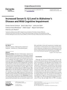
may be an indication of the function of the soluble form of p97 to shuttle iron and other metals across the BBB, from the blood into the neuropil, thereby transporting the iron necessary for normal physiology. Additionally, our results show that p97 is highly expressed in reactive microglia around senile plaques in the AD brain. Although the precise mechanism remains unclear, the secreted form of p97 expressed in reactive microglia might be carried out of the neuronal parenchyma into the peripheral blood in AD causing the level of serum p97 to increase (Fig. 1). Experiments are currently underway to examine the relationship between microglial activation and expression of p97, and the role of p97 in the transcytosis of metals across the BBB.
5. p97 serum level in AD patients With the finding that p97 is detected on the BBB and in the reactive microglia associated with the senile plaques of AD patients [19], it was postulated that p97 may represent a biological marker for AD. Our initial studies [23] investigated this putative relationship and found evidence that the soluble form of p97 is elevated in the serum taken from AD patients as compared to controls (n = 17, 43.8 ± 11.6 ng/ml and n = 15, 7.04 ± 3.28 ng/ml, respectively). All of these assays were conducted with a quantitative fluorescence immuno-assay, termed Pandex assay. It relies on the use of two non-competitive monoclonal antibodies, L235 against p97 [17] and Hybridoma C, and is similar to traditional ELISA or radioimmunoassay. Further more regression analysis of this data revealed that there was no correlation between p97 levels and age. To eliminate the possibility that environmental factors may result in the increased levels observed, serum samples from AD patients and their cognitively normal spouses (n = 10) were obtained [23]. We found that the levels of p97 from patients compared to their spouses were significantly elevated thereby suggesting that environmental factors have no influence on serum p97 levels. We also found that storage conditions and temperature changes could affect the apparent concentration of p97 determined in this assay. The finding that serum concentration of p97 in AD patients is elevated using the established two stage fluorescence immuno-assay was confirmed by a follow-up study with a different cohort using a quantitative RIA based on the monoclonal antibodies which were used in the first study [28]. The subjects consisted of 21 “probable”, 4 “possible” AD patients (as diagnosed by the
341
National Institute of Neurological and Communicative Disorders and Alzheimer’s Disease and Related Disorders Association criteria) and 7 age-matched control without dementia. For this blinded study, 19 ng of p97 per 1 ml of serum was used as the cutoff point with 71% sensitivity and specificity. Statistical analysis using Kruskal-Wallis test and ANOVA both showed a significant increase in the level of p97 in the “probable” group (mean concentration of 50.2 ± 2.1 ng/ml) when compared with ”possible” and control groups (mean concentration of 13.8±5.5 ng/ml and 12.8±2.1 ng/ml, respectively. These two published studies consistently noted an elevated serum level of p97 in AD patients suggesting that p97 has potential as a biomarker. A subsequent study using the RIA and a larger cohort of subjects (n = 123) showed that there is a 2 fold significant increase in serum p97 levels from patients with AD compared to non-AD controls (41 ng/ml versus 20 ng/ml, p < 0.001) [Feldman et al., in press]. In this double-blinded study, the data were compiled and analyzed by an independent consulting group (SERC) which was assigned the task of combining the identity of the patients with the estimates of serum p97 levels. Little correlation between serum p97 concentration and age, gender and severity of the disease was noted in the cohort of 123 subjects included in this study (52 “probable” and “possible” AD patients and 71 controls). By segregating control subjects according to age, it was found that in subjects over 60 years of age the mean p97 concentrations for AD subjects was double that of controls with average levels of 40.9 ng/ml and 19.2 ng/ml respectively. While there was some overlap in p97 concentrations, a cutoff level of 23 ng/ml equalized the specificity and sensitivity (72% and 71% respectively). We also detected a subgroup of approximately 5% of both AD and controls which had exceptionally high backgrounds in this assay, although the reason for this is not clear. In addition, p97 serum levels in patients who were taking anti-inflammatory agents including nonsteroidal anti-inflammatory drugs (NSAID) and aspirin were measured. There appeared to be a lack of association between NSAID or aspirin use and p97 levels. However, since the number of subjects were limited and use and dose of NSAID were not properly monitored, it is too early to conclude whether or not a relationship exists between p97 and anti-inflammatory agents. In addition to our laboratory, another group of investigators have also performed studies demonstrating the putative role of p97 as a biomarker for AD. The findings made by Kim et al. [25] agree with those previ-
342
W.A. Jefferies et al. / Assessing p97 as an Alzheimer’s disease serum biomarker
p97 Transferrin p97 receptor Fig. 1. Hypothetical model of the relationship between p97 and AD. A pathological hallmark of the AD brain, senile plaque, with associated reactive microglia is shown in this diagram. The mechanism of iron transport across the BBB is unclear. Although Tf does play a role, it does not appear to cross the abluminal surface of the BBB effectively. p97 is capable of transcytosing bi-directionally through the BBB (A). In the AD brain, reactive microglia associated with senile plaques express high levels of p97. As an explanation for the elevated level of p97 found in the serum of AD patients, we hypothesize that p97 expressed by microglia are exocytosed across the BBB into the bloodstream, possibly transporting iron out of the brain (B).
ously established by our laboratory and also add some new and important evidence to further substantiate the correlation between p97 levels and AD. In contrast to the studies conducted in our lab [23,28], the p97 assay utilized by Kim et al. [25] was based on a quantitative immuno blot analysis using L235, one of the p97 monoclonal antibodies used in our studies. p97 serum concentrations were determined by densometric scanning of autoradiographs of immuno blots of patient serum samples. A standard curve of p97 concentrations was calculated which allowed the quantification of p97 levels. Moreover, this study involved 211 subjects: 71 patients with AD, 56 patients with non-AD type dementia and 84 normal control subjects. Their findings indicate that there was a 3 to 4 fold significant increase in the serum p97 levels in AD cases compared to non-AD cases of dementia and controls (15.00 pg/µl, 2.85 pg/µl and 3.20 pg/µl respectively) with a specificity of 91.4% and a sensitivity of 91.5%. Kim et al. [25] noted no correlation between p97 serum levels and age. In ad-
dition, there appeared to be no trend with the levels of p97 and severity of disease. Serum p97 levels were elevated in early AD (14.45 pg/µl), however, there was no subsequent increase of serum levels in late AD cases (15.00 pg/µl). Finally, ApoE allelic frequencies were analyzed with respect to p97 serum levels. From the data obtained it was determined that ApoE genotypes, in particular the presence of ε4, had no significant impact on the serum p97 levels. Collectively these are compelling findings which independently corroborate the potential of serum p97 levels as a biomarker of AD.
6. AD biomarkers associated with iron metabolism Although there are several candidate biomarkers for AD, none have previously been compelling enough to replace cognitive testing or act as an aid to cognitive diagnosis. The use of tau and amyloid β [3,38] as CSF biomarkers have been suggested to aid in AD diagno-
W.A. Jefferies et al. / Assessing p97 as an Alzheimer’s disease serum biomarker
sis. Obtaining CSF via lumbar punctures, however, is impractical due to the invasiveness of the procedure, especially in the elderly population. Analysis of blood, urine or saliva samples is simple and would be preferable to extraction of CSF or biopsy if a reliable serum biomarker can be found. Since increased concentration of iron in the AD brain is well-documented [32], many researchers have focused their attention on assessing the potential usefulness of iron-related molecules as biomarkers for the disease. In addition to p97, heme oxygenase 1 (HO-1) is being tested as another possible serum biomarker for AD. Research by Schipper et al. [36] has demonstrated that the free iron and carbon monoxide particles that are generated from heme oxygenase 1 (HO-1) mediated catabolism may contribute to the abnormal patterns of iron deposition and metabolism seen in AD patients. Early studies based on ELISA assays have demonstrated that plasma levels of HO-1 protein is significantly decreased in patients who are affected with early sporadic AD compared to age matched controls [36]. Thus the measurement of HO-1, similar to p97, in serum may provide a new method for the diagnosis of sporadic AD in its early stages and thus help in early disease intervention and treatment.
References [1]
[2]
[3]
[4]
[5]
[6]
[7]
[8]
7. Conclusion In summary, the four studies conducted to date by two independent groups support the use of serum levels of p97 as an aid in the diagnosis of AD. Future studies in our laboratory are being directed at establishing the connection between p97, reactive microglia and AD. Additionally, new quantitative assays which are easily automatable and unperturbed by sample preparation are currently being designed for use in clinical testing laboratories. If direct correlation between serum levels of p97 and progression of AD can be supported, p97 assays might have useful application in evaluating the effectiveness of new drugs for AD. Finally, the potential utilization of p97 testing as a screening tool for AD must be investigated in large-scale prospective studies. We and others have demonstrated that p97 could have potential value as a biomarker for AD and its clinical merit should be further investigated.
[9]
[10] [11]
[12]
[13]
[14] [15] [16]
Acknowledgement We thank our past collaborators for their contribution and the Vancouver Foundation, Synapse Technology and CIHR for funding these studies.
343
[17]
R. Alemany, M.R. Vila, C. Franci, G. Egea, F.X. Real and T.M. Thomson, Glycosyl phosphatidylinositol membrane anchoring of melanotransferrin (p97): apical compartmentalization in intestinal epithelial cells, Journal of Cell Science 104 (1993), 1155–1162. E.N. Baker, H.M. Baker, C.A. Smith, M.R. Stebbins, M. Kahn, K.E. Hellstrom and I. Hellstrom, Human melanotransferrin (p97) has only one functional iron-binding site, FEBS Letters 298 (1992), 215–218. K. Blennow and E. Vanmechelen, Combination of the different biological markers for increasing specificity of in vivo Alzheimer’s testing, Journal of Neural Transmission Supplement 53 (1998), 223–235. J.P. Brown, R.M. Hewick, I. Hellstrom, K.E. Hellstrom, R.F. Doolittle and W.J. Dreyer, Human melanoma-associated antigen p97 is structurally and functionally related to transferrin, Nature 296 (1982), 171–173. J.P. Brown, R.G. Woodbury, C.E. Hart, I. Hellstrom and K.E. Hellstrom, Quantitative analysis of melanoma-associated antigen p97 in normal and neoplastic tissues, Proceedings of the National Academy of Sciences of the United States of America 78 (1981), 539–543. A. Crowe and E.H. Morgan, Iron and transferrin uptake by brain and cerebrospinal fluid in the rat, Brain Research 592 (1992), 8–16. T.K. Dickinson and J.R. Connor, Cellular distribution of iron, transferrin, and ferritin in the hypotransferrinemic (Hp) mouse brain, Journal of Comparative Neurology 355 (1995), 67–80. W.G. Dippold, K.O. Lloyd, L.T. Li, H. Ikeda, H.F. Oettgen and L.J. Old, Cell surface antigens of human malignant melanoma: definition of six antigenic systems with mouse monoclonal antibodies, Proceedings of the National Academy of Sciences of the United States of America 77 (1980), 6114–6118. G. Emilien, K. Beyreuther, C.L. Masters and J.M. Maloteaux, Prospects for pharmacological intervention in Alzheimer disease, Archives of Neurology 57 (2000), 454–459. D.A. Evans, Estimated prevalence of Alzheimer’s disease in the United States, Milbank Q 68 (1990), 267–289. J.B. Fishman, J.B. Rubin, J.V. Handrahan, J.R. Connor and R.E. Fine, Receptor-mediated transcytosis of transferrin across the blood-brain barrier, Journal of Neuroscience Research 18 (1987), 299–304. M.F. Folstein, Differential diagnosis of dementia. The clinical process, Psychiatric Clinics of North America 20 (1997), 45– 57. P.T. Francis, A.M. Palmer, M. Snape and G.K. Wilcock, The cholinergic hypothesis of Alzheimer’s disease: a review of progress, Journal of Neurology, Neurosurgery and Psychiatry 66 (1999), 137–147. S.R. Gambert, Is it Alzheimer’s disease? Postgraduate Medicine 101 (1997), 42–52. D.S. Geldmacher and P.J. Whitehouse Jr., Differential diagnosis of Alzheimer’s disease, Neurology 48 (1997), S2–S9. I. Grundke-Iqbal, J. Fleming, Y.C. Tung, H. Lassmann, K. Iqbal and J.G. Joshi, Ferritin is a component of the neuritic (senile) plaque in Alzheimer dementia, Acta Neuropathology 81 (1990), 105–110. A.N. Houghton, M. Eisinger, A.P. Albino, J.G. Cairncross and L.J. Old, Surface antigens of melanocytes and melanomas. Markers of melanocyte differentiation and melanoma subsets, Journal of Experimental Medicine 156 (1982), 1755–1766.
344 [18]
[19]
[20]
[21]
[22]
[23]
[24]
[25]
[26]
[27]
[28]
[29]
[30]
[31]
W.A. Jefferies et al. / Assessing p97 as an Alzheimer’s disease serum biomarker W.A. Jefferies, M.R. Brandon, S.V. Hunt, A.F. Williams, K.C. Gatter and D.Y. Mason, Transferrin receptor on endothelium of brain capillaries, Nature 312 (1984), 162–163. W.A. Jefferies, M.R. Food, R. Gabathuler, S. Rothenberger, T. Yamada, O. Yasuhara and P.L. McGeer, Reactive microglia specifically associated with amyloid plaques in Alzheimer’s disease brain tissue express melanotransferrin, Brain Research 712 (1996), 122–126. K.A. Jellinger, The neuropathological diagnosis of Alzheimer disease, Journal of Neural Transmission Supplements 53 (1998), 97–118. Y. Kaneko, T. Kitamoto, J. Tateishi and K. Yamaguchi, Ferritin immunohistochemistry as a marker for microglia, Acta Neuropathology 79 (1989), 129–136. A.M. Kazee, T.A. Eskin, L.W. Lapham, K.R. Gabriel, K.D. McDaniel and R.W. Hamill, Clinicopathologic correlates in Alzheimer disease: assessment of clinical and pathologic diagnostic criteria, Alzheimer Disease Associated Disorders 7 (1993), 152–164. M.L. Kennard, H. Feldman, T. Yamada and W.A. Jefferies, Serum levels of the iron binding protein p97 are elevated in Alzheimer’s disease, Nature Medicine 2 (1996), 1230–1235. M.L. Kennard, D.R. Richardson, R. Gabathuler, P. Ponka and W.A. Jefferies, A novel iron uptake mechanism mediated by GPI-anchored human p97, Embo Journal 14 (1995), 4178– 4186. D.K. Kim, M.Y. Seo, S.W. Lim, S. Kim, J.W. Kim, B.J. Carroll, D.Y. Kwon, T. Kwon and S.S. Kang, Serum melanotransferrin, p97 as biochemical marker of Alzheimer’s disease, Neuropsychopharmacology 25 (2001), 84–90. W.J. Krall, J.J. Sramek and N.R. Cutler, Cholinesterase inhibitors: a therapeutic strategy for Alzheimer disease, Annals of Pharmacotherapy 33 (1999), 441–450. D.A. Loeffler, J.R. Connor, P.L. Juneau, B.S. Snyder, L. Kanaley, A.J. DeMaggio, H. Nguyen, C.M. Brickman and P.A. LeWitt, Transferrin and iron in normal, Alzheimer’s disease, and Parkinson’s disease brain regions, Journal of Neurochemistry 65 (1995), 710–724. I. Moroo, T. Yamada, R. Gabathuler, M.L. Kennard, J. Nurminen and W.A. Jefferies, Use of p97 in the diagnosis of Alzheimer’s disease, Alzheimer’s Reports 2 (1999), 353–358. T. Ohgami, T. Kitamoto, R.W. Shin, Y. Kaneko, K. Ogomori and J. Tateishi, Increased senile plaques without microglia in Alzheimer’s disease, Acta Neuropathology 81 (1991), 242– 247. G.D. Plowman, J.P. Brown, C.A. Enns, J. Schroder, B. Nikinmaa, H.H. Sussman, K.E. Hellstrom and I. Hellstrom, Assignment of the gene for human melanoma-associated antigen p97 to chromosome 3, Nature 303 (1983), 70–72. D.L. Price, R.E. Tanzi, D.R. Borchelt and S.S. Sisodia, Alzheimer’s disease: genetic studies and transgenic models, Annual Review of Genetics 32 (1998), 461–493.
[32]
[33]
[34]
[35]
[36]
[37]
[38]
[39]
[40]
[41]
[42]
[43]
Z.M. Qian and Q. Wang, Expression of iron transport proteins and excessive iron accumulation in the brain in neurodegenerative disorders, Brain Research Reviews 27 (1998), 257–267. F.X. Real, K.S. Furukawa, M.J. Mattes, S.A. Gusik, C. Cordon-Cardo, H.F. Oettgen, L.J. Old and K.O. Lloyd, Class 1 (unique) tumor antigens of human melanoma: identification of unique and common epitopes on a 90-kDa glycoprotein, Proceedings of the National Academy of Sciences of the United States of America 85 (1988), 3965–3969. F.X. Real, A.N. Houghton, A.P. Albino, C. Cordon-Cardo, M.R. Melamed, H.F. Oettgen and L.J. Old, Surface antigens of melanomas and melanocytes defined by mouse monoclonal antibodies: specificity analysis and comparison of antigen expression in cultured cells and tissues, Cancer Research 45 (1985), 4401–4411. D. Richardson and E. Baker, The uptake of inorganic iron complexes by human melanoma cells, Biochimica et Biophysica Acta 1093 (1991), 20–28. H.M. Schipper, H. Chertkow, K. Mehindate, D. Frankel, C. Melmed and H. Bergman, Evaluation of heme oxygenase-1 as a systemic biological marker of sporadic AD, Neurology 54 (2000), 1297–1304. R. Sciot, R. de Vos, P. van Eyken, K. van der Steen, P. Moerman and V.J. Desmet, In situ localization of melanotransferrin (melanoma-associated antigen P97) in human liver. A lightand electronmicroscopic immunohistochemical study, Liver 9 (1989), 110–119. M. Shoji, E. Matsubara, M. Kanai, M. Watanabe, T. Nakamura, Y. Tomidokoro, M. Shizuka, K. Wakabayashi, Y. Igeta, Y. Ikeda, K. Mizushima, M. Amari, K. Ishiguro, T. Kawarabayashi, Y. Harigaya, K. Okamoto and S. Hirai, Combination assay of CSF tau, A beta 1-40 and A beta 1-42(43) as a biochemical marker of Alzheimer’s disease, Journal of Neurological Science 158 (1998), 134–140. G.W. Small, Treatment of Alzheimer’s disease: current approaches and promising developments, American Journal of Medicine 104 (1998), 32S–38S. M.A. Smith, P.L. Harris, L.M. Sayre and G. Perry, Iron accumulation in Alzheimer disease is a source of redox-generated free radicals, Proceedings of the National Academy of Sciences of the United States of America 94 (1997), 9866–9868. M.E. Strahan, A. Crowe and E.H. Morgan, Iron uptake in relation to transferrin degradation in brain and other tissues of rats, American Journal of Physiology 263 (1992), R924–R929. R.G. Woodbury, J.P. Brown, S.M. Loop, K.E. Hellstrom and I. Hellstrom, Analysis of normal neoplastic human tissues for the tumor-associated protein p97, International Journal of Cancer 27 (1981), 145–149. T. Yamada, Y. Tsujioka, J. Taguchi, M. Takahashi, Y. Tsuboi, I. Moroo, J. Yang and W.A. Jefferies, Melanotransferrin is produced by senile plaque-associated reactive microglia in Alzheimer’s disease, Brain Research 845 (1999), 1–5.