Clin. Otolaryngol. 2003, 28, 411±416
Audit-derived guidelines for training in endoscopic sinonasal surgery (ESS) ± protecting patients during the learning curve M.L. MONTAGUE, A. KISHORE & G.W. MCGARRY Department of Otolaryngology, Glasgow Royal In®rmary, Glasgow, UK Accepted for publication 19 March 2003 M O N TA G U E M . L . , K I S H O R E A . & M C G A R RY G . W .
(2003) Clin. Otolaryngol. 28, 411±416
Audit-derived guidelines for training in endoscopic sinonasal surgery (ESS) ± protecting patients during the learning curve The objective of the present study is to propose guidelines to ensure safe practice in teaching centres while allowing endoscopic sinonasal surgery (ESS) training to proceed. A prospective complications audit of ESS procedures was undertaken over a 5-year period (January 1996±December 2000). The results have been used to form speci®c guidelines for safe and effective ESS training. A total of 500 patients underwent ESS during the 5-year period. The senior author was the main surgeon in 55% of cases with the trainee observing or assisting. A supervised trainee was the main surgeon in 45% of cases. The overall complication rate was 1.2% (n 6) (i.e. 0.7% for the 815 procedures performed). These were all minor complications. We encountered no major complications in 500 patients over the 5-year period. This audit shows that training need not compromise patient safety provided it is phased and structured. We propose appropriate phases and suggest the minimum requirements for units involved in ESS training. Keywords
endoscopic sinus surgery
complications training
The introduction of techniques of endoscopic sinonasal surgery (ESS) in 1985 by Kennedy1 has resulted in a rapid proliferation in the number of surgeons performing endoscopic nasal surgery in the UK. ESS is now widely accepted as the surgical treatment of choice for chronic and recurrent sinusitis. Its safety and ef®cacy have been reported in large personal series2±5 and in the results of national surveys.6,7 Mosher pointed out the potential dangers of the ethmoid when he stated in 1929, ``If it were placed in any other part of the body, it would be an insigni®cant and harmless collection of bony cells. In the place where nature has put it, it has major relationships so that diseases and surgery of the labyrinth often lead to tragedy. Any surgery in this region should be simple, but it has proven one of the easiest ways to kill a patient.''8 Complications occurring as a direct result of ESS continue to be one of the greatest sources of litigation facing otolaryngologists today in the USA.9 Complications are particularly likely during the training phase, and therefore there exists a
Correspondence: G.W. McGarry, Department of Otolaryngology, Glasgow Royal Infirmary, Glasgow G4 0SF, UK (e-mail: marie. o'
[email protected]). # 2003 Blackwell Publishing Ltd
con¯ict between training, service provision and the need to minimize complications. With these observations in mind, it is not surprising that the ability to train adequately and safely otolaryngologists in ESS has been and continues to be a dif®cult challenge for trainers. The UK Intercollegiate Board in Otolaryngology requires trainees to be familiar with ESS procedures for accreditation, but the methods for training are not well de®ned or formally outlined. In particular, guidelines on how to minimize complications in teaching centres while allowing ESS training to proceed have not yet been forged. This prospective complications audit of ESS procedures over a 5-year period (January 1996±December 2000) was undertaken in the setting of a university teaching hospital with a dedicated consultant rhinologist responsible for training (senior author). On the basis of the results, an attempt has been made to formalize speci®c guidelines for safe and effective ESS training.
Methods An audit of ESS complication rates was undertaken. The medical records of 500 patients who underwent ESS on the
411
412 M.L. Montague et al.
senior author's list at the North Glasgow University Hospitals Trust between January 1996 and December 2000 were reviewed. The intention was to analyse what factors, if any, contributed to complications so that they could be avoided in the future. The database included details of patient age and sex, indication for surgery, preoperative investigations, procedures performed, type of anaesthesia, principal and assisting surgeons and their training status, endoscope(s) used, use of power tools, and intraoperative visibility technique. Throughout the study period, a prospective standard data set was used within the operative record to include surgeons (principal and assisting), diagnosis, procedure(s) performed, side(s) of procedure, type of anaesthesia, endoscope(s) used, use of power tools and intraoperative visibility technique. All patients were identi®ed from this data set, and 100% of medical records were accessed. Complications, if they occurred, were classi®ed as major (e.g. death, cerebrospinal ¯uid leak, meningitis, other intracranial complications, visual loss, permanent diplopia, and intraorbital haematoma), or minor (e.g. periorbital surgical emphysema, adhesion formation, epistaxis, ecchymosis around the medial part of the orbit, injury to the lacrimal apparatus and infection).
Results A total of 500 patients underwent ESS during the 5-year period. The average age of patients undergoing ESS was 50 years (range 14±93 years). Two hundred and ®fty-six were male (51%), and 244 were female (49%). Follow-up was between 1 and 5 years. i n d i c at i o n f o r s u r g e ry The majority of patients undergoing ESS had chronic sinusitis (47%) or polyposis (38%). Twelve per cent of patients were referred with epiphora, and the remaining 3% of patients had miscellaneous diagnoses such as mucocele, epistaxis, juvenile nasopharyngeal angio®broma and other neoplasms. The indications for surgery are summarized in Table 1.
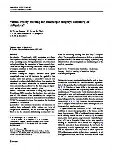
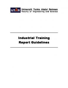
assess endoscopic access for the planned surgical procedure. A computed tomography (CT) scan was performed in all patients undergoing functional endoscopic sinus surgery (FESS) (n 428, 100%). CT scans were obtained in coronal section with complementary axial cuts with bony window settings. CT scanning was not performed in those patients with lacrimal disease in whom endoscopic dacrocystorhinostomy (DCR) was planned (n 58). An additional 12 patients had CT scans performed with three-dimensional (3D) reconstruction after being deemed appropriate candidates for intraoperative 3D CT localization with the LandmarxTM image guidance system. Magnetic resonance imaging with contrast enhancement was undertaken in three patients where a diagnosis of juvenile nasopharyngeal angio®broma had been made. pro c e du r e s pe r f or m e d A total of 500 patients underwent a total of 815 procedures (185 (37.0%) unilateral, 315 (63.0%) bilateral). Primary surgery was undertaken in 413 patients (83%). Eightyseven patients (17%) had revision ESS. Of those undergoing revision surgery, 45 patients (52%) were internal revisions and 42 patients (48%) were referred from other centres. The speci®c procedures performed are summarized in Fig. 1. The miscellaneous procedures performed included resection of mucocele as sole procedure, tumour resection, resection of juvenile nasopharyngeal angio®broma and sphenopalatine artery ligation. The extent of FESS performed is shown in Fig. 2. Anterior FESS comprised uncinectomy, middle meatal antrostomy, excision of concha bullosa if required and anterior ethmoidectomy. Posterior FESS included the addition of posterior ethmoidectomy with or without sphenoidotomy. Septal surgery was indicated to improve access for ESS in 65 patients (13%) and comprised submucous resection or formal sep-
p r e o p e r at i v e i n v e s t i g at i o n s All patients undergoing surgery underwent rod lens nasendoscopy in the outpatient clinic to con®rm the diagnosis and to Table 1. Indications for surgery Indication for surgery
Number of patients
%
Chronic sinusitis Polyposis Epiphora Miscellaneous (including neoplasia)
235 193 58 14
47 38 12 3
Figure 1. Procedures performed in 500 patients.
# 2003 Blackwell Publishing Ltd, Clinical Otolaryngology, 28, 411±416
Audit-derived guidelines for ESS 413
i n t r a o p e r at i v e v i e w
Figure 2. Extent of FESS performed.
toplasty. Intranasal adhesions were divided in 10 patients (2%). anaesthesia The vast majority of patients (96%, n 482) had surgery under general anaesthesia. Only 18 patients (4%) had surgery performed under local anaesthesia with intravenous sedation under the supervision of an anaesthetist. su rg e o n g r a de The senior author was the main surgeon in 275 patients (55%) with the trainee either observing or assisting. In 225 patients (45%), a trainee in the specialist registrar grade was the main surgeon with the senior author supervising and assisting. It is departmental policy for all ESS performed by trainees to be closely supervised by their consultant trainer. The senior author undertook all revision surgery with the trainee involved at stages as appropriate. e n d o s c o p e (s) u s e d Zero-degree, 30-degree and 70-degree rigid nasendoscopes were available throughout surgery in all 500 patients. The 0degree nasendoscope was used exclusively in 482 patients (96%). It was used in conjunction with the 30-degree nasendoscope in only 18 cases (4%). The 70-degree nasendoscope was not used. us e of pow e r to ols A microdebrider was used in 287 patients (57%). This was used by the consultant in 163 patients (57%) and by the trainee under consultant supervision in 124 patients (43%). It was used in all cases where polyp surgery was performed (n 193). The microdebrider supplemented standard FESS instrumentation in a further 94 non-polyp patients undergoing ethmoid surgery.
Endoscopic sinonasal surgery can be performed with the main surgeon viewing `down the scope' or with use of a video monitor ± `off-the-screen viewing'. Off-the-screen viewing was used throughout in all procedures. Off-the-screen viewing is essential to allow the main surgeon and assistant to remain fully orientated throughout. A single- and/or triple-chip camera in a sterile sleeve was used to display the operative ®eld on an appropriately placed monitor. In 12 preselected patients (2%), an intraoperative 3-D image guidance system was employed. c o m p l i c at i o n s The overall complication rate was 1.2% (n 6). These were all minor complications. They included primary haemorrhage requiring the procedure to be terminated (0.2%), secondary haemorrhage (0.6%), periorbital surgical emphysema (0.2%) and postoperative infection requiring antibiotics (0.2%). No patient required blood transfusion. The single case of periorbital surgical emphysema occurred in a patient in whom a recognized breach of the lamina papyracea had occurred intraoperatively. Postoperatively, the patient blew her nose and the emphysema ensued. The patient was managed conservatively, and the long-term outcome was good. Care is now taken to ensure that all patients are instructed not to blow the nose for 48 h postoperatively. No major complications were recorded in the 500 patients over the 5-year period.
Discussion Functional endoscopic sinus surgery describes ethmoid surgery on the osteomeatal complex while ESS encompasses the wider range of endoscopic procedures including endoscopic DCR and tumour resection. The complications of ESS can be divided into major and minor.10 Major complications include death, cerebrospinal ¯uid leak, meningitis, other intracranial complications (e.g. pneumoencephalocele and brain abscess), visual loss, permanent diplopia and intraorbital haematoma. Minor complications include periorbital surgical emphysema, adhesion formation, epistaxis, ecchymosis around the medial part of the orbit, injury to the lacrimal apparatus and infection. The overall complication rate in this series of 500 patients was 1.2% (i.e. 0.7% for the 815 procedures performed). We encountered no major complications in 500 patients over a 5year period (1996±2000). Of the six minor complications, three (two secondary haemorrhages and one postoperative infection) occurred in patients operated upon by the trainee under supervision. The other three (primary haemorrhage, periorbital emphysema and secondary haemorrhage) occurred
# 2003 Blackwell Publishing Ltd, Clinical Otolaryngology, 28, 411±416
414 M.L. Montague et al. Table 2. Review of major reported personal consecutive series Authors
Number of cases
Incidence of major complications (%)
Vleming et al. (1992)11 Lazar et al. (1993)4 May et al. (1994)5 Cumberworth et al. (1994)7 Present series
1235 773 2108 551 500
0.3 0.39 0.85 0.54 0.00
in patients operated on by the consultant. The complication rates reported in this series can be compared favourably with those from large personal series (Table 2) and national surveys (Table 3). Periorbital surgical emphysema following orbital penetration is a recognized complication of FESS. Its management is conservative with close observation of any change in visual acuity. The swelling may take up to 1 week to resolve completely. All patients in this series were advised against nose blowing in the postoperative period to minimize the risk of periorbital surgical emphysema. In one case in which this complication occurred, a breach of the lamina papyracea was evident intraoperatively. At no stage was visual acuity or globe movement compromised. Administration of antibiotics was commenced. The emphysema resolved within 24 h, and the ®nal postoperative outcome was unaffected. There was no difference in complication rates between consultant and trainee operations in this series. Nevertheless, the hazards to the trainee have been highlighted by Wigand13 who reported no major complications from a personal series of 220 patients whilst a series of similar size from trainees in his clinic included complications such as meningitis and brain abscess with subsequent death and blindness caused by postoperative intraorbital bleeding. This is undoubtedly related to the `learning curve' phenomenon that is reported for skill acquisition in many ®elds of surgery. Stankiewitz described it speci®cally in relation to FESS. He reported a 5% incidence of major complications for his ®rst 90 operations.14 This was reduced to 0.7% for the subsequent 90 operations.15 All patients in this series had a coronal CT scan preoperatively. This allows appreciation of the surgical anatomy, particularly at the level of the osteomeatal complex. Complementary axial views were obtained at the level of the
frontal sinuses and orbits to reduce the risk of complications by visualizing potentially dangerous anatomical abnormalities. Only 84% of surgeons in a UK survey undertaken in 2001 routinely performed a CT scan before surgery.6 This ®gure is concerning given that a preoperative CT scan has been described as `mandatory' in the UK.16 In 12 cases, a 3D image guidance system was used given the complex nature of the surgery to be undertaken and the need for precise disease localization. Proponents of intraoperative localization believe that it enhances surgical precision and reduces complications. Intraoperative 3D CT is useful for the con®rmation of key anatomical structures during endoscopic sinus surgery, especially when landmarks are distorted or absent. It may therefore be particularly useful for dif®cult and revision cases. It cannot, however, substitute for thorough anatomical and surgical training. Ultimately, the system should be readily available for any case, but re®nements to currently available systems will be required before they are accepted for widespread use.17 The authors believe that the use of `off-the-screen' operating is essential for training in ESS. Off-the-screen operating is also a practical and effective method of documenting cases. It allows documentation of anatomy and disease of deep structures of the nose and paranasal sinuses, teaching delicate surgical procedures, instantaneous high-quality hard copies of video images for teaching and patient records, instant replay and repeated viewing by a slow motion or frame-byframe analysis, simultaneous viewing by a small or large audience, and image digitalization for storage in the computer. Furthermore, review of video material is useful as a debrief following dif®cult cases and allows a video library to be built up. It will always play a vital role in ESS training, and there is probably room for expansion of its role in error avoidance.18 The 0-degree endoscope was used exclusively in 96% of patients in this series. Operating with it is safe, but caution is required with use of 30- and 70-degree endoscopes when it is easier to become disorientated.19,20 One hundred and twenty-four patients (25% of all patients, 43% of power tool operations) had their surgery performed by a trainee using a power tool. Such instrumentation is perceived to be advanced and associated with high risk. However, our data suggests that suitably supervised trainees can operate with power tools without compromising patient safety.
Authors
Number of cases
Overall incidence of complications (%)
Incidence of major complications (%)
Sharp et al. (2001)6 Cumberworth et al. (1994)7 Harkness et al. (1997)12
84493 15399 1064
0.69 ± 1.41
0.25 0.23 0.00
Table 3. Review of major reported national surveys
# 2003 Blackwell Publishing Ltd, Clinical Otolaryngology, 28, 411±416
Audit-derived guidelines for ESS 415 Table 4. Phases and structure for ESS training Phase
Aim
Method
Clinical setting
I
Attendance at a basic sinus surgery course
II
Development of hand±eye coordination, tissue- and instrument-handling skills Consolidation of phase I skills
Assisting at early stages of ESS (e.g. nasal preparation and injection, and uncinectomy) Anterior FESS procedures
III
Advanced instrument handling
IV
Supervised advanced training
Performance of minor and intermediate procedures under supervision Attendance at an advanced endoscopic sinonasal surgery dissection course Fellowships and clinical attachments
Among patients who have unsuccessful primary FESS, some eventually require revision FESS for management of their sinusitis. In this series, revision was performed in 17% of patients. Although the techniques and principles of revision surgery are essentially the same as those of primary FESS,21 it can be more dif®cult to perform because of missing anatomical landmarks, increased bleeding, adhesions and less experience of the surgeon with revision surgery than that with primary FESS. It is for these reasons that the senior author performed all revision procedures. In the setting of a university teaching hospital, experienced instruction by operating consistently with the same consultant trainer may accelerate the learning process and reduce complication rates. Attendance at a recognized cadaveric dissection course may also reduce the risk of complications. Wigand emphasized this fact when describing the increased major complication rates among his trainees, as opposed to his personal series of patients.13 A recent survey of trainees in the North (East) Thames Region revealed that almost half the trainees had started performing FESS without prior attendance at a recognized course or performing cadaveric dissection.22 The great availability of FESS training courses should overcome this problem. Stanley23 has suggested establishing endoscopic sinus laboratories akin to temporal bone laboratories. In a survey of fellows of the American Academy of Otolaryngology±Head and Neck Surgery in 1994, physicians generally did not rate their residency training in endoscopic ethmoidectomy high,24 and just over 50% had no residency ethmoid dissection experience. The reporting trainee's estimate of the quality of training in ethmoidectomy during residency affected complications (lower rates with better training), and the effect of the number of courses attended had a bene®cial effect on the incidence of orbital complications. Training in ESS, ideally, should be phased. The need for a structured approach to general ENT training for Specialist
Supervised performance of intermediate FESS and ESS procedures Experience of the extended role of ESS (e.g. orbital and frontal recess surgery, and surgery for neoplasia)
Registrars in the out-patient clinic and theatre since the implementation of the Calman Report has already been highlighted.25 See Table 4 for our proposed phases and structure for ESS training.
Conclusion This audit suggests that training need not compromise patient safety. Particular attention to an atraumatic technique, an intimate knowledge of anatomy and excellent haemostasis are key to avoiding complications. Additional factors important in the setting of a teaching centre are the need for constant supervision, adherence to preoperative protocols and `off-thescreen' operating. We suggest the following minimum requirements for units involved in ESS training: 1. Preoperative investigation protocol to include mandatory endoscopy and CT scanning. 2. Close direct operative supervision of trainee by consultant trainer at all times. 3. 0-degree rod lens endoscope is the instrument of choice. 4. Supervised operating with power tools to be introduced at an appropriate training phase (e.g. Phase III). 5. Off-the-screen operating essential for supervised training. 6. Formal phased (Phases I±IV) ESS training including cadaveric work to be recorded in the trainee's logbook. 7. Continuous audit of the surgical team's complication rates so that accurate informed consent can be obtained from patients. This is an audit of complications. The outcome of the surgery in terms of bene®t in this group of patients has been studied separately and is currently in press.
Acknowledgements The authors wish to thank Jennifer Mills of the Surgical Audit Department at Glasgow Royal In®rmary for her assistance with this audit project.
# 2003 Blackwell Publishing Ltd, Clinical Otolaryngology, 28, 411±416
416 M.L. Montague et al. References 1 KENNEDY D.W., ZINREICH S., ROSENBAUM A.E. et al. (1985) Functional endoscopic sinus surgery: theory and diagnostic evaluation. Arch. Otolaryngol. 111, 576±582 2 STAMMBERGER H. & WOLF G., (1988) Headaches and sinus disease: the endoscopic approach. Ann. Otol. Rhino Laryngol. 97, 3±23 3 WIGAND M.E. & HOSEMANN W.G., (1991) Results of endoscopic surgery of the paranasal sinuses and anterior skull base. J. Otolaryngol. 20, 385±390 4 LAZAR R.H., YOUNIS R.T. & LONG T.E. (1993) Functional endonasal sinus surgery in adults and children. Laryngoscope 103, 1±5 5 MAY M., LEVINE H., MESTER S. et al. (1994) Complications of endoscopic sinus surgery: analysis of 2108 patients ± incidence and prevention. Laryngoscope 104, 1080±1083 6 SHARP H.R., CRUTCHFIELD J.M., ROWE-JONES J.M. et al. (2001) Major complications and consent prior to endoscopic sinus surgery. Clin. Otolaryngol. 26, 33±38 7 CUMBERWORTH V.L., SUDDERICK R.M. & MACKAY I.S. (1994) Major complications of functional endoscopic sinus surgery. Clin. Otolaryngol. 19, 248±253 8 MOSHER H.P. (1929) The surgical anatomy of the ethmoidal labyrinth. Ann. Otol. Rhinol. Laryngol. 38, 869±901 9 BOLGER W. & KENNEDY D. (1993) Complications of surgery of the paranasal sinuses. In Complications in Head and Neck Surgery, Chapter 50, EISELE D. (ed.), pp. 458±470. Mosby-Year Book, St. Louis 10 SWIFT A.C. (1996) Functional endoscopic sinus surgery. Br. J. Hosp. Med. 55, 54±558 11 VLEMING M., MIDDELWEERD M.J. & DE VRIES N. (1992) Complications of endoscopic sinus surgery. Clin. Otolaryngol. 17, 460 12 HARKNESS P., BROWN S., FOWLER S. et al. (1997) A national audit of sinus surgery. Results of the Royal College of Surgeons of England comparative audit of ENT surgery. Clin. Otolaryngol. 22, 147±151
13 WIGAND M.E. (1990) Results of endoscopic sinus surgery. In Endoscopic Surgery of the Paranasal Sinuses and Anterior Skull Base, WIGAND M.E. (ed.), Chapter 7, pp. 134±141. Thieme, Stuttgart and New York 14 STANKIEWITZ J.A. (1987) Complications of endoscopic intranasal ethmoidectomy. Laryngoscope 97, 1270±1273 15 STANKIEWITZ J.A. (1989) Complications in endoscopic intranasal ethmoidectomy: an update. Laryngoscope 99, 686±690 16 LUND V.J., WRIGHT A. & YIOTAKIS J. (1997) Complications and medicolegal aspects of endoscopic sinus surgery. J. R. Soc. Med. 90, 422±428 17 ROTH M., LANZA D.C., ZINREICH J. et al. (1995) Advantages and disadvantages of three-dimensional computed tomography intraoperative localization for functional endoscopic sinus surgery. Laryngoscope 105, 1279±1286 18 CARR-LOCKE D.L. (1990) Videoendoscopy in clinical application. Impact on teaching. Endoscopy 22, 19±21 19 STAMMBERGER H. (1991) Surgical technique and options. In Functional Endoscopic Sinus Surgery ± the Messerklinger Technique, DECKER B.C. (ed.), pp. 290±319. Mosby, Philadelphia, PA 20 KANG S.K., WHITE P.S., LEE M.S.W. et al. (2002) A randomised control trial of surgical task performance in frontal recess surgery: zero degree versus angled telescopes. Am. J. Rhinol. 16, 33±36 21 LAZAR R.H., YOUNIS R.T., LONG T.E. et al. (1992) Revision functional endonasal sinus surgery. Ear Nose Throat J. 71, 131±133 22 MCFERRAN D.J., GRANT H.R., INGRAMS D.R. et al. (1998) Endoscopic sinus surgery: are junior doctors being properly trained? Ann. Royal Col. Surg. Eng. 80, 359±363 23 STANLEY R.E. (1992) Letter. Singapore Med. J. 33, 532±533 24 KENNEDY D.W., SHAMAN P., HAN W. et al. (1994) Complications of ethmoidectomy: a survey of fellows of the American Academy of Otolaryngology±Head and Neck Surgery. Otolaryngol. Head Neck Surg. 111, 589±599 25 DRAKE-LEE A. (2002) Structured training of ENT Specialist Registrars in the out-patient clinic and theatre. Clin. Otolaryngol. 27, 396±402
# 2003 Blackwell Publishing Ltd, Clinical Otolaryngology, 28, 411±416