and H reflex changes in antagonistic lower limb muscles. P.J. Delwaide *, B. Schepens. Unitiersity Department of Neurology, HSpital de la Citadelle, Bd. du 12â ...
EISEVIER
Electroencephalography
and clinical Neurophysiology
97 (1995) 416-423
Auditory startle (audio-spinal) reaction in normal man: EMG responses and H reflex changes in antagonistic lower limb muscles P.J. Delwaide
*, B. Schepens
Unitiersity Department of Neurology, HSpital de la Citadelle, Bd. du 12” de Ligne I, 4000 LiPge, Belgium Accepted for publication:
6 June 1995
Abstract The audiospinal reaction (ASR) to a 30 msec tone of 90 dB has been studied in 66 seated healthy volunteers. Electromyographic bursts induced in trapezius (Tra), soleus (Sol) and tibialis anterior (TA) have been looked for. Facilitation of Sol H reflex has been measured in 21 subjects in terms of delays from delivery of the sound. Among the subjects, I I had a stable TA H reflex whose facilitation was compared to that of Sol H. Effects of selective isometric voluntary contraction of either Sol or TA were assessed both on EMG responses and H reflex facilitation. At rest, only 36% of subjects exhibited a response in Sol at a mean latency of 123 msec and 39% in TA at a latency of 119 msec. Responses were seen in antagonist muscles in 74% of subjects. Incidence in Tra was 96%. During voluntary contraction, the results were not significantly changed either in the contracted muscle or its antagonist. H reflex facilitation of both TA and Sol started 50 msec after the sound to peak after 75-125 msec and returned to baseline values after 250 msec. Extent of Sol H reflex facilitation remained similar during voluntary contraction of Sol and TA. It was observed that EMG responses were more frequent in subjects with brisk reflexes but not necessarily in those who exhibited the largest H reflex facilitation. The results are in agreement with assumptions that ASR is mediated through reticula-spinal pathways but do not support the view that it corresponds to a flexor reaction. In addition to providing quantitative data for comparisons in pathological cases, they suggest differences in the mode of activation of motoneurones by the motor cortex and subcortical nuclei. Keywords:
Startlereaction; EMG responses; H reflex; Tibialis anterior; Soleus
1. Introduction The auditory startle reaction (ASR) is the motor response evoked by a sudden sound of adequate intensity. This response depends on subcortical activity with the nucleus reticularis pontis caudalis (NRPC) as the last supraspinal relay (Davis et al., 1982). From that nucleus, a facilitatory message reaches motoneurones via reticulo-spinal tracts. ASR has been studied both in animals and humans (Paillard, 1955; Rossignol, 1975; Rude11 and Eberle, 1985; Wu et al., 1988; Brown et al., 1991a,b,c; Chokroverty et al., 1992; Delwaide et al., 1993). In man, two methods have been used: on the one hand, recording by surface electrodes of electromyographic responses of various muscles (Rossignol, 1975; Wilkins et al., 1986; Brown et al., 1991a,b,c; Chokroverty et al.,
??
Corresponding author.
0924-980X/95/$09.50 D 1995 Elsevier Science Ireland Ltd. All rights reserved SSDI 0013-4694(95)00136-O
1992) and, on the other, changes in amplitude of the Hoffmann (H) reflex at various delays after the sound (Paillard, 1955; Melvill Jones et al., 1973; Rossignol and Melvill Jones, 1976; Rude11 and Eberle, 1985). The first method studies many muscles, in both upper and lower limbs, flexors and extensors, and reflects a behavioural reaction. The second method, which is applicable to the lower limb antagonist muscles soleus (Sol) and tibialis anterior (TA) presents several advantages: quantification of effects, definition of a precise time-course of facilitation and disclosure of infraliminal effects (inhibitory or facilitatory). Although the Sol H reflex is facilitated by an auditory stimulus (Melvill Jones et al., 1973; Rude11 and Eberle, 1985), the concept that ASR corresponds basically to a generalized flexor reaction (Landis and Hunt, 1939) is generally accepted. Rossignol (1975), for example, has shown that responses in TA predominate over those in extensors (gastrocnemius). However, Brown et al. (1991al
EEM 9470 I
P.J. Delwaide, B. Schepens / Electroencephalography and clinical Neurophysiology 97 (I 995) 416-423
observed that EMG responses were equally distributed in flexor and extensor muscles of the leg. The question of ASR distribution and possible predominance of one group over the other is still open. Influence of voluntary contraction upon ASR is not clear. Rossignol (1975) reported that previous or ongoing EMG activity in a muscle favoured the incidence of responses while Brown et al. (1991a) did not observe any facilitatory effect. However, these observations appear anecdotal and no systematic study has been conducted of the influence of voluntary contraction, especially using EMG recording and H reflex facilitation together. In this study, we investigated ASR simultaneously in TA and Sol. We used both EMG recording and recovery curves of monosynaptic reflexes (H reflexes) in order to appreciate whether a muscle was more influenced than its antagonist. For that reason, we chose leg muscles where it is sometimes possible to obtain stable H responses both in extensor and flexor muscles. In addition, we studied the influence of selective voluntary contraction.
2. Subjects and methods 2.1. Population Sixty-six healthy volunteers (32 males; 34 females; age range: from 19 to 67 years) were included in the experimental protocol. They were all normal on neurological examination. Intensity of patellar and Achilles tendon jerks was scored as +, + + or + + +. All patients gave oral informed consent to the experimental procedure which had been approved by the local Ethical Committee. The subjects were comfortably seated in an armchair with the knee extended (170”) and the ankle at 90- 100”. Experiments were performed in a quiet room. In all subjects, EMG activity was recorded from trapezius (Tra), tibialis anterior (TA) and soleus (Sol) muscles. As an H reflex cannot be elicited in TA in all subjects, 11 out of the 66 subjects were selected in whom it was possible to record a stable H reflex in TA as well as in Sol. The Sol H reflex was studied alone in an additional 10 subjects. Thus, in 21 subjects, comparisons between possible EMG activity and corresponding changes in Sol H reflex amplitude were possible. 2.2. Conditioning
auditory stimulation
The auditory stimulus, delivered through Sansui S22 binaural earphones, consisted of a square 30 msec tone burst at 700 Hz with an intensity of 90 dB. 2.3. Experimental
procedure
In each subject, induced EMG responses were sought in the 3 muscles under study, in 3 situations: seated at rest
417
and with Sol or TA voluntary contraction. Selective isometric voluntary contraction was performed by asking the subject to develop between 15 and 25% of maximal isometric force with the help of audio- and visual feedback of its EMG activity. The sound was delivered only once in each situation to avoid habituation. Thereafter, a possible stable H reflex in TA was looked for. If it was present, audio-spinal facilitation of TA and Sol H reflex (see below) was studied at rest and during selective voluntary contraction of either Sol or TA. 2.4. EMG responses Bipolar electrodes (silver/silver chloride) were fixed 2 cm apart over muscle bellies of Tra, TA and Sol. Muscle potentials were filtered (10 Hz-3 kHz), amplified (Digitimer D150, UK) and visualized on an oscilloscope (Tektronix S223, USA) before being stored on floppy disks for further analysis (C.E.D. 1401, Cambridge Electronics, UK). Special attention was paid to possible crosstalk between activity in one leg muscle and recording from electrodes fixed over its antagonist. Cross-talk resulting from both anatomical and technical reasons was recognized by symmetry of responses from the 2 muscles. To check this possibility, subjects were asked to contract selectively one muscle group. If EMG activity was recorded from over the antagonist muscle, the subject was not included in the studied population. Sound may or may not evoke a visible EMG activity. In the resting conditions, incidence, latency and duration of these EMG bursts were measured for the 3 muscles under study. During voluntary contraction, sound may be followed by a superimposed burst on tonic background EMG activity. These bursts, detected by visual inspection, were considered provided their peak-to-peak amplitude reached at least 3 times that of the background activity. The latency to onset of reflex EMG activity was measured using the initial voltage sustained above the background level of EMG activity (Brown et al., 1991b). Incidence and duration were also measured. 2.5. Conditioned
H reflexes
Amplitude changes of H reflexes recorded from TA and Sol were used to quantify effects of the auditory stimulation. H reflexes were evoked at various delays after the sound in steps of 25 msec from 0 to 300 msec. Sol H reflex was obtained by bipolar posterior tibia1 nerve stimulation using the classic technique (Desmedt, 1973). For TA H reflex, stimulating surface electrodes were fixed over the peroneal nerve at the fibular head. Amplitude of the test H reflexes was adjusted to half maximum (H,,,/2). Control H reflexes were evoked alternately with those conditioned by auditory stimulation, these latter being separated by a time interval of at least 2 min (from 2 to 5 min). TO verify
418
P.J. Delwaide,
B. Schepens / Electroencephalography
and clinical
Neurophysiology
97 (19951416-423
B.
A. tt t 1 I I I
Sol
/ . . ii is‘ 1 15o / 501/ x
/ I
k
I”
I
ms
5 100
-
.
P l-
r = 0.91
V
I I
.
00
50 100150ms
Sol latency
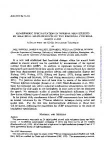
100 ms Fig. 1. A: example of electromyographic bursts in 3 distinct muscles following a brief auditory stimulus. Response in Tra precedes those in Sol and TA, which occur concomitantly. B: correlation between response latencies in TA and Sol. The correlation is linear with a coefficient of 0.91.
that nerve stimuli were kept equal between rest and contraction, larger responses with an M component were elicited and M amplitude should have been stable in these 2 situations. We observed that after such delays habituation of H reflex facilitation - contrary to EMG responses - was absent or very feeble. At each interval after the sound, a minimum of 5 responses was measured peak-topeak and averaged. Conditioned responses were expressed as percentage of control values and plotted against delays after the sound. 2.6. Statistical
analysis
Statistical analysis was performed using the MannWhitney U test to compare mean values. Following others (Rossignol, 1975), significance was considered for P < 0.005. In addition, Student’s t test for paired data (rest vs.
contraction) Correlation software.
has been used with significance for P < 0.05. coefficients were determined by using Slide
3. Results 3.1. Incidence tion
of EMG responses
aj?er auditory stimula-
In 24 out of 66 subjects (36%). the sudden sound was followed by a clearly identifiable response in Sol. It appeared after a mean latency of 123 msec and lasted a mean of 55 msec. A response was found in TA in 26 out of 66 subjects (39%). Its mean latency and duration were 119 msec and 56 msec respectively. The values of latency and duration
Table 1
Incidence
Mean latency *SD. Mean duration It S.D.
Sol TA Tra Sol TA Tra Sol TA TRA
At rest
Sol voluntary contraction
TA vohmtary contraction
37% 39% 96% 123 f 29.7 119k23.8 48* 15.1 55 rf: 28.7 56 + 24.7 35 + 15.2
41% 39% 96%
39% 43% 94% 116k33.7 113 f 25.3 52 f 18.2 5Orf: 29.5 54 f 24.3 361t 14.1
msec msec msec msec msec msec
112+ 112 f 49* 45 rt 43 f 32 f
20.4 17.5 12.1 25.7 22.4 10.3
msec msec msec msec msec msec
msec msec msec msec msec msec
PJ. Delwaide.
B. Schepens/
Electroencephalography
and clinical Neurophysiology
97 (I 995) 416-423
419
measured from TA did not differ significantly from those obtained from Sol. In Tra, responses were found in 96% of subjects with a mean latency of 48 msec and mean duration of 35 msec. Fig. 1A illustrates responses appearing in the 2 antagonist muscles and in Tra. Table 1 gives the mean of individual values with S.D. In 74% of subjects, when a response was identifiable in one leg muscle, a concomitant response was also seen in its antagonist. A linear regression was found between Sol and TA latencies with r = 0.91 and an angular coefficient of 0.98 (Fig. 1B).
significantly different from corresponding values obtained at rest (Mann-Whitney U test and Student’s t test for paired data). Latency of EMG activity measured in Tra with leg muscles at rest (48 msec) was not significantly modified by voluntary contraction in the lower limb (49 msec for Sol voluntary contraction and 52 msec for TA voluntary contraction). Incidence of the responses was also similar at rest (94%) and during voluntary contraction of one of the leg muscles.
3.2. EfSect of voluntary contraction
3.3. H reflex changes after auditory stimulation
on the EMG responses
During constant selective voluntary contraction of Sol (15-25% of the maximal isometric force>, 27 (41%) identifiable EMG bursts were observed in Sol after auditory stimulation and 26 (39%) in TA. In 69% of cases, bursts were concomitant in Sol and TA. Mean latency of responses during Sol voluntary contraction was 112 msec both for Sol and TA. Mean duration of the burst was 45 msec in Sol and 43 msec in TA. There was no significant difference in latency or EMG burst duration between the values measured at rest and the corresponding values during contraction, even with the Student’s t test for paired data. When TA was voluntarily contracted, 26 clearly definable EMG bursts were recorded in Sol (39%) and 28 in TA (42%) after auditory stimulation. In 80%, bursts appeared in both muscles concomitantly. Mean latency was 116 msec for Sol and 113 msec for TA and mean duration was 50 and 54 msec respectively. These results were not
B.
A. Individual
H (%) 250
250 1
200
Sol
150
150
,*+-kZ)__ o---*
.H
100
100
L._.-._._.‘%%~
0
50
100
150
200
250
50
100
150
200
250
ms
H (%)
250
250
TA
1
50 50
100
150
200
TA
1
’ 0
0
ms
H (%)
50
Mean curves
examples
H (%) 200
Facilitation of Sol H reflex after an auditory stimulus has already been extensively documented (Melvill Jones et al., 1973; Rude11 and Eberle, 1985). In the present study, special attention was paid to the quantitative aspects of that facilitation. In 11 subjects, both TA and Sol H reflexes were studied and, in 10 additional subjects, H Sol only. Fig. 2A illustrates typical‘examples. At a delay of 50 msec after the conditioning sound, Sol H reflex facilitation appeared and reached a maximum at 75 msec delay. Following that, there was either a short plateau (empty circles) or a decrease in amplitude followed by a second peak of facilitation at 125 msec (filled circles). Thereafter, the amplitude progressively declined to return to control values after 250 msec. In these examples, EMG burst appeared after 94 (filled circles) and 103 msec (empty circles) respectively. Facilitation exhibiting 2 distinct peaks was found in 63% of subjects. Every patient showed a
250
ms
’
0
50
100
150
200
250ms
Fig. 2. Recovery curves of Sol H reflex (upper row) and TA H reflex (lower row) respectively. In A, examples of individual curves. In B, mean curves: n = 21 for Sol and 11 for TA. In ordinate, facilitation is expressed as percentage of the control values. In abscissa, delays from delivery of the sound
P.J.
420
Delwnide.
A. Schepens/
Elecrroencephalography
facilitation with at least one value higher than control values plus 3 S.D. However, there was clear variability in the amount of facilitation. If consideration is given to the start and the end of facilitation, the individual curves have a similar time-course although the peak of facilitation may vary from subject to subject between 75 and 150 msec. The mean curve (n = 21; Fig. 2B) indicates that facilitation is maximum from 75 (178%) to 125 msec (175%) delay. The audio-spinal response using TA H reflex has not been studied previously, probably because a monosynaptic response can be evoked only in a small number of subjects. In 11 of our subjects it was possible to evoke it. As seen in two illustrative examples (Fig. 2A), auditory facilitation starts after a delay of 50 msec, reaches its maximum at 100 msec and 125 msec respectively and then progressively decreases to reach baseline values after 250 msec. In these examples, an EMG burst is seen after 122 (empty circles) and 130 msec (filled circles) respectively. Magnitude of the facilitation varies from subject to subject and 55% exhibited a double peak of facilitation. Using the mean curve (Fig. 2B), the maximum is seen at a delay of 75 msec (195%). 3.4. Effect of a voluntary facilitation of Sol H reflex
contraction
clrr~~cnl Neurophysroiogy
97
1/WY) 416423
A. n=15 n=30 n=21
JZI
/
II-
+ Tendon
1
++
jerk
. :
. :
i
’
.
??
contraction
r=0.07
I__--.__~_++
+
Tendon
jerk
+++
intensity
. .
!
.
1 i
voluntary
+++
intensity
:
.
on the auditory
During Sol constant isometric contraction, the amplitude of Sol H,,,/2 reflex is increased by 15-20%. The H&2 value is further facilitated in all subjects after auditory stimulation. The mean curve (Fig. 3) indicates that facilitation starts 50 msec after the conditioning sound, reaches its maximum (179%) after 75 msec and returns to
H Sol during
and
No burst
i
Burst
Fig. 4. A: percentage of subjects exhibiting an EMG response after auditory stimulation. The subjects are classified in terms of tendon jerk intensity: + , + + or + + + The number of patients in each group is 21, 30 and 15 respectively. B: peak of Sol H reflex facilitation in terms of tendon jerk intensity in the 21 subjects in whom recovery curves have been plotted. C: amplitude of peak facilitation of Sol H reflex in subjects exhibiting or not an EMG burst after similar auditory stimuli. The results show that appearance of an EMG response is not closely linked with intensity of H reflex facilitation seen in the recovery curves.
H (%) 250
Sol contraction
200
:::t-i^‘\--50
’ 0
50
100
150
200
250
ms
H (%)
TA contraction
control values at 250 msec. In 65% of subjects, facilitation evolves in 2 successive peaks. During constant isometric contraction of TA, the amplitude of Sol H reflex is reduced by 20%. The H,,,/2 value is, however, facilitated after auditory stimulation, as shown in Fig. 3. At 100 msec facilitation reaches 179%. All individual curves show a significant facilitation, sometimes with a double peak. 3.5. Relationships
50
’ 0
50
100
150
200
250
ms
Fig. 3. Mean curve of Sol H reflex auditory facilitation under selective isometric contraction of soleus (top) and tibialis anterior (bottom). In ordinate, facilitation is expressed as percentage of the values obtained during contraction. In abscissa, delays from delivery of the sound.
with reflex intensity
The results have been analysed in terms of intensity of the tendon jerks (scored +, + + or + + + >. Fig. 4A indicates the incidence of Sol EMG responses. The numbers at the top of each column give the partition of the 66 subjects in 3 groups in terms of reflex briskness. The columns give for each group the percentage of subjects in whom an EMG burst was identified. With feeble tendon
P.J. Delwaide. B. Schepens/ Electroencephalography
jerks ( +), the incidence of EMG bursts is only 24% whereas with brisk reflexes (+ + + ), this value amounts to 60%. Fig. 4B illustrates the relationship between tendon jerk briskness and peak of Sol H reflex facilitation in the 21 subjects studied. There is a clear overlap of the values corresponding to the 3 groups and it cannot be said that peak facilitation is higher with brisk reflexes. Correlation coefficient is very poor (r = 0.07). 3.4. Relationships
between occurrence
of EMG bursts and
intensity of H rejlex facilitation
In the 21 subjects in whom a recovery curve of Sol H reflex was plotted, it was looked for whether occurrence of a sound-induced EMG burst was related to the magnitude of H reflex facilitation. Only 7 out of these 21 (33%) showed an EMG response. They are represented in Fig. 4C (column “Burst”) and compared to the 14 without any response (“No burst”) in terms of peak of H reflex facilitation. H reflex facilitations of more than 200% may be seen in the absence of EMG response whereas facilitations of less than 150% are observed in subjects exhibiting EMG responses. So, occurrence of an EMG burst is not closely linked to Sol motoneurone pool facilitation as revealed by recovery curves.
4. Discussion With our experimental conditions (auditory stimulus at 90 dB) not all but only 36-39% of healthy volunteers responded with detectable EMG activity in lower limb muscles after an unexpected sound. Brown et al. (1991a) reported a percentage of responses varying from 22 to 25%. These percentages contrast with a higher incidence in Tra (96%) as if there was a rostro-caudal gradient of facilitation. EMG responses were seen equally in both antagonist lower limb muscles without a preferential activation of either flexor or extensor. Surprisingly, although voluntary contraction increases excitability of a motoneurone (MN) pool, it does not affect significantly the incidence of responders. In other words, increased excitability of motoneurones does not seem determinant to explain why some subjects exhibit an EMG burst and others not. However, briskness of tendon jerks seems to favour appearance of a response but this factor not only depends on MN excitability but is also conditioned by gamma innervation and the level of presynaptic inhibition acting on Ia afferents. As far as latency of EMG responses is concerned, our results agree with those previously reported (Wilkins et al., 1986; Brown et al., 1991a,b,c; Chokroverty et al., 1992) and clearly indicate that they appear after a longer delay than those evoked by cortico-spinal tract activation. Values
and clinical Neurophysiology 97 (I 995) 416-423
421
for TA and Sol were not found significantly different in our study. Wilkins et al. (1986) and Brown et al. (1991a) reported also typical synchronous activations of antagonist muscles whereas Rossignol (1975) found a shorter latency in Sol than in TA. Durations of EMG bursts were also similar in TA and Sol and comparable with the results of Rossignol(1975). It might be objected that similarity observed in the two muscles would in fact reflect an artefact, for example cross-talk between electrodes fixed over antagonist muscles. This possibility has been considered but ruled out after previous careful examination of records during selective voluntary contraction of only one of these muscles. If EMG responses are only seen in a limited percentage of subjects, all those in whom an H reflex was elicited demonstrated increased facilitation both of TA and Sol MN pools as reflected by various degrees of H reflex amplitude increase. Our results concerning auditory facilitation of the Sol H reflex correspond to those reported earlier: Melvill Jones et al. (1973) and Rossignol and Melvill Jones (1976) observed that the auditory conditioned Sol H reflex was increased after a delay of 60 msec, that facilitation lasted a mean of 200 msec and had an amplitude almost doubled around 100 msec after the sound. More recently, Rude11 and Eberle (1985) also showed that the extensor H reflex was facilitated at intervals ranging from 60 to 210 msec after the sound. There is no previous reference in the literature to facilitation of the TA H reflex. Time-course of facilitation in that motoneurone pool is similar to that seen in Sol. It is more critical to compare the magnitude of facilitation because the proportion of neurones within the motor pool involved in the monosynaptic response is not the same for TA and Sol. Nevertheless, TA H reflex is increased by a mean of 195% while Sol H reflex by 178%. On the basis that a startle response appeared in TA 3 times more frequently than in Sol (Rossignol, 1975) it has been claimed that audio-spinal responses are part of a flexor reflex and this view remains popular. However, the similar incidence of responses, latencies, durations and H reflex facilitations in TA and Sol indicates that audio-spinal responses can no longer be considered as a flexor reflex, at least when the subject is studied in the sitting position. As flexor and extensor MN pools are equally influenced after the same latency, it is very likely that both nuclei receive the same descending message which is not directed preferentially toward one nucleus of the pair, extensor or flexor. Selective voluntary contraction of one muscle out of an antagonist pair has a limited influence on audio-spinal responses, either in the active muscle or in its antagonist. For the active muscle, our results are in agreement with those of Brown et al. (1991a) but contradict those of Rossignol(1975) who reported facilitation of ASR after or during voluntary contraction. As far as the antagonist is concerned, reciprocal inhibition is no longer appreciated
422
P.J. D&wide,
B. Schepens/
Electroencephalography
either at rest or during active contraction as EMG responses appear with the same incidence and at the same delay: the ASR response is not inhibited by voluntary contraction of the antagonist muscle although Ia inhibitory intemeurones are known to be activated in that situation. So, the mode of activation of an MN pool by ASR seems to differ from that which is brought about by cortico-spinal stimulation. It also differs from what is observed when a spinal reflex is evoked during voluntary contraction: facilitation when the MN is activated and reduction or disappearance when reciprocal inhibition is acting. What descending pathway is involved in ASR? The long latency of responses suggests that its conduction velocity is not comparable to that of the fastest fibres of the cortico-spinal tract. In fact, conduction velocity of the descending message can be measured by comparing the latency of the H reflex auditory facilitation at the level of the flexor carpii radialis (FCR) and soleus muscles. By subtracting the peripheral conduction times of FCR and Sol H reflex, we took into account the moment at which motor nuclei began being facilitated. With this difference and the measure of the distance over the spine between these 2 MN pools, it was possible to calculate the conduction velocity of the descending facilitatory wave, which travels at a conduction velocity of around 20 m/set. This value is confirmed by latency differences found between EMG responses in Tra and Sol after measurements of the peripheral times obtained by magnetic stimulation of the corresponding roots (Maertens de Noordhout, 1991). This conduction velocity is in agreement with the view that the audio-spinal response is mediated through a reticula-spinal pathway (Baldissera et al., 198 1) as was proposed by Davis et al. (19821, Wu et al. (1988) and Iwamato and Sasaki (1990) in animals. They demonstrated that the reticular formation - especially the reticularis pontis caudalis (NRPC) - is the last supraspinal relay in the genesis of ASR. It is likely that the reticula-spinal tract impinges directly on the MNs, as described in animals. Peterson et al. (1979) have shown in cat that stimulation of the NRPC produced monosynaptic excitation of MN innervating limb distal parts. This does not exclude additional polysynaptic access to MN as mentioned by Takakusaki et al. (1989) who described mono- and polysynaptic connections (facilitatory and inhibitory) to motoneurones. If 15 msec is allowed for impulses to reach the spinal cord after sciatic nerve stimulation, then the latency of H reflex facilitation corresponds to that of EMG bursts (about 95-105 msec). Such an observation is also valuable for TA. Synchronous appearance of EMG bursts and peak facilitation of the monosynaptic reflex suggests that an EMG response might appear only in subjects in whom H reflex facilitation is very marked and above motoneurone discharge threshold. However, as shown in Fig. 4C, there is no good correlation between appearance of an EMG burst and magnitude of H reflex facilitation. This seems surprising. However, one could speculate on the origin of
and clinical
Neurophysrology
97 (199.i) 416-423
this lack of correlation by considering that the reticulo-spinal tract, at the same time it facilitates MN, also activates intemeurones responsible for presynaptic inhibition acting on Ia afferents. In other words, H reflex amplitude could be influenced in two opposite directions: a facilitation at the motoneuronal level, responsible for the EMG response, and a reduced efficacy of the test Ia discharge attenuated by increased presynaptic inhibition. Our study provides quantitative data permitting comparisons of audio-spinal responses in lower limb antagonist muscles. They will facilitate recognition of modified responses in other functional situations or of abnormal responses in pathology. Moreover, the results illustrate some pecularities in the mode of activation of motoneurones by the reticula-spinal tract.
Acknowledgements The active contribution of P. Gerard and V. De Pasqua are gratefully acknowledged. This work was supported by grants from the Fonds Facultaire de Medecine, University of Liege, Belgium, the Fondation Charcot, Belgium and Contract CEE ERBCHRX-CT93-0205 (Human Capital and Mobility. TREAD: Technology for Rehabilitation and Autonomy of Motor Disabled).
References Bald&era, F., Hultbom, H. and Illert, M. Integration in spinal neuronal systems. In: V. Brooks (Ed.), Handbook of Physiology. Section 1. The Nervous System, Vol. 2. American Physiological Society, Bethesda, MD, 1981: 509-595. Brown, P., Day, B.L., Thompson, P.D. and Marsden, C.D. The effect of posture on the normal and pathological auditory startle reflex. J. Neural. Neurosurg. Psychiat., 1991a, 54: 892-897. Brown, P., Rothwell, J.C., Thompson, P.D., Britton, T.C., Day, B.L. and Marsden, C.D. New observations on the normal auditory startle reflex in man. Brain, 1991b. 114: 1891-1902. Brown, P., Rothwell, J.C., Thompson, P.D., Britton, T.C., Day, B.L. and Marsden, C.D. The hyperreflexias and their relationship to the normal startle reflex. Brain, 1991~. 114: 1903-1928. Chokroverty, S., Walczak, T. and Hening, W. Human startle reflex: technique and criteria for abnormal response. Electroenceph. clin. Neurophysiol., 1992, 85: 236-242. Davis, M., Gendelman, D.S., Tischler, M.D. and Gendelman, P.M. A primary acoustic startle circuit: lesion and stimulation studies. J. Neurosci., 1982, 2: 791-805. Delwaide, P.J., Pepin, J.L. and Maertens de Noordhout, A. The audiospinal reaction in parkinsonian patients reflects functional changes in reticular nuclei. Ann. Neurol., 1993, 33: 63-69. Desmedt, J.E. A discussion of the methodology of the triceps surae T and H reflexes. In: J.E. Desmedt (Ed.), Human Reflexes. Pathophysiology of Motor Systems. Methodology of Human Reflexes. Karger, Basel, 1973: 773-780. Iwamato, Y. and Sasaki, S. Monosynaptic excitatory connexions of reticulospinal neurones in the nucleus reticularis pontis caudalis with dorsal neck motoneurones in the cat. Exp. Brain Res., 1990, 80: 277-289.
P.J. Delwaide.
B. Schepens/EIecrroencephalography
Landis, C. and Hunt, W.A. The Startle Pattern. Farrar and Rinehart, New York, 1939. Maertens de Noordhout, A. Stimulation Percutante du Cortex Moteur chez 1’Homme. Don&es Physiologiques et Utilisation Clinique. These. d’A&gation. Mardaga, Litge, 1991: 232 pp. Melvill Jones, Cl., Watt, D.G.D. and Rossignol, S. Eight nerve contributions to the synthesis of the locomotor control. In: R.B. Stein et al. (Eds.), Control of Posture and Locomotion. Plenum Press, New York, 1973: 579-597. Paillard, J. RCflexes et RCgulations d’origine Proprioceptive chez I’Homme. Amette, Paris, 1955. Peterson, B.W., Pit&, N.G. and Fukushima, K. Reticulospinal connections with limb and axial motoneurons. Exp. Brain Res., 1979, 36: l-20. Rossignol, S. Startle responses recorded in the leg of man. Electroenceph. clin. Neurophysiol., 1975, 39: 389-397. Rossignol, S. and Melvill Jones, G. Audio-spinal influence in man
and clinical Neurophysiology
97 (1995) 416-423
423
studied by the H-reflex and its possible role on rhytmic movements synchronized to sound. Electroenceph. clin. Neurophysiol., 1976, 41: 83-92. Rudell, A. and Et&e, L. Acoustic facilitation of the Hoffmann reflex. Exp. Neurol., 1985, 189: 592-602. Takakusaki, K., Ohta, Y. and Mori. S. Single medullary reticulospinal neurons exert postsynaptic inhibitory effects via inhibitory intemeurons upon alpha-motoneurons innervating cat hind leg muscles. Exp. Brain Res., 1989, 74: 11-23. Wilkins, D.E., Hallett, M.and Wess, M.M. Audiogenic startle reflex in man and its relationship to startle syndromes. A review. Brain, 1986, 109: 561-573. Wu, M.F., Suzuki, S.S. and Siegel, J.M. Anatomical distribution and response patterns of reticular neurons active in relation to acoustic startle. Brain Res., 1988, 457: 399-406.