Augmented, Pulsating Tactile Feedback Facilitates Simulator Training of Clinical Breast Examinations Gregory J. Gerling, University of Virginia, Charlottesville, Virginia, and Geb W. Thomas, University of Iowa, Iowa City, Iowa Haptic training devices can facilitate tactile skill development by providing repeatable exposures to rare stimuli. Extant haptic training simulator research primarily emphasizes realistic stimuli representation; however, the experiments reported herein suggest that providing augmented feedback can improve training effeetiveness, even when the feedback is not natural. A novel clinical breast examination training device uses inllated balloons embedded in silieone to simulate breast lumps. Oscillating the balloon water pressure makes the lumps pulsate. The pulsating lumps are easier to detect than the static lumps used in current simulators, and this manipulation seems to effectively introduce trainees to small, deep lumps that are initially difficult to perceive. A study of 48 medical students indicates that training with the dynamic breast model increased the number of lumps detected, f {1,47) = 9.34. p = .004, decreased the number of false positives. /'(I. 47) = 5.78. p = .020, and improved intersimulator skill transfer, F{\. 47) - 26.56. p < .001. The results suggest that at least in this case, auginented, tactile feedback increases training effectiveness, despite the faet that the feedback does not attempt to mimic any physical phenomenon present in the natural stimulus. Applications of this research include training techniques and tools for improved detection of palpable cancers. INTRODUCTION Each year breast cancer kills 40.000 women in the United States, with approximately 211,240 new cases estimated for 2005 (|emal et a!,, 2005). Fortunately, if tumors are treated before reaching 2.0 cm in maximum diameter, the 5year survival rate exceeds 98%. Because of high breast cancer mortality rates worldwide, research has focused on early detection as one possible means of saving lives. A clinical breast exam (CBE) is a common component of many breast cancer screening protocols, often used as a complement to mammography. To conduct a CBE. a health care professional methodically palpates the patient's breast, pressing the tissue against the patient's rib cage with his or her finger pads, feeling for tissue irregularities. The critical skill in this exam is tactile perception, the elicitation and perception of nonuniform pressures across the finger
pad surface resulting from the variable stiffness of the underlying material. The potential benefit of CBE is currently limited by sensitivity ranges of 39% to 59% (Shen & Zclcn. 2001). This limited sensitivity may be caused by inadequate training or training procedures. Many physicians self-report low CBE confidence and skill levels, possibly because they never received formal training or validated their CBE skills (Fletcher, O'Malley, & Bunco. 1985; Pilgrim. Lannon, Harris. Cogburn. & Eletcher, 1993; Wiecha & Gann, 1993). Forty-three percent of residents, faculty, and nurse practitioners lack confidence in their CBE skills (Wiecha & Gann, 1993), and most surveyed physicians acknowledge a need to increase their CBE competence (Ereund, 2001). Practitioners may underutilize CBE if they do not feel proficient (Korn. 1998). Training tactile skills is difficult for several reasons. Tasks such as distinguishing tumors
Address correspondence to Gregory ). Gerling, University of Virginia. Department of Systems and Information Engineering, P.O. Box 400747, 151 Engineer's Way. Chariotte.sville, VA 22904;
[email protected]. HUMAN FACTORS, Voi. 47. No. 5. Fail 2003, pp. 670-681. Copyright © 2005. Human Factors and Ergonomius Society. All rights reserved.
PULSATING FEEDBACK AIDS TACTILE TRAINING
671
from normal breast tissue nodularity demand discrimination of subtle differences. Consistent task perfomiance requires tbe simultaneous application of multiple skills, which include maintaining a preci.sely controlled pressure, moving with a consistent frequency and duration, and visualizing tbe tbree-dimensional tissue volume. Although 0,2- to 1.0-em lumps are palpable (Bloom. Criswell. Pennypaeker, Catania. & Adams, 1982: Wolfe, 1974), small, deep tumors are initially very difficult to find witbout gradual learning and practice, advancing from larger to smaller tumors.
witb low-cost breast models resulting from tbe researcb. Tbe team initially empbasized training procedures for discrimination, feedback, and attaining a fixed performance criterion (Hall et al., 1980) and later focused on performance proficiency and maintenance improvement (Pennypaeker et al.. 1982). Tbis researeb led to the development of tbe current static CBE breast models, wbich are flattened silicone hemispheres embedded witb five hard lumps (i.e., artificial tumors) and covered with an opaque, flexible skin. The trainee palpates the breast model to discover the lumps. After the search is completed, the gel may be turned over and a cloth backing removed to reveal the position of tbe lumps within the translucent silicone.
Training can substantially improve performance, and effective training tools can improve training success {Bennett et al., 1990; Campbell. Fletcber, Pilgrim, Morgan, & Lin, 1991; Hail et al., 1980). Current training approacbes emphasize a thorough searcb pattern, adequate pressure, proper finger positioning, and the ability to discriminate a solid mass from normal breast tissue, including nomial, potentially confusing structures within tbe breast, sucb as milk ducts (Coleman & Heard. 2001; Pennypaeker et al,. 1982), Tbese skills are typically introduced with live patient volunteers, artificial breast models, and training videos. Tbe tactile discrimination skills can be trained witb simulators, live patients with benign breast tumors, or both. Breast model training can provide a 44% to 66% skill improvement (Bennett ct al., 1990; Clarke & Savage, 1999; Hall et al.. 1980) and allow trainees to detect tumors as small as 2 or 3 mm in diameter (Adams. Hall, & Pennypaeker, 1976; Bloom et al., 1982). One consistent limitation of CBE breast model training researcb. bowever, is the increase in false positives after training, which suggests that breast model training may increase a healtb care practitioner's willingness to diagnose more breast anomalies as tumors (Bennett et al., 1990; Campbell et al,, 1991; Lee, Dunlop. & Dolan, 1998). Current CBE breast model training techniques, developed in the late 1970s and 1980s, emphasize realistic stimulus representation. Mucb of this work is reported in a series of nine papers published by Pennypaeker. Hall. Goldstein, and colleagues (e.g., Adams et al., 1976; Bloom et a!., 1982; Hall. Goldstein, & Stein, 1977). a team that founded tbe Mammatech Corporation to provide tbe medical community
Salas and Cannon-Bowers (2001) noted the recent reverse trend toward using low-fidelity simulators, sucb as tbcse silicone breast models, to train complex skills. They suggested tbat these somewhat less sophisticated displays do well in representing tbe knowledge, skills, and attitudes to be trained as well as facilitating transfer of training. Breast models made of rubber-like materials can avoid tbe technical limitations of mechanical resolution, update rate, and repeatability associated witb haptic displays based on progi'ammabie force-feed back devices. Developers of haptic simulators seeking greater device flexibility, generality, and programmability have recently focused on developing electromechanical devices tbat simulate palpated tissue or tbe interaction between tissue and a surgical instrument. Such simulators are being developed for laproscopic surgery (Tendick et ai., 2000), spinal needle biopsy (Ra et al., 2001), endoscopic sinus surgery (Yagel et al., 1996). epidural anesthesia (Stredney, Sessanna. McDonald, Hiemenz. & Rosenberg. 1996). and prostate cancer exams (Burdea, Patounakis. Popescu, & Weiss, 1999). Mucb of this researcb has focused on the realistic and efficient modeling of tbe mechanics and dynamics of soft tissues and tool-tissue interaction (Berkley et al., 1999; Picinbono, Lombardo, Delingette, & Ayachc, 2000). Combining the realism of tbe low-fidelity, njbber-like materials witb the Hexibility of the electromecbanicai instrumentation could lead to training simulators that provide relatively natural haptic feedback wbilc incorporating a wide variety of programmable stimuli conditions.
672 To explore this design opportunity, we developed a clinical breast exam training device that presents lumps by inflating one or more balloons embedded in a breast-sbaped silieonc matrix (Gerling, 2001), This approach provides the advantage of facilitating extended practice by allowing tbe position of active lumps to be reconfigured between trials. Wbile developing tbe model, we observed that oscillating tbe water pressure in tbe balloons seemed to help trainees localize and detect subtle lumps. The pulsation, which relies on the training device's unique design, is a novel form of augmented task feedback. Tbis feedback may simplify higb-difficulty tasks by allowing the trainee to focus on the perception subtask ratber than the judgment subtask. Current training procedures witb a CBE training device are easily modified to incorporate tbe augmented, pulsating feedback. Training witb botb tbe static and dynamic training devices begins by presenting one or more stimuli (a silicone breast with one or more lumps) to the trainee. Tbe trainee palpates tbe breast model, searcbing for and reporting the location of each suspected lump. After each trial, the trainer provides post performance feedback to tbe trainee, explaining whether or not a tumor was present at a designated location. Witb traditional CBE training devices, this procedure may be repeated just once or twice before tbe trainee memorizes tbe fixed positions of tbe tumors, after wbich the trainee gains little benefit from the postperformance feedback. Tbe dynamic training device, however, allows the procedure to be repeated indefinitely because each lump may be independently activated, offering a new configuration of lumps to trainees and allowing valuable postperformance feedback to aid and speed skill acquisition. Wbcn the trainee misses a lump, tbe dynamic training device can also provide augmented feedback by oscillating the water pressure in tbe lumps. Providing tbis feedback directly after a trial witb a missed lump helps the trainee locate the missed lump and identify the previously bidden stimulus. Previous researcb suggests tbat this additional feedback provided on missed trials is likely to improve training effectiveness. Various researcbers bave demonstrated, for example, tbat frequent feedback quickly improves performance
Fall 2005 - Human Factors and consistency (Schmidt, Young, Swinnen, & Sbapiro. 1989; Winstein & Schmidt, 1989; VVulf & Scbmidt. 1989). Eeedback that is customized to tbe trainee's needs, sucb as providing frequent feedback when a task is first introduced and tben reducing its frequency as the trainee becomes more competent, also increases its effectiveness and supports long-term retention (Swinnen, 1996). An interesting aspect of tbe pulsating feedback is that it is not realistic. It is a caricature of the realistic stimulus presentation, using exaggeration to direct the trainee's attention toward specific aspects of the perceptual stimuli. Nevertheless, tbe dynamic breast model's augmented haptic presentation may belp the trainee to develop critical discrimination skills necessary in tbe real task environment. The following experiment compares tbe training effectiveness of the feedback facilitated by tbe dynamic and static training devices. A betweensubjects experimental design was chosen to balance the clinical relevance and benchmarking provided by tbe well-known static training device and the novel capabilities of tbe dynamic training device. To emphasize tbe effect of the feedback ratber than tbe effect of tbe testing device (static or dynamic), care was taken to ensure that training conditions presented by the two devices were as similar as possible, although this somewhat limited the full capabilities of tbe dynamic simulator. Eor example, the testing involved only 5 of the 15 lumps available in the dynamic model, matching as well as possible tbe stimuli presented by the static model. Also, the experimental protocol suggested that several static models were available. To do tbis, the static model was discreetly rotated to change the absolute lump positions, and tbe model remained bidden from sight between tests. Altbough tbese measures limited the participants' opportunity to memorize tbe static model's lump positions, no advantage was provided to the dynamic trainees to compensate for bow static trainees could have learned the tumor locations. The experimental objective was to determine whether providing the additional feedback facilitated by the dynamic training device, in particular the pulsating lump feedback, would improve clinical breast exam training effectiveness for medical students.
PULSATING FEEDBACK AIDS TACTILE TRAINING
EXPERIMENT The experiment compares training with the dynamic versus static training models on lump detection accuracy, false positive rate, and intersimulator skill transfer. Two hypotheses were tested: (a) that training with the dynamic breast model leads to higher lump detection without increasing false positives, and (b) that lump detection skills learned on the dynamic breast model transfer to the static breast model.
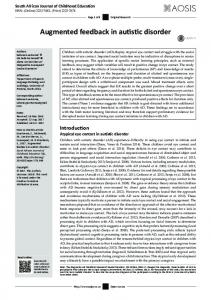
Method Participants. Forty-eight first- through thirdyear medical students at the University of Iowa, a population similar to that used in previous studies {Coleman, Coon. & Fitzgerald. 2001). participated in the experiment, which followed a protocol approved by our institutional review board. The 50 women and 18 men were between 22 and 40 years old with a mean age of 25. Using variance estimates from a pilot study, we selected this sample size so that a betweensubject difference of one lump detected could be tested at the 5% confidence level. Apparatus. Three breast models were used in the experiment: the dynamic model, the Mammaearc firm model (CPM-E). and the Mammacare soft model (CPM-S; Mammatech Corp.. Gainesville, FL). Participants trained with either the CPM-E model or the dynamic model. The dynamic breast model (Figure 1) is a breast-shaped silieone matrix embedded with a scries of handmade balloons that, when pres-
673 surized with water, create simulated lumps in the breast. The hardness of each lump may be independently controlled, including dellating the balloon entirely, which makes the lump undetectable. The silieone matrix is opaque and has a hard silieone backing with embedded ribs and entry points of thin tubes leading to the balloons. The balloons and tubes are constructed by heat fusing a pair of polyethylene sheets. The silieone matrix is made with a general purpose, highstrength, tin-based silieone polymer (B|B Enterprises TC-5005 with 8 5 % cross-linker). It is fairly homogeneous with little nodularity. The silieone fomiula was selected so that its stress/ strain properties were similar to the properties of live breast tissue reported by Krouskop. Wheeler, Kallcl. Garra. and Hall (1998) and Gerling. Thomas, and Weissman (2003). Simulated ribs fabricated with a ceramic-like sculpturing compound. Super Sculpey. He below the silieone matrix. Below these, a hard silieone backing made of a translucent, platinum-based silieone rubber (B|B Enterprises TC-5030) supports the structure and simulates the inteirib muscle structures. The dynamic breast model includes 15 lumps that may be individually inflated to different hardness (Table 1). The inflated lumps vary in size (0.3-1.25 cm), depth of placement (shallow, medium, and deep), fixedness (fixed and mobile), and hardness (20-50 durometers). The lump sizes of 0.3 cm (4 lumps), 0.5 cm (4 lumps), 1.0 cm (5 lumps), and 1.25 cm (2 lumps) are similar to those in other CBE palpation training devices (Bennett et al. 1990; Bloom et al., 1982;
Figure I. Breast model comparison, static: (left) and dynamic (right), with a 12-inch (30.48-cm) ruler
674
Fall 2005 - Human Factors
TABLE 1: Lump Characteristics in the Dynamic and Static Breast Models Overall Comparison
Lump ID
Size (cm)
Hardness (Durometers, Shore A)
Mobility
Depth
Fixed Fixed Fixed Fixed Mobile Mobile Fixed Mobile Fixed Mobile Mobile Mobile Fixed Mobile Fixed
Medium Deep Deep Deep Medium Medium Deep Medium Deep Shallow Medium Shallow Deep Shallow Deep
Fixed Mobile Mobile Mobile Fixed
Deep Medium Deep Deep Deep
Fixed Fixed Mobile Mobile Fixed
Deep Shallow Deep Medium Deep
Dynamic Model Weight: 830.8 g Hardness: 3 dur Dimensions L19cm W 11.5 cm H 9 cm Nodularity: no
2b
3 4 5^ 6
7' 8 9 10^ 11^ 12 13^ 14b 15"
1.25 1.25 1.0 1.0 1.0 1.0 1.0
0.5 0.5 0.5
0.5 0.3
0.3 0.3 0.3
0-45 0-45 0-45 0-45 0-45 0-45 0-45 0-45 0-45 0-45 0-45 0-45 0-45 0-45 0-45
(40) (40)
(35, 30) (40) (40) (40) (40) (35) (35)
CPM-F Models Weight: 298.3 g Hardness: 2 dur Dimensions L, W 13 cm H 3.5 cm Nodularity: yes
1 2
3 4 5
40 60 60 60 40
1.0
0.3 0.5 0.7 1.0
CPM-S Model Weight: 298.0 g Hardness: 1 dur Dimensions L, W 1 3 c m H 3.5 cm Nodularity: yes
1 2
1.0 0.3
3 4 5
0.5 0.7 1.0
40 60 60 60 40
^Pretest and training; posttest;
Campbell et al.. 1991; Pilgrim ct al.. 1993). The exact lump size varies with water pressure; however, the range of inflation pressures used increased the physical dimensions by less than 5%, as measured along the direction perpendicular to each lump's major axis (Gerling, 2001). These variations are small relative to the variance in clinical tumor size estimation. The depth of lump placement ranges from just under the outer silicone surface to between the ribs. Lumps arranged along the back wall of the dynamic model are fixed, whereas lumps farther out into the silicone matrix are more mobile. Cancerous tumors have a hardness of between 0 and 60 durometers (Bloom et al., 1982); the
dynamic model lumps have a hardness of between 0 and 45 durometers. Lump hardness varies linearly with water pressure over the range of interest [R- values ranged between .832 and .998); the exact relationship depends on lump size. A regression of water pressure (in pounds per square inch, or psi) versus hardness (Shore A durometer scale) provides separate slopes and offsets for each lump size (Gerling, 2001). Large lumps need less pressure to reach high hardness values than do small lumps. An external pressure system delivers between 20 and 45 psi (137.9-310.26 kPa) to lumps selected by opening and closing valves. Once the lump is inflated, its pressure is maintained by closing
PULSATING
FEEDBACK AIDS TACTILE TRAINING
its valve. A relief valve protects any balloons from experiencing excessive water pressures. The two static breast models used as experimental controls (Mammatech Corp.. CPM-S and CPM-F) have nearly hemispherical shape, are made of soft silieone with a tough skin, and have a flexible, square backing (Figure I; Gerling et al., 2003). F.ach static model contains five lumps made of fibrous cotton wound tightly into a cylinder. The lumps vary in size (0.3-1.0 cm), hardness (20. 40. and 60 durometers). depth of placement (shallow, medium, and deep), and mobility (fixed and mobile). Lump size, hardness, position, and depth are fixed and equal in both models. The models simulate a low amount of glandular nodularity with a slightly lumpy silieone surface underneath the skin and small air pockets within the silieone matrix. Table I summarizes the physical differences among the CPM-S, CPM-F, and dynamic breast models. To match the five static lumps in the CPM-F model, five lumps of similar size, hardness, and depth were used in the dynamie breast model for the pretest, posttest. and training. The specific lumps used are indicated in Table 1. Experimental design. The repeated-measures experiment included two pretests, with both the dynamic and CPM-F models; a training session; a break; and three posttests. with the dynamie and CPM-F models, followed by the CPM-S model. The 48 participants were randomly assigned to eight experimental cohorts (A1-A4 and B1-B4) balanced by gender and year in medical school, factors that reflect prior opportunities to praetice breast examination skills. Cohorts A| through A4 trained with the CPM-F model, whereas Cohorts B] through B4 trained with the dynamic model. The subscripts indicate each variation of the four possible orderings of the pretests and posttests. For example. A, indicates pretesting with the CPM-F followed by the dynamie model, training with the CPM-F. postesting with the dynamic model followed by CPM-F, and then testing with the CPM-S. The between-subjects independent variables were the training device (static or dynamie). the order of the pretest (statie then dynamic or dynamie then statie). and the order of the posttest (statie then dynamic or dynamic then static). Six within-subject dependent variables were defined: static model detection improvement.
675 dynamie model deteetion improvement, composite detection improvement, dynamic model false positive improvement, composite false positive improvement, and intersimulator skill transfer. Static and dynamic detection improvement is measured by the number of lumps found after training minus the number of lumps found on the same device before training (two measures per participant, one for CPM-F and one for the dynamie model training). Composite detection improvement is the sum of the static and dynamie detection improvement seores for eaeh participant. Composite false positive improvement was seored as "worse" if the number of combined false positives in the dynamic and static posttests was larger than the combined pretest false positives; as "same" if the posttest and pretest false positive sums were equal; as "better" if the posttest sum was smaller; and as "NA" if there were no false positives in any of the first four tests. Intersimulator skill transfer is the within-model improvement for dynamic model tests when training with the CPM-F model or the within-model improvement for CPM-F model tests when training with the dynamie model. Procedure. During the pretest and posttest sessions, each participant was provided a 2-min interval to examine each breast model, consistent with the typical time spent in a breast examination (Campbell et al.. 1991; Fletcher et al., 1985). For the dynamic model, all five lumps were simultaneously inflated and the water pressure was kept constant during the test. The participant reported the presence or absenee of identified lumps to the research assistant. The location of each lump discovered by the participant was recorded on a diagram. Immediately following the participant's set of tests, the diagrams were scored. A trial was scored as correct if the participant noted the lump's presence in the correct position, as a miss if the participant failed to note the presence of the lump, or as a false positive if the partieipant claimed to detect a lump where no balloon had been inflated. Neither tumor size nor depth eonsisteney was scored. The 15-min training session for all breast models covered search pattern, finger pressure, part and number of fingers used, finger motion. nodularity effects, breast area coverage, and
Fall 2005 - Human Factors
676 lump properties on either the dynamic or CPM-F model. The research assistant provided training according to detailed, written inslructions. Of the five lumps available, three lumps of different sizes were used for the training practice, starting with the largest and moving to the smallest. With the dynamic breast model, lumps could be tumed on and off and pressure could be oscillated while the participant applied fmger pressure. If the participant reported difficulty in detecting the stimulus, the water pressure was oscillated until the participant reported that he or she could detect the stimulus. Water oscillation was necessary in nearly all cases for the middle- and small-sized tumors. Oscillation was induced for approximately 10-s periods while the trainee palpated the model. Up to about five such periods would occur for each lump as necessary or until the trainee found the lump. Once the lumps were located, the assistant could covertly inflate or deflate the tumor to retest and validate the participant s ability to perceive the stimulus. With the static model, participants alternately palpated areas with and without lumps, for lump and no-lump conditions. After the 20-min rest, posttraining scores were gathered on each of the two models and the third model (CPM-S) following pretest instructions. Before the posttest. the CPM-F mode! was rotated 90° to change the positions of the lumps. Between experimental stages, the static models were removed from the trainee's sight and placed in a box with other static models to
create the illusion that multiple static models were employed. Results
Preliminaiy analysis of variance {ANOVA) of the six dependent variables showed no significant difference for pretest and posttest training order, so Cohorts A| through A4 and B, through B4 were collapsed in the final reported analysis. Table 2 provides a summary of the significant results for the six dependent variables. Training had a significant effect on composite detection improvement, f (1. 47) = 9.54. p = .004. Training with the dynamic model resulted in an average composite detection improvement of 1.35 lumps (SD = 0.92), as compared with 0.60 lumps iSD - 0.96) for the static model (Figure 2). Training also had a significant effect on the number of lumps detected in the CPM-S posttest. /-(1, 47) = 2.94, p = .093, with an average of 3.04 lumps found (SD ^ 1.12) after dynamic training, as compared with 2.54 {SD = 0.88) after static training. The dynamic model training improved lump detection on both the dynamic and static models, whereas static model training improved lump detection perfonnance only for the static model {Figure 3). Both types of training increased the number of lumps detected on the static training device with approximately the same effectiveness: 1.04 (SD = 1.4) and 1.17 (SD = 1.09) for the static and dynamic models, respectively. F( 1. 47) = 0.12, p = .731. Only the dynamic model
TABLE 2: Summary of Results for the Six Dependent Variables
Composite detection improvement Lumps detected in CPM-S posttest Static model detection improvement Dynamic model detection improvement Composite false positive improvement False positives in CPM-S posttest *p